ABSTRACT
ORIGINAL AR
TICLE
Juliane Garcez Musacchio
Maria da Glória da Costa Carvalho
José Carlos Oliveira de Morais
Nathalie Henriques Silva
Adriana Scheliga
Sérgio Romano
Nelson Spector
Detection of free circulating
Epstein-Barr virus DNA in plasma
of patients with Hodgkin’s disease
Hematology Service, Universidade Federal do Rio de Janeiro and Oncology
Service, Instituto Nacional do Câncer, Rio de Janeiro, Brazil
CONTEXT AND OBJECTIVE: Free circulating Ep-stein-Barr virus (EBV) DNA is often present in the plasma of Hodgkin’s disease patients. The aim here was to evaluate the prevalence of this fi nd-ing, its correlation with the immunohistochemical expression of LMP-1 (latent membrane protein 1) and the infl uence of other clinical factors. DESIGN AND SETTING: Prospective study in two public tertiary institutions: Hematology Service, Universidade Federal do Rio de Janeiro, and Oncology Service, Instituto Nacional do Câncer, Rio de Janeiro.
METHODS: A cohort of 30 patients with newly diagnosed Hodgkin’s disease was studied. The control group consisted of 13 healthy adult volun-teers. EBV DNA was determined by conventional polymerase chain reaction (PCR).
RESULTS: The median age was 28 years, and 16 patients were women. Advanced disease was present in 19 patients, and six were HIV-positive. EBV DNA was present in the plasma of 13 patients and one control (43% versus 8%, p = 0.03). EBV DNA prevalence was higher in HIV-posi-tive patients (100% versus 29%, p = 0.0007) and those with advanced disease (63% versus 9%, p = 0.006). Among HIV-negative patients alone, EBV DNA prevalence remained higher in those with advanced disease. EBV DNA was found in 10/11 patients with LMP-1 expression in the lymph nodes, and in 3/19 without LMP-1 expression (kappa coeffi cient = 0.72). CONCLUSION: EBV DNA was present in 91% of patients with EBV-associated Hodgkin’s disease, and in all patients with HIV-associated Hodgkin’s disease. EBV DNA prevalence was higher in patients with advanced disease, irrespective of HIV status.
KEY WORDS: DNA. Epstein-Barr virus. Hodgkin disease. Lymphoma. Polymerase chain reaction.
INTRODUCTION Epstein-Barr virus (EBV), a lymphotropic herpesvirus that is widespread in the human population, is associated with approximately 40% of Hodgkin’s disease cases. Its prevalence is higher in developing countries, where a higher proportion of Hodgkin’s disease cases are subclassifi ed as mixed cellularity.1
The demonstration that EBV genomes are present in the Hodgkin-Reed Sternberg (H-RS) cells of a proportion of Hodgkin’s disease cases provided important new evidence of its role in the pathogenesis of the disease.2 In patients with EBV-associated Hodgkin’s disease, H-RS cells express high levels of latent membrane protein 1 (LMP-1), which functions as a constitutively activated tumor necrosis factor receptor. Many of the growth transforming effects of LMP-1 derive from its ability to activate various signaling pathways, including nuclear factor κB.3
Small amounts of free EBV DNA circu-late in the plasma of both healthy individu-als and cancer patients.4 Some studies have reported the presence of EBV DNA in the peripheral blood of patients with EBV-as-sociated malignancies, such as nasopharyn-geal carcinoma, NK/T cell lymphoma and post-transplant lymphoproliferative disease (PTLD),4-8 but there is still only limited experience of EBV in Hodgkin’s disease. Gal-lagher et al. reported that the conventional polymerase chain reaction (PCR) detected 90% of known EBV-positive Hodgkin’s dis-ease cases.9 Quantitative PCR appeared to be less sensitive but more specifi c. In two other studies, a reduction in plasma viral load to low or undetectable levels was observed in patients who obtained complete remission, whereas in poorly responding patients, disease progres-sion was associated with rapidly increasing DNA levels.10,11 Thus, analysis of plasma EBV DNA might be of value for diagnostic
and prognostic evaluation and follow-up of Hodgkin’s disease patients.
OBJECTIVE
The aim of this study was to confi rm whether free circulating EBV DNA can be consistently detected in patients with EBV-associated Hodgkin’s disease, and to verify possible associations with such patients’ clini-cal characteristics.
METHODS
Patients
Patients
Between November 2001 and November 2003, plasma samples from 30 consecutive pa-tients with newly diagnosed Hodgkin’s disease were collected before their treatment started at the Hematology Service of Universidade Federal do Rio de Janeiro and the Oncology Service of Instituto Nacional do Câncer (two public tertiary institutions). Their diagnoses and histological classifi cations were confi rmed in a pathological review performed by two of the authors together (SR and JCM). The pres-ence of EBV in tumor cells was assessed by the immunohistochemical expression of the LMP-1 protein, using a cocktail of monoclonal antibod-ies (CS1-4, Dako®), as described previously.12
Patient evaluation included a complete history, physical examination, complete blood count with differential count, biochemical profi le, HIV serology, chest radiography, com-puted tomography of the chest and abdomen, and bone marrow biopsy. Blood samples were collected for EBV DNA analysis.
Blood samples from 13 healthy volunteers were also analyzed. They were all health work-ers at our hospital, their median age was 29 years, and seven were women.
The study was approved by the Institutional Review Boards of the two institutions. Informed consent was obtained from all patients.
155
DNA extraction from
DNA extraction from plasma samples plasma samples
DNA extraction was performed using the QIAamp Blood Kit (Qiagen, Uniscience, Bra-zil). The steps were performed according to the supplier’s protocol and samples were resuspended in 200 µl H2O. DNA was stored at – 80º C.
Conventional polymerase
Conventional polymerase chain reaction
chain reaction
Initially, EBV DNA polymerase chain reaction (PCR) was carried out using prim-ers derived from the BamH1 W repeat sequence of EBV, as previously described.13 The PCR routine was performed using 2 µl of the DNA extracted from plasma. PCR reactions were performed with 2.5 units/µl Amplitaq (Perkin-Elmer, Branchburg, New Jersey), 1.5 mM MgCl2, 25 mM dNTPs, 10 x buffer and 10 µM of each primer, in a 50 µl reaction mix. A negative water control and a positive control consisting of DNA from the EBV-positive Raji cell line were included in each assay. Thermal cycling was performed using the following conditions: initial dena-turation at 95º C for fi ve minutes, followed by 40 cycles of ramping to 94º C over one min; 94º C for 30 sec; cooling to 55º C over two min; 55º C for 10 sec; heating to 72º C over
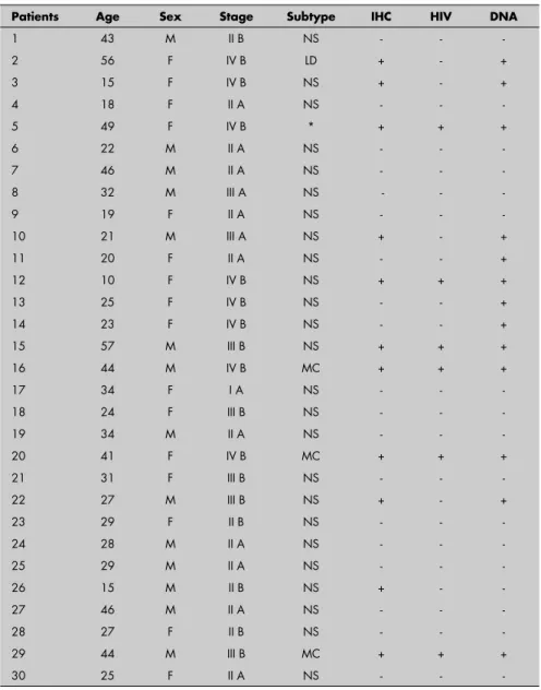
Table 1. Clinical characteristics and plasma Epstein-Barr virus (EBV) DNA of 30 patients with Hodgkin’s disease
Characteristics n (%)
Median age 28
Range 10-57 years
Sex
Female 16 (53)
Male 14 (47)
Stage (Ann Arbor method)*
Localized 11 (37)
Disseminated 19 (63)
HIV-positive Status 6 (20)
Histopathology subtype†
Nodular sclerosis 25 (86)
Mixed cellularity 3 (11)
Lymphocytic depletion 1 (3) LMP-1 protein
Positivity 11 (37)
Plasma EBV DNA
Detectable 13 (43)
EBV = Epstein-Barr virus.*Localized disease: stages IA, IB and IIA; disseminated disease: stages IIB, IIIA, IIIB, IVA and IVB; †One patient unclassifi ed; diagnosis from bone
marrow specimen.
Table 2. Clinical characteristics and plasma Epstein-Barr virus (EBV) DNA of each patient with Hodgkin’s disease
Patients Age Sex Stage Subtype IHC HIV DNA
1 43 M II B NS - -
-2 56 F IV B LD + - +
3 15 F IV B NS + - +
4 18 F II A NS - -
-5 49 F IV B * + + +
6 22 M II A NS - -
-7 46 M II A NS - -
-8 32 M III A NS - -
-9 19 F II A NS - -
-10 21 M III A NS + - +
11 20 F II A NS - - +
12 10 F IV B NS + + +
13 25 F IV B NS - - +
14 23 F IV B NS - - +
15 57 M III B NS + + +
16 44 M IV B MC + + +
17 34 F I A NS - -
-18 24 F III B NS - -
-19 34 M II A NS - -
-20 41 F IV B MC + + +
21 31 F III B NS - -
-22 27 M III B NS + - +
23 29 F II B NS - -
-24 28 M II A NS - -
-25 29 M II A NS - -
-26 15 M II B NS + -
-27 46 M II A NS - -
-28 27 F II B NS - -
-29 44 M III B MC + + +
30 25 F II A NS - -
-IHC = imunohistochemistry (for LMP-1); HIV = status of human immunodefi ciency virus; F = female; M = male; NS = nodular sclerosis; LD = lymphocytic depletion; MC = mixed cellularity; *diagnosis in bone marrow specimen.
one min; 72º C for 30 sec; followed by a fi nal extension step at 72º C for seven min.9 PCR products were analyzed by electrophoresis on 10% polyacrylamide gels. All plasma DNA samples were also subjected to PCR analysis for the p53 gene, which served as a control for the ability to amplify the plasma DNA.
Statistical analysis
Statistical analysis
Fisher’s exact test (two-sided) was used to as-sess the association between categorical variables. The kappa coeffi cient was calculated to estimate the concordance between EBV detection meth-ods. Statistical analysis was performed using the softwares Epi-Info 6.0 and PEPI 4.0.
RESULTS The patients’ characteristics, EBV status in tumor cells and pre-treatment plasma EBV DNA are summarized in Tables 1 and 2. Tissue samples were available from all 30 patients for evaluating their EBV status.
Prior to treatment, plasma EBV DNA was detected in 13 of the 30 patients (43%). In contrast, plasma EBV DNA was detected in only one of the 13 control subjects (p = 0.03; 95% CI for the difference between propor-tions = 0.12-0.58).
Circulating EBV DNA was found in 10 of the 11 LMP-1 positive patients, and in only three of the 19 patients without LMP-1
156
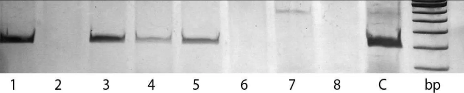
expression in the lymph nodes (kappa coef-fi cient = 0.72). Figure 1 shows the presence of EBV DNA in the serum of four patients.
According to the Ann Arbor staging method, advanced disease (defi ned as stages IIB-IV) was present in 19 patients and the frequency of EBV DNA was higher in this group (63% versus 9%, p = 0.006, 95% CI = 0.26-0.81). Plasma EBV DNA was found in all HIV-positive patients (100% versus 29%, p = 0.0007). When only the 24 HIV-negative patients were analyzed, the frequency of EBV DNA remained higher in the 13 patients with advanced disease (54% versus 9%, p = 0.03)
DISCUSSION The presence of tumor-derived DNA in the plasma and serum of cancer patients opens up new possibilities for detecting and monitoring cancer.
In the present study, 43% of the patients with Hodgkin’s disease had EBV DNA that was detectable by conventional PCR in the plasma prior to treatment, whereas only one healthy individual (8%) was positive for plasma EBV DNA (p = 0.03). Moreover, plasma EBV DNA was present in almost every patient (10/11, 91%) with proven LMP-1 in the lymph nodes, and also in 3/19 patients (16%) without LMP-1. Whether the latter were false-positive results from the PCR or false-negative results from the LMP-1 stain-ing method remains to be elucidated. In the present study, EBV encoded RNA (EBER) in situ hybridization was not performed, but the correlation with LMP-1 was usually good.14
Gallagher et al.9 observed similar results from patients selected retrospectively on the basis of their known EBV status: 91% of their patients with EBV-associated disease were positive by conventional PCR, while 26% of patients with non-EBV-associated Hodgkin’s disease were positive. Subsequently,
a subgroup of their patients was also studied by quantitative PCR. With this method, only 69% of the patients with EBV-associated disease and 10% of the patients with non-EBV associated Hodgkin’s disease were posi-tive. The authors speculated that this lower positivity might have been due to technical reasons, such as the smaller amount of DNA used in RT-PCR. However, as expected, the median amount of EBV DNA detected by RT-PCR was much higher in patients with EBV-associated Hodgkin’s disease.
The good correlation between the EBV status of the involved lymph nodes examined and the ability to detect EBV DNA in plasma suggests that H-RS cells are the most likely source of viral DNA. Support for this idea is provided by Kornacker et al.,15 who dem-onstrated identical immunoglobulin gene rearrangements in biopsy and blood samples from a patient with Hodgkin’s disease.
A strong correlation between plasma EBV DNA and HIV status was observed. Virtually all cases of Hodgkin’s disease in HIV-infected patients are EBV-associated,16,17 and in our series the prevalence of free EBV DNA was 100%. Free circulating EBV DNA could have clinical use in patients with HIV-related Hodgkin’s disease, both as an indicator of complete remission and as a tool for following up patients who are in complete remission.18
A strong correlation was also observed between the presence of free circulating EBV DNA and the presence of advanced Hodgkin’s disease. A similar correlation has been observed in patients with nasopharyn-geal carcinoma: plasma cell-free EBV DNA levels in advanced cases were signifi cantly higher than those in early-stage nasopha-ryngeal cases.19 These results again suggest that free EBV DNA is related to the tumor mass, and that its analysis might be useful as a molecular marker for the detection and follow-up of EBV-related diseases.
The control group had the same median age and sex distribution as the patients with Hodgkin’s disease. The clinical signifi cance of the presence of plasma EBV DNA in one healthy control is unclear. Such positive findings in control groups have been ob-served before in developing countries, with a prevalence of 7%-13%.19,20 However, in develo-ped countries, EBV DNA has never been detected in the plasma or serum of healthy, non-immunosuppressed children or adults.21 Therefore, this discrepancy might be related to yet undefi ned socioeconomic conditions.
In studies using large DNA banks, Hodgkin’s disease has been shown to de-velop as late as seven years after free EBV DNA was detected.22 For this reason, the only control volunteer with a positive result in our study is being monitored for clinical signs of EBV-associated diseases.
It is of interest that, in one study that detected plasma EBV DNA in controls, the levels of plasma EBV DNA in patients with nasopharyngeal carcinoma were much higher than those in controls with detect-able plasma EBV DNA.6 Thus, quantitative plasma EBV DNA might prove more useful as a screening method for EBV-associated diseases in high-prevalence areas.
CONCLUSIONS In summary, free circulating EBV DNA was frequently found in Hodgkin’s disease patients before treatment by conventional PCR, and its prevalence was especially high in patients with advanced-stage disease and HIV-related Hodgkin’s disease. Further lon-gitudinal studies are clearly needed in order to evaluate the diagnostic impact of this fi nding on the monitoring of treatment re-sponse and remission status among Hodgkin’s disease patients. The use of quantitative PCR in a larger population is likely to shed light on many of the questions raised by this study.
Figure 1. Detection of Epstein-Barr virus (EBV) DNA by polymerase chain reaction (PCR); electrophoresis on 10% polyacrylamide gels in buffer TBE 1x; bp: DNA size marker; C: positive control, DNA from Raji cell line. Samples were from: EBV-associated Hodgkin’s Disease, lanes 1, 3, 4 and 5; non-EBV-associated Hodgkin’s Disease, lanes 2 and 7; healthy individuals, lanes 6 and 8.
157
1. Glaser SL, Lin RJ, Stewart SL, et al. Epstein-Barr virus-associated Hodgkin’s disease: epidemiologic characteristics in international data. Int J Cancer. 1999;70(4):375-82.
2. Weiss LM, Strickler JG, Warnke R, Purtilo DT, Sklar J. Epstein-Barr viral DNA in tissues of Hodgkin’s disease. Am J Pathol. 1987;129(1):86-91.
3. Huen DS, Henderson SA, Croom-Carter D, Rowe M. The Epstein-Barr virus latent membrane protein-1 (LMP-1) mediates activation of NF-kappa B and cell surface phenotype via two effector regions in its carboxy-terminal cytoplasmic domain. Oncogene. 1995;10(3):549-60.
4. Anker P, Mulcahy H, Chen XQ, Stroun M. Detection of cir-culating tumour DNA in the blood (plasma/serum) of cancer patients. Cancer Metastasis Rev. 1999;18(1):65-73. 5. Yamamoto M, Kimura H, Hironaka T, et al. Detection and
quantification of virus DNA in plasma of patients with Epstein-Barr virus-associated disease. J Clin Microbiol. 1995;33(7):1765-8.
6. Lo YM, Chan LY, Chan AT, et al. Quantitative and temporal correlation between circulating cell-free Epstein-Barr virus DNA and tumor recurrence in nasopharyngeal carcinoma. Cancer Res. 1999;59(21):5452-5.
7. Lei KI, Chan LY, Chan WY, Johnson PJ, Lo YM. Diagnostic and prognostic implications of circulating cell-free Epstein-Barr virus DNA in natural killer/T-cell lymphoma. Clin Cancer Res. 2002;8(1):29-34.
8. Limaye AP, Huang ML, Atienza EE, Ferrenberg JM, Corey L. Detection of Epstein-Barr virus DNA in sera from transplant recipients with lymphoproliferative disorders. J Clin Microbiol. 1999;37(4):1113-6.
9. Gallagher A, Armstrong AA, MacKenzie J, et al. Detection of Epstein-Barr virus (EBV) genomes in the serum of pa-tients with EBV-associated Hodgkin’s disease. Int J Cancer. 1999;84(4):442-8.
10. Lei KI, Chan LY, Chan WY, Johnson PJ, Lo YM. Quantitative analysis of circulating cell-free Epstein-Barr virus (EBV) DNA levels in patients with EBV-associated lymphoid malignancies. Br J Haematol. 2000;111(1):239-46.
11. Wagner HJ, Schlager F, Claviez A, Bucsky P. Detection of Ep-stein-Barr virus DNA in peripheral blood of paediatric patients with Hodgkin’s disease by real-time polymerase chain reaction. Eur J Cancer. 2001;37(15):1853-7.
12. Rowe M, Evans HS, Young LS, Hennessy K, Kieff E, Rickinson AB.. Monoclonal antibodies to the latent membrane protein of Epstein-Barr virus reveal heterogeneity of the protein and inducible expression in virus-transformed cells. J Gen Virol. 1987;68(Pt 6):1575-86.
13. Deacon EM, Matthews JB, Potts AJ, Hamburger J, Bevan IS, Young LS. Detection of Epstein-Barr virus antigens and DNA in major and minor salivary glands using immunocytochem-istry and polymerase chain reaction: possible relationship with Sjogren’s syndrome. J Pathol. 1991;163(4):351-60. 14. Gulley ML, Glaser SL, Craig FE, et al. Guidelines for
interpret-ing EBER in situ hybridization and LMP1 immunohistochemi-cal tests for detecting Epstein-Barr virus in Hodgkin lymphoma. Am J Clin Pathol. 2002;117(2):259-67.
15. Kornacker M, Jox A, Vockerodt M, et al. Detection of a Hodgkin/Reed-Sternberg cell specifi c immunoglobulin gene rearrangement in the serum DNA of a patient with Hodgkin’s disease. Br J Haematol. 1999;106(2):528-31.
16. Audouin J, Diebold J, Pallensen G. Frequent expression of Epstein-Barr virus latent membrane protein-1 in tumour cells of Hodgkin’s disease in HIV-positive patients. J Pathol. 1992;167(4):381-4. 17. Herndier BG, Sanchez HC, Chang KL, Chen YY, Weiss LM.
High prevalence of Epstein-Barr virus in Reed-Sternberg cells of HIV-associated Hodgkin’s disease. Am J Pathol. 1993;142(4):1073-9.
18. Fan H, Kim SC, Chima CO. Epstein-Barr viral load as a marker of lymphoma in AIDS patients. J Med Virol. 2005;75(1):59-69. 19. Lo YM, Chan LY, Lo KW, et al. Quantitative analysis of cell-free
Epstein-Barr virus DNA in plasma of patients with nasopha-ryngeal carcinoma. Cancer Res. 1999;59(6):1188-91. 20. Shotelersuk K, Khorprasert C, Sakdikul S, Pornthanakasem
W, Voravud N, Mutirangura A. Epstein-Barr virus DNA in serum/plasma as a tumor marker for nasopharyngeal cancer. Clin Cancer Res. 2000;6(3):1046-51.
21. Wagner HJ, Wessel M, Jabs W, et al. Patients at risk for development of posttransplant lymphoproliferative disorder: plasma versus peripheral blood mononuclear cells as material for quantifi cation of Epstein-Barr viral load using real-time quantitative polymerase chain reaction. Transplantation. 2001;72(6):1012-9.
22. Lin L, Yang J, Levin L, et al. Viral DNA in serum and plasma of patient with Epstein-Barr (EBV)-associated Hodgkin’s lymphoma may be tumor derived and appears years before diagnosis. In: 44th Annual Meeting of the American Society of Hematology; 2002 Dec 6-10. Philadelphia: American Society of Hematology; 2002. [abstract 753].
Acknowledgments: We thank Marcelo S. M. Silva for excellent laboratory assistance.
Place where the work was presented: The results of this paper were partially presented at the International Symposium on Hodgkin’s Lymphoma, Cologne, Germany, in September 2004.
Sources of funding: Financial support was provided by Conselho Nacional de Desenvolvimento Científi co e Tecnológico (CNPq) 301.346/1996-8 and Funda-ção de Apoio à Pesquisa do Rio de Janeiro (Faperj) 26/152.021/2004.
Confl icts of interest: Not declared Date of fi rst submission: July 29, 2005 Last received: May 17, 2006 Accepted: May 29, 2006
REFERENCES
AUTHOR INFORMATION
Juliane Garcez Musacchio, MD, PhD. Hematology Service, Faculdade de Medicina and Hospital Universitário, Univer-sidade Federal do Rio de Janeiro, Rio de Janeiro, Brazil.
Maria da Glória da Costa Carvalho, MD, PhD. Biophysics Institute, Universidade Federal do Rio de Janeiro, Rio de Janeiro, Brazil.
José Carlos Oliveira de Morais, MD, PhD. Pathology Service, Faculdade de Medicina and Hospital Universitário, Univer-sidade Federal do Rio de Janeiro, Rio de Janeiro, Brazil.
Nathalie Henriques Silva. MD, PhD. Biophysics Institute, Universidade Federal do Rio de Janeiro, Rio de Janeiro, Brazil.
Adriana Scheliga, MD. Oncology Service, Instituto Nacional do Câncer, Rio de Janeiro, Brazil.
Sérgio Romano, MD. Pathology Service, Instituto Nacional do Câncer, Rio de Janeiro, Brazil.
Nelson Spector, MD, PhD. Hematology Service, Faculdade de Medicina and Hospital Universitário, Universidade Federal do Rio de Janeiro, Rio de Janeiro, Brazil.
Address for correspondence:
Juliane Garcez Musacchio
Rua Senador Vergueiro, 157/703 — Flamengo Rio de Janeiro (RJ) — Brasil — CEP 22230-001 Tel. (+55 21) 2553-5364
E-mail: [email protected]
Copyright © 2006, Associação Paulista de Medicina
RESUMO Detecção do DNA livre circulante do vírus Epstein-Barr no plasma de pacientes com Doença de Hodgkin CONTEXTO E OBJETIVO: O DNA do vírus Epstein-Barr (EBV) está freqüentemente presente no sangue periférico de pacientes com doença de Hodgkin. O objetivo deste estudo foi avaliar a prevalência deste achado, e correlacioná-lo com a expressão imunoistoquímica da LMP-1 (latent membrane protein 1) e a presença de outros fatores clínicos.
TIPO DE ESTUDO E LOCAL: Estudo prospectivo realizado no Serviço de Hematologia da Universidade Federal do Rio de Janeiro e no Serviço de Oncologia do Instituto Nacional do Câncer, Rio de Janeiro, Brasil. MÉTODOS: Trinta pacientes com doença de Hodgkin recém-diagnosticada foram estudados, assim como um grupo controle composto por 13 indivíduos saudáveis. O DNA do EBV no plasma foi determinado pela reação em cadeia da polimerase (PCR) convencional.
RESULTADOS: A idade mediana foi 28 anos e 16 pacientes eram do sexo feminino. A doença dissemi-nada esteve presente em 19 pacientes e seis eram HIV+. O DNA do EBV foi detectado no plasma de 13 pacientes e um controle (43% versus 8%, p = 0,03). A prevalência do DNA do EBV foi maior nos pacientes HIV+ (100% versus 29%, p = 0,0007) e naqueles com doença disseminada (63% versus 9%, p = 0,006). Quando somente os pacientes HIV-negativos foram analisados, a prevalência do DNA do EBV permaneceu maior nos pacientes com doença disseminada. A prevalência do DNA do EBV variou de acordo com o subtipo histológico: foi de 32% nos pacientes com esclerose nodular e de 100% nos pacientes com celularidade mista e depleção linfocítica (p = 0,02). O DNA do EBV foi encontrado em 10/11 pacientes com a expressão da LMP-1 em linfonodos, e em 3/19 pacientes sem a expressão da LMP-1 (coefi ciente de kappa = 0,72).
CONCLUSÕES: O DNA circulante do EBV foi detectado no plasma de 91% dos pacientes com doença de Hodgkin associada ao EBV, e em todos os pacientes com doença de Hodgkin associada ao HIV. A prevalência do DNA circulante do EBV foi maior nos pacientes com doença avançada, independentemente do status para o HIV.
PALAVRAS-CHAVE: DNA. Vírus Epstein-Barr. Doença de Hodgkin. Linfoma. Reação em cadeia da polimerase.