ABSTRACT
C
A
SE REPOR
T
INTRODUCTION Papillary squamous cell carcinoma is an entity characterized by highly atypical squa-mous cell proliferation. Stromal invasion, however, is rarely seen.1,2The evaluation of an exophytic or papil-lary lesion should always include differential diagnosis in relation to other lesions present-ing the same growth pattern, such as verrucous carcinoma, but without the high count of atypical cells found in papillary squamous cell carcinoma. Squamous papillomatosis may present with a high count of atypical cells,1 but
also with a high count of koilocytic cells,1
and should be differentiated from papillary squamous cell carcinoma. The importance of the differential diagnosis lies in the different prognoses for these lesions.
Recent studies have demonstrated better prognosis for papillary squamous cell carci-noma with a papillary pattern than for papillary squamous cell carcinoma with the exophytic pattern or for conventional squamous cell carcinoma.2 On this basis, a modifi cation to
the therapy for this kind of tumor has been put forward, with regard to its aggressiveness.2
In this report, the authors present a case of papillary squamous cell carcinoma with papil-lary patterns that had an unusual malignant evolution, for which radical surgery, adjuvant chemotherapy and radiotherapy were used in the treatment.
Case report A 42-year-old white female was referred to our clinic with a complaint of dysphonia and pain in the right cervical area. The patient had no complaints of dyspnea, dysphagia or odinophagia. She had no previous history of tobacco or alcohol use, and no symptoms related to gastroesophageal refl ux.
Fiberoptic examination of the upper airway was carried out and this showed an Adriano Santana Fonseca
Carlos Takihiro Chone Agrício Nubiato Crespo Albina Altemani
CONTEXT: According to the literature, laryn-geal papillary carcinoma is rare and has a benign prognosis.
CASE REPORT: In this report we present a sur-prising case with nodal metastasis at the time of diagnosis. Computed tomography showed infi ltration of the lesion and metastatic lymph nodes. The resected specimen was submitted to histopathological study that confi rmed the diag-nosis of papillary squamous cell carcinoma. KEY WORDS: Laryngeal neoplasms. Papillary carcinoma. Radiotherapy. Prognosis. Lym-phatic metastasis.
exophytic verrucous lesion occupying the pos-terior third of the left vocal and vestibular fold. It had a submucous and infi ltrative pattern, and there was decreased mobility of the left vocal fold. A lymphatic metastasis of 5.0 cm was found at the right level IV, featuring hard consistency and adherence to the deep tissues. The lesion was clinically staged as laryngeal, cT2N2cM0. Biopsy was performed on the posterior laryngeal area and fi ne-needle as-piration on the enlarged cervical mass. The specimen was also sent for hybridization in situ and electron microscopy.
The histopathological fi ndings from the laryngeal specimen indicated well-differenti-ated squamous cell carcinoma. Fine-needle aspiration biopsy on the right cervical mass showed atypical squamous cells compatible with squamous cell carcinoma, upon histo-pathological evaluation.
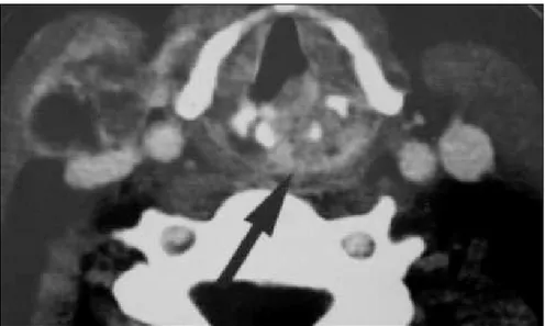
The computed tomography scan showed that the lesion had infi ltrated the posterior third of the vocal fold, left arytenoid cartilage and interarytenoid portion and destroyed the upper posterior half of the cricoid cartilage. The lesion was also occupying the left para-glottic space. In addition, images of metastatic lymph nodes at left level II and right level IV were noticed (Figure 1).
The patient underwent total laryngec-tomy with bilateral modifi ed radical neck dissection and right anterior compartment neck dissection. A primary tracheoesopha-geal puncture for voice rehabilitation was also performed.
The evaluation of the surgical specimen using histopathological study confi rmed the diagnosis of papillary squamous cell carcinoma with a papillary pattern (Figure 2), involving the posterior half of the left vocal and vestibular fold, the posterior half of the left ventricle, and the left arytenoid cartilage. There was one positive lymph
Laryngeal papillary carcinoma
with unexpected evolution:
case report
Department of Otolaryngology and Head and Neck Surgery,
Universidade Estadual de Campinas, Campinas, São Paulo, Brazil
159
node on the right side at level IV, measur-ing 4 x 2.8 cm in size with extracapsular spreading. On the left side, one lymph node demonstrated metastatic disease at level II, measuring 2.8 x 2.3 cm, also with extracapsular spreading.
The patient underwent adjuvant treat-ment using chemotherapy, consisting of three cycles of 5-fl uoracil and cisplatin, and radio-therapy with a total dose of 5000 cGy.
The hybridization in situ revealed no evidence of human papillomavirus in any of the specimens obtained.
DISCUSSION Papillary squamous cell carcinoma is a variant of squamous cell carcinoma. The latter accounts for up to 90% of all the malignant lesions found in the larynx.3
The histological variants of squamous cell carcinoma include verrucous carcinoma, sarcomatoid carcinoma, basaloid carcinoma, adenocarcinoma and cystic adenoid car-cinoma.2,4 These may also be divided into
papillary or exophytic types, according to their growth pattern. The exophytic pattern pres-ents rounded and caulifl ower-like projections, whereas the papillary pattern resembles more delicate fi ngerlike projections. The classifi ca-tion as exophytic or papillary should follow the dominant pattern that corresponds to at least 70% of the tumor size.2 The diagnosis of
papillary carcinoma is made through histologi-cal examination when there is a high count of atypical cells and low rate of koilocytosis.
Conclusion In our report we have described a case of papillary squamous cell carcinoma in a young patient, at an unusual site in the larynx, with positive lymph nodes and extracapsular spread-ing. The observed outcome was the opposite of what is presented in the current literature, which describes a good prognosis for this lesion.1-5
Figure 1. Axial computed tomography scan with lesion infi ltrating posterior half of vocal fold, arytenoid cartilage and surrounding tissues (black arrow). Note the lymph node metastasis at level IV (right side).
Figure 2. Black arrow shows fi ngerlike projections of the papillary pattern of papillary squamous cell carcinoma, in histopathological study of surgical specimen resected from the larynge of a 42-year-old woman.
160
RESUMO Carcinoma papilífero de laringe com evolução inesperada: relato de caso
CONTEXTO: Segundo a literatura, o carcinoma pilífero de laringe é raro e tem um bom prognóstico, quando comparado aos outros carcinomas espinocelulares.
RELATO DE CASO: Neste relato, apresentamos um caso já avançado ao diagnóstico, com acometimento linfonodal e invasão extracapsular bilateral. Tomografi a computadorizada mostrou infi ltração da lesão e metástase em linfonodos. Após cirurgia, o espécime ressecado foi submetido a estudo histopatológico, que confi rmou o diagnóstico de carcinoma papilífero.
PALAVRAS-CHAVE: Neoplasias laríngeas. Carcinoma papilar. Radioterapia. Prognóstico. Metástase linfática.
AUTHOR INFORMATION
Adriano Santana Fonseca, MD. Assistant professor of Depart-ment of Otolaryngology and Head and Neck Surgery, Santa Casa de Misericórdia, Hospital Santa Izabel, Salvador, Bahia, Brazil.
Carlos Takihiro Chone, MD, PhD. Assistant professor of Department of Otolaryngology and Head and Neck Sur-gery, Universidade Estadual de Campinas, Campinas, São Paulo, Brazil.
Agrício Nubiato Crespo, MD, PhD. Head of Department of Otolaryngology and Head and Neck Surgery, Universidade Estadual de Campinas, Campinas, São Paulo, Brazil.
Albina Altemani, MD, PhD. Assistant Professor of Department of Pathological Anatomy, Universidade Estadual de Campinas, Campinas, São Paulo, Brazil.
Address for correspondence:
Adriano Santana Fonseca
Rua das Patativas, 43 — Apto 1004 — Imbuí Salvador (BA) — Brasil — CEP 41720-110 Tel./ Fax. (+55 71) 8109-2769
E-mail: [email protected]
Copyright © 2006, Associação Paulista de Medicina 1. Crissman JD, Kessis T, Shah KV, et al. Squamous papillary
neoplasia of the adult upper aerodigestive tract. Hum Pathol. 1988;19(12):1387-96.
2. Thompson LD, Wenig BM, Heffner DK, Gnepp DR. Exo-phytic and papillary squamous cell carcinomas of the larynx: A clinicopathologic series of 104 cases. Otolaryngol Head Neck Surg. 1999;120(5):718-24.
3. Esteban Ortega F, Concha López A. Aspectos controvertidos del carcinoma verrucoso de laringe: etiologia viral, diagnóstico
y tratamiento. [Controversial aspects of verrucous carcinoma of the larynx: viral etiology, diagnosis and treatment]. Acta Otorrinolaringol Esp. 1991;42(6):443-9.
4. Ferlito A. Malignant laryngeal epithelial tumors and lymph node involvement: therapeutic and prognostic considerations. Ann Otol Rhinol Laryngol. 1987;96(5):542-8.
5. Ereno C, Lopez JI, Sanchez JM, Bilbao FJ. Papillary squamous cell carcinoma of the larynx. J Laryngol Otol. 2001;115(2):164-6.
Sources of funding: Not declared Confl ict of interest: Not declared Date of fi rst submission: August 6, 2004 Last received: May 31, 2006
Accepted: May 31, 2006
REFERENCES