232
Pakistan Veterinary Journal
ISSN: 0253-8318 (PRINT), 2074-7764 (ONLINE) Accessible at: www.pvj.com.pk
Effects of Trypsinization on Viability of Equine Chondrocytes in Cell Culture
B. C. Sutradhar, J. Park, G. Hong, S. H. Choi and G. Kim*
Laboratory of Veterinary Surgery, College of Veterinary Medicine, Chungbuk National University, Cheongju, Chungbuk 361-673, Korea.
*Corresponding author: [email protected]
A R T I C L E H I S T O R Y A B S T R A C T
Received: Revised: Accepted:
June 03, 2010 June 21, 2010 July 01, 2010
Key words:
Cell culture
Equine chondrocytes Trypsinization Viability
Trypsin is an essential reagent for routine cell culture work. In the cultivation of mammalian cells, it has been extensively used for cell isolation from tissues or cell dislodging in subculturing. It may damage the cell membrane in contact of cells during long trypsinization. However, there is no specific report on time-dependent effect of trypsinization on cells. In the present study, we investigated the time dependent effects of trypsinization on equine chondrocytes. Cell viability after trypsinization with 0.25% trypsin-EDTA for 5 to 60 minutes was quantified by trypan blue exclusion assay, propidium iodide-Hoechst double staining, flow cytometry analysis and XTT assay. The results showed that trypsin-EDTA decreased the proliferation of equine chondrocytes depending on the exposure time of trypsinization. After 20 and 60 minutes of trypsinization, the cell membranes were strongly affected and the percentages of viable cells reduced to 91% and 85% respectively detected by trypan blue exclusion assay. Similar results were observed both in flow cytometric evaluation and propidium iodide-Hoechst double staining. The XTT assay result also showed decreased cell viability with the extended time of trypsinization. In order to minimize the time dependant cytotoxicity of trypsinization, as minimum as short time exposure is suggestive that maximizes live cell isolation from tissue as well as subculture of equine chondrocytes or other cells. ©2010 PVJ. All rights reserved
To cite this article: Sutradhar BC, J Park, G Hong, SH Choi and G Kim, 2010. Effects of trypsinization on viability of equine chondrocytes in cell culture. Pak Vet J, 30(4): 232-238.
INTRODUCTION
Trypsin has a wide range of industrial and scientific uses. In the cultivation of mammalian cells, trypsin has been extensively used for cell dislodging in primary cell culture to obtain single cells from tissues and organs (Stewart et al., 1995). It has also been widely used for the subcultivation and scale-up of several cell lines, to detach cells from either static or carrier surfaces (Cruz et al., 1997). Moreover, it is used as spray or ointment in clinics for debridement of necrotic cells (Eneroth and van Houtum, 2008). However, it is well known that trypsin may injure the cell membrane in contact of cells (Collett
et al., 2007) and may damage molecules involved in the cellular reactions (Yanase et al., 2007); although time dependent quantitative effect of trypsinization was not arranged in literature, which is very essential for usual cell culture.
Trypsin is an endopeptidase produced by the gastro-intestines of mammals has an optimal operating pH of about 8 (Vestling et al., 1990) and an optimal operating temperature of about 37°C (Brown et al., 2007). In vivo,
the proteolytic enzymes, such as trypsin can break these bonds creating a single-cell suspension.
Usually in adherent cell culture, calcium and magnesium ions enhance the cell-to-cell adhesion and obscure the peptide bonds on which trypsin acts and separates the cells. So EDTA is frequently used with trypsin solutions as a chelating agent with gentle dissociative properties that acts to increase trypsin efficiency by neutralizing calcium and magnesium ions (Olsen et al., 2004). So in cell culture research and cell biology studies trypsin-EDTA is widely used to dissociate tissues and cell monolayer to maximize the yield of functionally viable, dissociated cells (Amano et al., 1996).
Enzymatic disaggregation, such as using trypsin-EDTA, is the most widely used technique in cell culture which allows rapid passaging for large scale cell expansion (Mitalipova et al., 2005). The timing of trypsinization may vary depending on the cell type, age of monolayer, cell density, serum concentration and other factors (Melican et al., 2005). Trypsinization for 2 hours at 37°C with 0.25% trypsin-EDTA is useful to collect equine epithelial cells from tissue sample (Shibeshi et al., 2008). The trypsin concentrations to detach different cells range from 0.05% to 0.5% (Brown et al., 2007), although trypsinization with 0.25% trypsin-EDTA for 30-60 minutes at 37°C is a common method for chondrocyte isolation used in cartilage tissue engineering (Hsieh et al., 2009).
Trypsinization is fast and reliable method but over-trypsinization is a common cause of subculture problem can damage the cell surface by digesting exposed cell surface proteins (Glade et al., 1996). Prolonged exposure to trypsin can damage cell membranes and cause lysis (Melican et al., 2005). Trypsin is known to degrade plasma membrane glycoproteins and repeated exposure to trypsin is likely to contribute to accelerate aging (Ryan, 1984).
This study for the first time clearly stated significantly non-viable data based effect of trypsin-EDTA at a commonly used dose in chondrocyte culture. The purpose of the present study was to investigate the effects of trypsin-EDTA on viability of equine chondrocytes, suggesting mainly on its cautious application in cell culture, especially in long trypsinization.
MATERIALS AND METHODS Equine chondrocyte isolation and culture
Cartilage was collected from articular surfaces of the distal metacarpal bones of two-year old Thoroughbred horse. The pieces of cartilage were washed with sterile physiological saline and cut into small pieces which were digested with 1mg/ml collagenase type-I (Sigma, St. Louis, MO, USA) in Dulbecco's Modified Eagle Medium (DMEM) (Welgene, Daegu, South Korea) that contains 63.5 µg/ml penicillin and 100µg/ml streptomycin, incubated for 18h at 37ºC in shaking water bath. Then the isolated chondrocytes were filtered with sterilized gauze and centrifuged at 200xg for 5 min and pellets were resuspended for culture at a density of 1x10⁶ cells/dish with DMEM containing 10% FBS in a 75 cm2 cell culture flask. Cultures were carried out at 37ºC in a 95% air and
5% CO2 humidified atmosphere changing media twice a week.
Treatment of the cells
Equine chondrocytes were seeded onto six-well flat-bottomed microculture plates (except in XTT assay) in DMEM medium with 10% FBS (1x10⁵ cells/well), and were incubated at 37oC for six days. The number of cells was measured by using trypan blue-exclusion assay. Chondrocyte monolayer was then washed twice with 10 mM phosphate buffered saline (PBS, pH 7.4) and treated with 0.25% trypsin-EDTA (Bovine Trypsin, Sigma, St. Louis, MO, USA; EDTA, DC Chemical Co. Ltd., Seoul, South Korea) for 5, 20 and 60 minutes at 37ºC in a 95% air and 5% CO2 humidified atmosphere. The trypsinization effect was neutralized with DMEM supplemented with 10% fetal bovine serum. The cells were collected after centrifugation at 1000 rpm for 5 minutes and washed with PBS for viability assays.
Trypan blue exclusion assay
Chondrocytes were treated with PBS, DMEM, and trypsin-EDTA separately for 5, 20 and 60 min and collected following PBS wash. Then, cells were incubated with 0.4% w/v trypan blue solution (Sigma, St. Louis, MO, USA) for 2-3 min in room temperature. Cell viability was determined by using a hemocytometer. The percentage cell survival for each treatment group was determined by counting the dead cells and the live cells. The number of viable cells was calculated by dividing the number of living cells with the total number of cells in percentage.
Propidium iodide (PI)-Hoechst double staining
After harvesting, the cells were washed once in PBS and viability was determined by PI-Hoechst double staining. Cells were stained with fresh Hoechst 33258 (5 µg/ml) and incubated at 37ºC for 15 minutes. Then PI (50 µg/ml; Sigma, St. Louis, MO, USA) was added to the cells and incubated at 37ºC for 5 minutes. A 20 µl volume of suspended cells was placed onto glass slides and mounted with glycerol. Nuclear morphology was then examined under a fluorescent microscope (S46; Microscopes, Inc. St. Louis, MO, USA) using blue and red filters for Hoechst and PI views respectively. At least two individuals were involved to count the PI- or Hoechst positive nuclei to prevent personal biasness. At least 5 vision fields were randomly selected for each slide to obtain mean number of surviving or dead cells, respectively. Normally 100-120 Hoechst-positive nuclei with normal nuclear morphology were obtained from each vision field in the control cultures. The mean values were calculated from combined results of at least four independent cultures. The “Death Index” was defined as the number of PI-positive nuclei divided by that of Hoechst-positive nuclei in the same vision field.
Flow cytometric analysis
equipped with an argon ion laser tuned at 488 nm wavelengths (Becton Dickinson FACS Scan, Heidelberg, Germany). Propidium iodide sensitive positive cells were considered as non-viable cells. Results were expressed as a percentage of non-viable PI+ cells in this experiment.
XTT assay
Cytotoxicity was tested by the XTT-formazan dye formation. Briefly, chondrocytes were seeded onto 96-well flat-bottomed microculture plates at a density of 2 x 10⁴ cell/well in 200µl of DMEM and allowed to adhere up to confluent within the CO2 incubator. Cells were treated with trypsin-EDTA for 5, 20 and 60min at 37ºC followed by PBS wash. The trypsinization effect was neutralized with DMEM supplemented with 10% fetal bovine serum. Fresh XTT and phenazine methosulfate were mixed together; 20µl of the mixed reagent was added to 100 µl of cell suspension in each well of the 96-well plates. The cells and reagent mixture were incubated at 37oC for 2 hours, and the sample’s absorbance was measured with a spectrophotometer (Emax, Molecular Device, Sunnyvale, CA, USA) at a wavelength of 450 nm.
Percentage cytotoxicity was calculated by the following formula:
AC- AT % cytotoxicity = A
C X 100
Where AC is the absorbance of the control wells and AT is the absorbance of the test wells.
Statistical analysis
Data were presented as the mean ± standard deviation. Statistical analyses were performed by one-way analysis of variance (ANOVA) with multiple range tests using the SPSS 11.5 statistical software (SPSS Inc. Chicago, IL, USA). Significant differences between groups were determined at P<0.05.
RESULTS
The cytotoxicity of trypsin-EDTA in equine chondrocytes was assessed as decrease of membrane integrity and decrease of cell viability. Trypsin-EDTA time dependently induced a significant loss of both membrane integrity and cell viability. The chondrocytes showed a rapid decrease of cell viability from 20 minutes to 60 minutes of exposure and morphologically changed to blebbing and lost their membrane integrity with time of trypsinization.
The cytotoxic effect of trypsin-EDTA was investigated by testing most commonly used concentration of trypsin-EDTA (0.25% trypsin with 0.02% EDTA) on equine chondrocytes at three time points (5, 20 and 60 min) whereas the 5 min was considered as a control. Viability was decreased from 20 minutes and continued up to 60 min in our experiments.
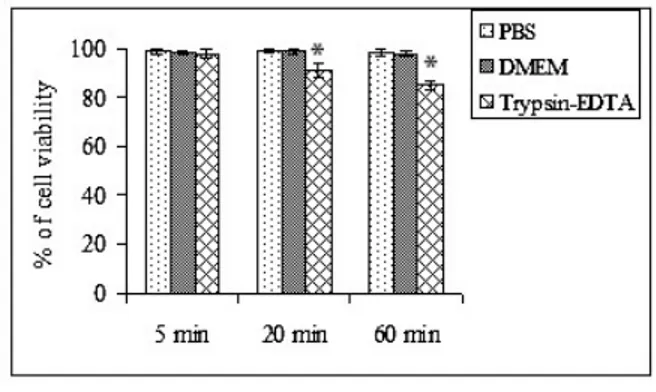
The treatment of equine chondrocytes with PBS and DMEM showed no cytotoxic effect up to 60 minutes whereas trypsin-EDTA decreased the viability with the time of trypsinization. At 20 and 60 min of trypsinization, the cell membranes were strongly affected and the percentages of viable cells were reduced to 91and 85% respectively evaluated by trypan blue dye-exclusion assay (Fig. 1). The cytotoxic effect started from 20 minutes was
statistically significant when compared with the control group. Extended exposure to trypsin-EDTA showed a potent cytotoxicity on equine chondrocytes. Trypsinization with 0.5% trypsin-EDTA was more potent to induce deleterious effect on chondrocytes viability compared to 0.25% trypsin-EDTA (Data not shown).
Fig. 1: Time dependent trypsin-EDTA effects on chondrocyte viability comparing to the PBS and DMEM. The chondrocytes were treated with PBS, DMEM and trypsin-EDTA for 5, 20 and 60 minutes at 37ºC in a humidified atmosphere in CO2 incubator. Subsequently,
cells were harvested and cytotoxic effect was investigated by trypan blue exclusion assay. The data were analyzed (*P<0.05) in one-way ANOVA with Tukey’s test.
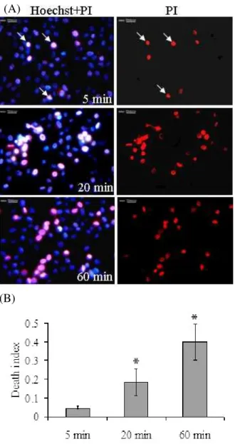
The percentage of PI+ cells varied 3-5% in control groups and increased gradually when trypsinization was extended to 20 and 60 min evaluated in Hoechst-PI double staining under fluorescent microscope. This staining revealed a “Death Index” of 0.18 and 0.40, corresponding to the death rate of 18 and 40% for 20 and 60 min, respectively. The blebbing and red fluorescent PI stained cells were significantly increased from 20 to 60 min of trypsinization (Fig. 2).
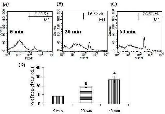
The results of the flow cytometric analysis of cell viability are shown in Fig 3. Flow cytometric analysis of trypsin-EDTA exposed equine chondrocytes revealed two subpopulations corresponding to damaged cells, i.e., cells that were PI stained, and to cells with an intact cytoplasmic membrane, which were not stained. There was a time-dependent decrease in cell viability, as revealed by a reduced percentage of cells excluding PI (20 and 27% after 20 and 60 min of trypsinization, respectively), in all groups of treatment.
Live cells reduce XTT to a soluble product XTT-formazan,which can be estimated spectrophotometrically as a measure of cell viability. Equine chondrocytes showed reduced cell viability in extended trypsinization. Fig. 4 depicts the percentage cytotoxicity of trypsinization which was increased in a time dependent manner.
DISCUSSION
Fig. 2: Time dependent trypsin-EDTA effects on chondrocyte viability in PI-Hoechst double staining. (A) Equine chondrocytes were treated with trypsin-EDTA (0.25%) for 5, 20 and 60 minutes at 37ºC in a humidified atmosphere containing 5% CO2 and 95%
air. The cells were subsequently subjected to PI-Hoechst double staining. The arrows denote PI-positive nuclei indicating ruptured plasma membrane permeable to PI. (B) Quantitative results based on PI-Hoechst double staining. The ‘‘Death Index’’ was defined as the number of PI-positive nuclei divided by the number of Hoechst-positive nuclei in the same vision field. *P<0.05 compared to control cells using one way ANOVA with Tukey’s test.
there was a time dependent effect of trypsin-EDTA on the viability of equine chondrocytes as a reduced viability in longer trypsinization. The effect of trypsin-EDTA on decreased chondrocytes viability appeared to be permanent and was significantly started from 20 minutes of trypsinization among the three selected times. The
reduction of cell viability was increased and continued up to 60 minutes and no significant change was observed in the groups treated for 5 minutes as a control.
Necrosis occurs after sudden severe injuries or noxious compound treatment and accounts for many destructive effects that represent only a passive consequence of pathologic damages. Morphologically, dramatic alterations of plasma membrane permeability occur. These events lead to cellular swelling and disintegration followed by mitochondrial disruption, cellular content release and chromatin flocculation. The partial analogy between apoptosis (especially in its late phase) and necrosis contributes to complicate the ascription of structurally altered cells to one or the other phenomenon (Foglieni et al., 2001). In our study, we assessed the necrosis including late apoptosis of equine chondrocytes following trypsinization with 0.25% trypsin-EDTA.
Microscopy cell viability assessment has been widely described (Foglieni et al., 2001). Previous studies have reported that detachment of cells with trypsin-EDTA may alter or even damage the cell population (Collett et al.,
2007). Light and fluorescent microscopy revealed as the damaged plasma membranes with stained cells were increased from 20 minutes of trypsinization in our experiment. The horse shoe shaped cells were changed to rounded and enlarged under microscope which were possibly attributed primarily to the changes in the level of cytoskeleton proteins involved in regulating the stability and elasticity of the cell membrane (Mohanty et al., 2010). The findings suggest that a short trypsinization should be used in cell culture area to maintain membrane integrity of the cells. Some other studies also reported the trypsin-induced changes in cultured keratinocytes (Umegaki et al., 2004) and endothelial cells (Lopes et al., 2001) which support our results. Although mild trypsinization of rat thymocytes in cell suspensiondid not cause appreciable nonspecific damage to the cells, sinceit did not change the ability of the cells to exclude dye in trypan blue assay (Segal and Ingbar, 1986).
For the detection of cell viability, Hoechst and PI are the most commonly used fluorescent dye in cell culture area (Foglieni et al., 2001). Both Hoechst and PI intercalate to DNA, but only Hoechst can permeate the active cell membrane (Nakajima et al., 1998). Therefore
we used these dyes jointly to differentiate the viable and nonviable cells. Our results showed that the trypsin-EDTA strongly reduced the viability of equine chondrocytes at extended time of trypsinization by inducing necrosis. The appearance of necrotic chondrocytes were swollen and lost their plasma membrane integrities demonstrated by PI-Hoechst double staining (Fig. 2). PI can enter only in late apoptotic and necrotic cultured cells, intercalating nucleic acids every 4–5 bp without sequence preference (Ertel et al., 1998). Swelling of cellular organelles and loss of plasma membrane integrity are the best way to describe necrotic cells under light or electron microscopes (Hotchkiss et al., 2009). Our study revealed that the red fluorescence after PI staining (distinctive for necrotic nuclei) was increased PI staining (distinctive for necrotic nuclei) was increased with prolonged trypsinization (Fig. 2).
(A)
Fig. 3: Influence of trypsin-EDTA on chondrocyte necrosis. Equine chondrocytes were treated with trypsin-EDTA (0.25%) for 5 minutes (A), 20 minutes (B) and 60 minutes (C) at 37ºC in a humidified atmosphere containing 5% CO2 and 95% air. Subsequently, cells were harvested, and cytotoxic effect was investigated
by flow cytometric analysis after PI staining (M1). Average peak values for the PI staining (D) were from at least three independent assays, showing a time-dependant increased cytotoxicity compared to the untreated cells. Asterisks indicate P<0.05 in one-way ANOVA with Tukey’s test.
Fig. 4: Cytotoxic effects of trypsin-EDTA on equine chondrocytes. Equine chondrocytes were treated with trypsin-EDTA (0.25%) for 5, 20 and 60 minutes at 37ºC in a humidified atmosphere containing 5% CO2 and 95% air.
Cell viability assay used a colorimetric method with a 2,3-bis {2-Methoxy-4-nitro-5-sulfo- phenyl}-2H-tetrazolium -5 carboxyanilide inner salt (XTT) assay. Vertical bars indicate the mean ± standard deviation (N=6). *P<0.05 was considered significant in one-way ANOVA with Tukey’s test.
Cells in native tissues adhere to the surrounding extracellular matrix (ECM) via cell membrane receptors (e.g. integrins) (Hynes, 1992) that specifically bind to ECM adhesion proteins such as fibronectin, vitronectin and laminin. But trypsin treated primary and passage 1 chondrocytes were reported as decreased in cell adhesivity (Tsai and Wang, 2005). Integrinslink ECM ligands to the cytoskeleton which provide strong attachmentto enable cell-shape change and tissue integrity (Delon and Brown, 2009). But trypsin can destroy the membrane proteins of the cells (Cruz et al., 1997) and reduce the cell’s ability to form adhesive bonds with adsorbed cell adhesion proteins by decreasing the number of functional integrins of the cell membrane (Brown et al., 2007). Hence we designed to study on membrane integrity of equine chondrocytes at various time points of trypsinization.
Flow cytometry (FCM) is a well-proven technique that allows cells, spores, and other particles to be individually analyzed. In addition, FCM can analyze large samples, thus enabling the collection of a set of statistically reliable results. FCM combined with a fluorescent technique can differentiate between biotic particles and abiotic particles (Chen and Li, 2005). Viability studies consist of nucleic acid dyes, such as propidium iodide (PI), which are excluded by viable cells with intact membranes. The fluorescence conferred by these probes indicates the degree of cell damage, cell permeability, and ultimately cell death (Amor et al., 2002). In this study, it was authenticated by flow cytometry that trypsinization with 0.25% trypsin-EDTA for 5 minutes at 37°C was the best method for detachment of intact chondrocytes from monolayer culture. Although from 20 minutes of trypsinization viability of equine chondrocytes were significantly decreased as like as the results of other assays.
Previous data suggest that prolonged exposure of cells to active trypsin might damage cell viability (Tsai et al., 2005). It can trigger the metabolic changes by
interaction with the cellular membrane which induces cellular death (Lepsch et al., 2009). Our XTT results also confirmed the gradual decrease in chondrocyte viability with the time of trypsinization. In another study it has been reported that some epitopes of the cell membrane were particularly sensitive to trypsin and were lost after trypsinization (Bolwell, 1986). Therefore the effect of trypsin is very important for starting a new experiment including its exposure which may differ with manufacturers and which is usually not mentioned throughout scientific literature. In this study, we found that trypsinization for 5 minutes with 0.25% trypsin-EDTA did not have any significant detrimental effect on the cell membrane and cell viability, although from 20 minutes of trypsinization, there was a potent deleterious effect on both cell membranes and viability of equine chondrocytes. Consequently prolonged incubation with trypsin-EDTA and more concentrated solutions decreased the number of viable cells significantly in our results.
In conclusion, our results showed that trypsin-EDTA had the cytotoxic effect on equine chondrocytes in monolayer culture. We confirmed that trypsin-EDTA affect the cell membrane integrity, cell viability and finally induce necrosis of the chondrocytes with extended exposure time. Concentration of trypsin-EDTA had also played an important role on cytotoxicity of the cells. However, additional molecular studies are needed to explain the specific genetic change of the cell on response to trypsin-EDTA. However, to our knowledge, time dependent effect of trypsinization on chondrocytes viability has not been demonstrated before. In order to minimize the time dependant cytotoxicity associated with trypsinization, as minimum as short time exposure may be useful to maximize live cell isolation from tissue or subculture of equine chondrocytes or from other cells. The use of trypsin-EDTA in cell culture should occur with awareness of the risk, especially in prolonged trypsinization.
REFERENCES
Amano H, R Kurosaka, M Ema and Y Ogawa, 1996. Trypsin promotes C6 glioma cell proliferation in serum and growth factor-free medium. Neurosci Res, 25: 203-208.
Amor KB, P Breeuwer, P Verbaarschot, FM Rombouts, ADL Akkermans, WM De Vos and T Abee, 2002. Multiparametric flow cytometry and cell sorting for the assessment of viable, injured, and dead bifidobacterium cells during bile salt stress. Appl Environ Microb, 68: 5209–5216.
Bolwell GP, 1986. Significance of a common epitope of plant and animal endomembranes. J Cell Sci, 82: 187-201.
Brown MA, CS Wallace, CC Anamelechi, E Clermont, WM Reichert and GA Truskey, 2007. The use of mild trypsinization conditions in the detachment of endothelial cells to promote subsequent endothelialization on synthetic surfaces. Biomaterials, 28: 3928–3935.
Chen PS and CS Li,2005. Bioaerosol characterization by flow cytometry with fluorochrome. J Environ Monit, 7: 950-959.
Collett J, A Crawford, PV Hatton, M Geoghegan and S Rimmer, 2007. Thermally responsive polymeric hydrogel brushes: synthesis, physical properties and use for the culture of chondrocytes. J R Soc Interface, 4: 117-126.
Cruz HJ, Y Yanase and MJT Carrondo, 1997. Cell-dislodging methods under serum-free conditions. Appl Microbiol Biotechnol, 47: 482-488.
Delon I and NH Brown, 2009. The integrin adhesion complex changes its composition and function during morphogenesis of an epithelium. J Cell Sci, 122: 4363-4374.
Eneroth M and WH van Houtum, 2008. The value of debridement and Vacuum-Assisted Closure.(V.A.C.) Therapy in diabetic foot ulcers.Diab Metab Res Rev, 24: 76-80.
Ertel W, M Keel, M Infanger, U Ungethum, U Steckholzer and O Trentz, 1998. Circulating mediators in serum of injured patients with septic complications inhibit neutrophil apoptosis through up-regulation of protein tyrosine phosphorylation. J Trauma, 44: 767–775.
Foglieni C, C Meoni and AM Davalli, 2001. Fluorescent dyes for cell viability: an application on prefixed conditions. Histochem Cell Biol,115: 223–229. Glade CP, BA Seegers, EF Meulen, CA van Hooijdonk,
PE van Erp and PC van de Kerkhof, 1996. Multiparameter flow cytometric characterization of epidermal cell suspensions prepared from normal and hyperproliferative human skin using an optimized thermolysin-trypsin protocol. Arch Dermatol Res, 288: 203-210.
Hotchkiss RS, A Strasser, JE McDunn and PE Swanson, 2009. Cell death. N Engl J Med, 361: 1570-1583. Hughes EN and JT August, 1981. Characterization of
Hsieh SC, NT Chen and SH Lo, 2009. Conditional loss of PTEN leads to skeletal abnormalities and lipoma formation. Mol Carcinog, 48: 545–552.
Hynes RO, 1992. Integrins: versatility, modulation, and signaling in cell adhesion. Cell, 69: 11-25.
Lepsch LB, CD Munhoz, EM Kawamoto, LM Yshii, LS Lima, MF Curi-Boaventura, TM Salgado, R Curi, CS Planeta and C Scavone, 2009. Cocaine induces cell death and activates the transcription nuclear factor kappa-b in pc12 cells. Mol Brain, 2: 3. doi: 10.1186/1756-6606-2-3.
Lopes AAB, TMS Peranovich, NY Maeda and SP Bydlowski, 2001. Differential effects of enzymatic treatments on the storage and secretion of von Willebrand factor by human endothelial cells. Thromb Res, 101: 291–297.
Melican D, R Butler, N Hawkins, LH Chen, E Hayden, M Destrempes, J Williams, T Lewis, E Behboodi, C Ziomek, H Meade, Y Echelard and W Gavin, 2005. Effect of serum concentration, method of trypsinization and fusion/activation utilizing transfected fetal cells to generate transgenic dairy goats by somatic cell nuclear transfer. Theriogenology, 63: 1549–1563.
Mitalipova MM, RR Rao, DM Hoyer, JA Johnson, LF Meisner, KL Jones, S Dalton and SL Stice, 2005. Preserving the genetic integrity of human embryonic stem cells.Nat Biotechnol,23: 19-20.
Mohanty JG, HD Shukla, JD Williamson, LJ Launer, S Saxena and JM Rifkind, 2010. Alterations in the red blood cell membrane proteome in Alzheimer's subjects reflect disease-related changes and provide insight into altered cell morphology.Proteome Sci,8: 11.
Mutin M, F George, G Lesaule and J Sampol, 1996. Reevaluation of trypsin-EDTA for endothelial cell detachment before flow cytometry analysis. Endothelium, 4: 289-295.
Nakajima T, M Yageta, K Shiotsu, K Morita, M Suzuki, Y Tomooka and K Oda, 1998. Suppression of adenovirus E1A-induced apoptosis by mutated p53 is overcome by coexpression with Id proteins.Proc Natl Acad Sci USA, 95: 10590–10595.
Olsen JV, SE Ong and M Mann, 2004. Trypsin cleaves exclusively C-terminal to arginine and lysine residues. Mol Cell Proteomics, 3: 608-614.
Ryan US, 1984. Isolation and culture of pulmonary endothelial cells. Environ Health Persp, 56: 103-114. Segal J and SH Ingbar, 1986. The effect of trypsinization
on the plasma membrane binding and action of 3,5,3'-triiodothyronine in rat thymocytes. Endocrinology, 118: 1863-1868.
Shibeshi W, G Abraham, C Kneuer, C Ellenberger, J Seeger, HA Schoon and FR Ungemach, 2008. Isolation and culture of primary equine tracheal epithelial cells. In Vitro Cell Dev Biol Anim, 44: 179-184.
Stewart GJ, Y Wang and S Niewiarowski, 1995. Methylcellulose protects the ability of anchorage-dependent cells to adhere following isolation and holding in suspension. Biotechniques, 19: 598–604. Tsai WB and MC Wang, 2005. Effect of an avidin-biotin
binding system on chondrocyte adhesion, growth and gene expression, Biomaterials, 26: 3141-3151.
Umegaki R, K-O Masahiro and M Taya, 2004. Assessment of cell detachment and growth potential of human keratinocyte based on observed changes in individual cell area during trypsinization. Biochem Eng J,17: 49–55.
Vestling MM, CM Murphy and C Fenselau, 1990. Recognition of trypsin autolysis products by high-performance liquid chromatography and mass spectrometry. Anal Chem, 62: 2391–2394.