ADULT CEREBELLAR MEDULLOBLASTOMA
CT and MRI findings in eight cases
Arnolfo de Carvalho Neto
1, Emerson L. Gasparetto
2, Sérgio E. Ono
3,
Guilherme A. Bertoldi
4, André F. Gomes
5ABSTRACT - Medulloblastoma is a brain tumor of neuroepithelial origin, which represents 15 to 30% of all pediatric brain tumors, and less than 1% of CNS adult neoplasms. We report the imaging findings of 8 adult patients with medulloblastoma. The mean age was 35 years, ranging from 20 to 65 years, and the male:female rate was 3:5. The tumors were predominantly lateral (63%), hyperdense on CT scans (83%), and on the MRI, hypointense on T1 (100%) and hyperintense on T2 (80%) weighted images. It was seen intratumoral necrosis and cysts in six cases and calcifications in three. Hydrocephalus was observed in 5 cases and brain stem invasion in four. The imaging findings of medulloblastomas in adults are different of those in child, and also nonspecific. Although these tumors are uncommon in adults, they must be considered in the differential diagnosis of cerebellar masses in the posterior fossa of this age group.
KEY WORDS: medulloblastoma, adults, computed tomography, magnetic resonance imaging.
Meduloblastoma cerebelar em adultos: achados de TC e RM em oito casos Meduloblastoma cerebelar em adultos: achados de TC e RM em oito casos Meduloblastoma cerebelar em adultos: achados de TC e RM em oito casos Meduloblastoma cerebelar em adultos: achados de TC e RM em oito casos Meduloblastoma cerebelar em adultos: achados de TC e RM em oito casos
RESUMO - O meduloblastoma é um tumor de origem neuroepitelial, que representa entre 15 e 30% dos tumores cerebrais em crianças e menos de 1% das neoplasias intracranianas em adultos. Apresentamos os achados de imagem de 8 pacientes adultos com meduloblastoma. A média de idade foi 35 anos, variando entre 20 e 65 anos, e a relação homem:mulher foi 3:5. Os tumores foram predominantemente laterais (63%), hiperdensos na tomografia computadorizada (83%), e, na ressonância magnética, hipointensos nas imagens ponderadas em T1 (100%) e hiperintensos nas ponderadas em T2 (80%). Necrose e cistos intratumorais foram observados em seis casos e calcificações em três. Cinco pacientes apresentavam hidrocefalia e quatro tinham invasão neoplásica do tronco cerebral. Os achados de imagem dos meduloblastomas em adultos são inespecíficos e diferentes daqueles que acometem crianças. Apesar destes tumores serem incomuns em adultos, eles devem sem considerados no diagnóstico diferencial de massas cerebelares na fossa posterior de pacientes neste grupo etário.
PALAVRAS-CHAVE: meduloblastoma, adulto, tomografia computadorizada, ressonância magnética.
1Assistant Professor of the Discipline of Diagnostic Radiology, University of Paraná, Curitiba PR, Brazil (UFPR); 2Resident of the Diagnostic
Radiology Section, Hospital de Clínicas, UFPR; 3Medical Student of the UFPR School of Medicine; 4Substitute Professor of the Discipline
of Diagnostic Radiology, UFPR; 5Attending of the DAPI - Diagnóstico Avançado Por Imagem - Curitiba PR, Brazil.
Received 31 July 2002, received in final form 11 November 2002. Accepted 13 November 2002.
Dr. Arnolfo de Carvalho Neto - Serviço de Radiologia Médica, Hospital de Clínicas, UFPR - Avenida General Carneiro 181 - 80060-900 Curitiba PR - Brasil. E-mail: arnolfo.carvalho@avalon.sul.com.br
Medulloblastoma accounts for 4-8% of primitive intracranial tumors of neuroepithelial origin. It re-presents 15 to 30% of all pediatric brain tumors and it is a rare entity in the adult age accounting to less than 1% of brain tumors in this age group. The peak of occurrence is between 5-7 years of age, with 70% of them occurring under 16 years. At the presenta-tion, truncal ataxia, intracranial hypertension syn-drome, lethargy, headache, and morning vomiting are the most frequent symptoms1-3.
On CT scans, these tumors are described as a well-defined, hyperdense and homogeneous midline mass
with marked enhancement after contrast injection. The MRI frequently shows a well-defined posterior fossa tumor, which presents low signal on T1 and high signal on T2 weighted images, with marked enhancement after gadolinium injection. When com-paring the imaging findings of medulloblastomas in children and adults, there are some contradictions, with several authors reporting the same pattern on this two populations, and others showing different imaging findings4-7.
medulloblasto-mas diagnosed since 1988 at the Hospital de Clíni-cas of the University of Paraná.
METHOD
We reviewed retrospectively the patients over 16 years of age with medulloblastoma diagnosed between 1988 and 2002 at the Hospital de Clínicas of the University of Paraná. All films (CT scan and MRI) of these patients were selected and reviewed by a neuroradiologist (ACN). The evaluation consisted of observation of tumor location, presence of necrosis and cysts, hydrocephalus,
calcifica-Fig 1. Axial CT scan before contrast injection showing a hyperdense right cerebellar mass, with a hypodense cystic portion.
females and three male patients. Three patients un-derwent CT scan and MRI, three only CT scan and in two patients only MRI was performed. The six CT scans showed that five (83%) were hyperdense and one hypodense before IV contrast injection. The MRI showed tumors predominantly hypointense on T1 (100%, n=5) and hyperintense on T2 (80%, n=4) weighted images. The tumors were most commonly located on the cerebellar hemispheres (63%), and only three were vermian tumors. It was seen intratumoral necrosis and cysts in six of them and calcifications in three cases. Hydrocephalia was pre-sent in 4 patients and invasion of the brain stem was observed in 4 cases (Figs 1-6). The data related to these images are presented in Tables 1 and 2.
DISCUSSION
Medulloblastomas arise from nests of germinal cells, which proliferate in the neuroepithelial roof of the fourth ventricle in fetal life and migrate upwards and laterally5. There are two other theories trying to
explain the histogenesis of this tumor1,3. One
sug-gests that it originates from the external granular layer of the cerebellum and the most recent theory suggests two possible origins: the classic medullo-blastoma is derived from the fourth ventricle and the desmoplastic medulloblastoma from the external granular layer2,8.
This malignant cerebellar tumor represents 15 to 30% of all pediatric brain tumors2,4,7-10 with an
inci-dence of 0.5 per 100.000 per year1 and accounts for
Table 1. Description of the CT scan findings of 6 adult patients with medulloblastoma.
Case Age Sex Location Necrosis Hydrocephalus Calcifications Density Brain stem
years / Cysts invasion
1 65 F RCH yes no no hypodense no
4 20 F vermis yes yes yes hyperdense yes
5 24 F RCH yes yes no hyperdense yes
6 29 M vermis yes no no hyperdense yes
7 22 M LCH yes yes yes hyperdense yes
8 48 M vermis no yes yes hyperdense no
4-8% of primitive intracranial tumors of neuroepi-thelial origin10. Medulloblastomas are rare in adults,
accounting for less than 0.005 per 100000 annually8
and representing less than 1% of central nervous system tumors in this age group2,4,7,10. There is a
pre-dominance of males, with a male:female distribution of 2:15,9,11. Its peak of incidence is around 5 to 7
years of age, 70% of them occurring under 16 years1,9. Al-Mefty et al.1 reviewed 75 cases in Saudi
Arabia and reported a similar number, with 79% occurring in children. In adulthood, 80% occur bet-ween 21 and 40 years. The mean age of our study group was 35 years, and we observed predominance of female patients (63%)7.
The imaging findings of cerebellar medullo-blastoma in children is well established on CT images as a well defined, hyperdense and homogeneous midline mass with marked enhancement after IV con-trast injection4,5,11. Nevertheless, atypical features,
which include calcification, cystic or necrotic regions, ill-defined margins and lack of enhancement, should not be considered against the diagnosis of medullo-blastoma in the first decade of life5.
Several authors stressed the imaging features of medulloblastomas in adult patients2,4,7,8,10. Hubbard
et al. reported the Mayo Clinic casuistic and found 62% of these tumors located in the cerebellar hemis-pheres4. Bourgouin et al. studied 28 cases of
medul-Fig 2. (A) Axial T1 weighted MRI before contrast injection revealing a hypointense homogeneous left cerebellar mass. (B) After gadolinium injection, the lesion showed mild enhancement.
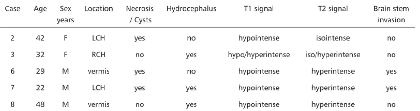
Table 2. Description of the MRI findings of 5 adult patients with medulloblastoma.
Case Age Sex Location Necrosis Hydrocephalus T1 signal T2 signal Brain stem
years / Cysts invasion
2 42 F LCH yes no hypointense isointense no
3 32 F RCH no yes hypo/hyperintense iso/hyperintense no
6 29 M vermis yes no hypointense hyperintense yes
7 22 M LCH yes yes hypointense hyperintense yes
8 48 M vermis no yes hypointense hyperintense no
Fig 3. Axial CT scan showing a midline cerebellar mass, with enhancement after contrast injection. Areas of calcification are also observed.
Fig 4. Coronal T1 weighted MRI after contrast injection revealing an enhanced midline cerebellar mass. Hydrocephalus is also observed.
loblastoma in adults and observed a similar
percen-tage of vermian (50%) and lateral (50%) tumors4.
Maleci et al. compared the imaging features of adult and children with medulloblastoma and found that 50% of the lesions were lateral in the first group, compared to 30% in the second. In our study we
found predominance of lateral tumors (63%)7.
Hubbard et al. observed that vermian medullo-blastomas closely resembled the childhood tumors, and paramedian lesions have the same characteristics of the midline tumors in density and contrast enhan-cement pattern. Regarding the lateral tumors, they found a pattern slight different from the children tumors, with hyperdensity and contrast enhan-cement being more marked.Thery also concluded that coronal CT scans and MRI were more helpful in defining the intra-axial location of the cerebellar medulloblastoma than were axial CT slices5.
All the studies concerning medulloblastomas in adult patients had concluded that hyperdensity is a common feature in these tumors2,4-7,10. Hubbard et
al. found hyperdense lesions in 66% of the patients and in the Bourgouin et al casuist all the tumors were hyperdense before intravenous contrast injection2.
Maleci et al. observed on CT scans that 89% of the medulloblastomas were hyperdense in the adults, compared to 63% in the children. In our casuistics 83% (5 of 6) of the medulloblastomas presented as hyperdense lesions on CT scans7.
Atypical findings in medulloblastomas of adult patients were also stressed by Bourgouin et al.2. Low
density areas, consistent with intratumoral cysts and necrosis were seen in 23 patients (82%), calcifications
were observed in 2 patients (7%) and edema was present in 21 tumors (75%). Hydrocephalus of va-rious degrees of severity was seen in 26 patients (93%). These authors concluded that medulloblas-toma in adults shows a high prevalence of intratu-moral low-density areas caused by cystic and necrotic degeneration. The prevalence of calcifications, ede-ma, hydrocephalus and the size of tumors are com-parable in adults and children. In our casuistics, low density areas compatible with necrosis and/or cysts were observed in 75% of the tumors (n=6), hydro-cephalus in 63% (n=5) and calcifications in three of six cases (50%).
Regarding the MRI features, Koci et al.6 evaluated
12 MRI of adult patients with medulloblastoma. Preoperative T1 weighted images showed hypoin-tense tumors in 8 cases (67%), isoinhypoin-tense to gray matter in three cases (25%) and a hyperintense mass
in one case. T2-weighted images revealed five hyperintense tumors (42%), three slightly hyperin-tense (25%), and three isoinhyperin-tense tumors (25%); one lesion had a dominant markedly hyperintense cystic component. Nine patients presented enhancement after gadolinium injection. Five of our patients un-derwent MRI and all presented with hypointense sig-nal on T1 and 80% showed hyperintense sigsig-nal on T2 weighted images.
A literature review regarding medulloblastomas in adults shows that the CT scans do not follow a consistent pattern like the well defined, homoge-neous, hyperdense, median mass seen in the me-dulloblastoma of children. The tumors in adults have a variety of presentations, but a tendency of being more frequently located laterally and presenting with atypical images of necrosis and cysts is noted. Enhan-cement on CT scans is not always seen after contrast injection. For these reasons, the differential diagnosis of masses in the posterior fossa of an adult must include the hypothesis of a medulloblastoma.
REFERENCES
1. Al-Mefty O, Jinkins JR, El-Senoussi M, et al. Medulloblastomas: a review of modern management with a report on 75 cases. Surg Neurol 1985; 24:606-624.
2. Bourgouin PM, Tampieri D, Grahovac SZ, Léger C, Del Carpio R, Melançon D. CT and MR imaging findings in adults with cerebellar medulloblastomas: comparison with findings in children. AJR 1992; 159:609-612.
3. Giangaspero F, Bigner S H, Kleihues P, Pietsch T, Trojanowski JQ. Medulloblastoma. In Kleihues P, Cavanee WK (eds). World Health Organization classification of tumours pathology and genetics: Tumours of the nervous system. 2.Ed. Oxford: Oxford Univ Press, 2000:123-127. 4. Haie-Meder C, Song PY. Medulloblastoma: differences in adults and children: regarding Frost et al., IJROBP 32:951-957; 1995 and Prados et al., IJROBP 32:1145-1152; 1995. Int J Radiation Oncology Biol Phys 1995;32:1255-1257.
5. Hubbard JL, Scheithauer BW, Kispert DB, et al. Adult cerebellar medulloblastomas: the pathological, radiographic, and clinical disease spectrum. J Neurosurg 1989;70:536-544.
6. Koci M, Chiang F, Mehringer CM, et al. Cerebellar medulloblastoma: imaging features with emphasis in MR findings. AJNR 1993;14:929-939. 7. Maleci A, Cervoni L, Delfini R. Medulloblastoma in children and in
adults: a comparative study. Acta Neurochir 1992;119:62-67. 8. Reis JS Filho, Gasparetto EL, Faoro LN, et al. Medulloblastomas: clinical,
epidemiological and pathological findings in 28 cases. Arq Neuro-psiquiatr 2000;58:76-80.
9. Shandu A, Kendall B. Computed tomography in management of medulloblastomas. Neuroradiology 1987;29:444-452.
10. Juhl JH, Crummy AB, Kuhlman JE. Paul and Juhl´s essentials of radiologic imaging. 2.Ed. Philadelphia: Lippincott Williams & Wilkins, 1998:338-339.
11. Tortori-Donati P, Fondelli MP, Rossi A, et al. Medulloblastoma in children: CT and MRI findings. Neuroradiology 1996,38:352-359