267
CLINICS 2006;61(3):267-70
aDivision of Musculoskeletal Radiology, Department of Radiology,
Massachusetts General Hospital and Harvard Medical School - Boston, MA, USA.
bDepartment of Orthopaedics, Medical Sciences School, State University of
Campinas - Campinas/SP, Brazil.
cDepartment of Pathology, Medical Sciences School, State University of
Campinas - Campinas/SP, Brazil. Email: [email protected]
LETTER TO THE EDITORS
MAGNETIC RESONANCE IMAGING OF LOW-GRADE
FIBROMYXOID SARCOMA
Martin Torriani,a Mauricio Etchebehere,b Eliane M. I. Amstalden,c and Hugue Ouellettea
Low-grade fibromyxoid sarcoma (LGFMS) is a rare soft-tissue tumor with a deceptively benign histologic ap-pearance affecting predominantly young adults during the
fourth decade of life1. Low-grade fibromyxoid sarcoma has
a predilection for involving deep soft tissues of the thigh, inguinal region, or chest wall, affecting less frequently the
shoulder or axilla1. Local postsurgical recurrence and
metastases to lungs and bone are frequently seen1. The
sur-gical management, histopathologic findings, and biologi-cal behavior of LGFMS have been outlined in the litera-ture1,2. There are very few reports on the magnetic
reso-nance (MR) imaging features of LGFMS3,4. We present the
MR imaging features of a surgically confirmed case of LGFMS affecting the shoulder.
A 30-year-old man presented with a 20-year history of a painless slow-growing mass in the right shoulder. Mag-netic resonance images were obtained on a 2.0T scanner (Elscint, Haifa, Israel), demonstrating a well-defined soft-tissue mass measuring 12.0 x 7.0 x 9.0 cm located between the deltoid muscle, rotator-cuff muscles, and proximal hu-merus. No peri-tumoral edema in adjacent subcutaneous tis-sues or muscles was noted. The signal intensity (SI) of the bone marrow was normal. The mass had an intermediate, heterogeneous SI on T1-weighted images [repetition time (TR) / time to echo (TE), 600/25] (Fig. 1a). Heterogene-ous low to high SI with multiple hypointense intralesional nodules was seen on T2-weighted fast spin-echo images (TR/TE, 3500/90) (Fig. 1b). The mass enhanced heteroge-neously on T1-weighted fat-suppressed images (TR/TE, 720/20) after intravenous injection of gadolinium (Fig. 1c), predominantly in corresponding T2-weighted hyperintense areas.
The lesion was surgically removed with negative mar-gins. The surgical specimen consisted of a soft-tissue tumor, measuring 14.5 x 9.5 x 5.0 cm and weighing 290 g. The cut sections showed a firm, grossly circumscribed, and lobulated mass, with a yellow-white color and glistening appearance secondary to the accumulation of myxoid ground substance (Fig. 2a). Microscopically, the tumor had low to moderate cellularity composed of bland spindle-shaped cells with a fibroblast pattern, arranged mainly in
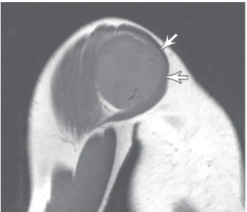
Figure 1a – Sagittal T1-weighted MR image of the shoulder shows a well-defined mildly heterogeneous soft tissue mass of intermediate signal intensity displacing the deltoid muscle (arrows)
268
CLINICS 2006;61(3):267-70 Magnetic resonance imaging of low-grade fibromyxoid sarcoma
Torriani M et al.
a whorled fashion and amidst a variable fibrous and myxoid stroma. The myxoid zones alternated abruptly with the fi-brous zones (Fig. 2b). The cells had indistinct pale eosi-nophilic cytoplasm and showed mild nuclear pleomorphism with low mitotic activity. Immunohistochemically, the neoplastic cells stained strongly and diffusely for vimentin and did not express immunoreactivity to muscle-specific actin, cytokeratins, S-100 protein, or CD34. A histopatho-logical diagnosis of LGFMS was made. The patient had
no evidence of local recurrence or metastases after 2 years of follow-up.
Low-grade fibromyxoid sarcoma was first recognized by Evans in 1987, when he described a bland fibromyxoid neoplasm arising in the deep soft tissues of 2 young women
with subsequent development of metastases5. Although this
is a rare entity, it is reasonable to speculate that it is prob-ably more common than reported in the medical literature due to the likelihood of misdiagnosis with other low-grade fibrous or myxoid soft-tissue neoplasms.
Low-grade fibromyxoid sarcoma occurs more frequently in young to middle-aged adults; however, cases have been reported in patients between 6 to 65 years of age. Males
are affected more commonly than females.1,6 The usual
presentation is a slow-growing painless mass. Low-grade fibromyxoid sarcoma is frequently surrounded by skeletal muscle1,6 and has a predilection for involving deep soft
tis-sues of the thigh, inguinal region, or chest wall. Less fre-quently, it can involve the shoulder or axilla.7 Low-grade
fibromyxoid sarcoma frequently recurs and can metastasize, especially to the lungs,1 with patients surviving between
10 and 15 years after detection of metastases1,6. Due to the
rarity of this neoplasm, the role of chemotherapy and ra-diotherapy remains unknown.
The gross findings of LGFMS include a well-circum-scribed mass with a mean tumor size at diagnosis of 9.5
cm.1 On cut section, LGFMS has a yellow-white or
grey-white appearance with focal areas of gelatinous myxoid tis-sue, rarely exhibiting hemorrhage or necrosis. Histologically, although a pseudocapsule surrounds the tumor, focal microscopic infiltration into the adjacent soft tissues has been reported.8 Histologically, LGFMS is of low
or moderate cellularity, with swirling fibrous and myxoid areas with deceptively benign-appearing fibroblastic spin-dle cells. Mitotic figures are uncommon, and nuclear
pleo-morphism is usually absent or rare.1
Immunohistochemi-cally, the tumor cells stain strongly and diffusely for vimentin, and smooth muscle actin or muscle-specific ac-tin may be present.6,9 Supernumerary ring chromosomes are
a hallmark of LGFMS.10
A prior report on the imaging characteristics of LGFMS described a heterogeneously hyperechogenic multi-nodu-lar sonographic appearance, a heterogeneous MR imaging appearance with low to slightly high SI on T1-weighted images, heterogeneously low to high SI on T2-weighted
images, and heterogeneous postcontrast enhancement.3 In
our case, MR imaging showed a well-defined mass with heterogeneous intermediate to low SI on T1-weighted im-ages, and heterogeneous high SI on T2-weighted images with hypointense nodular areas. Postcontrast T1-weighted images showed heterogeneous enhancement mostly in the
Figure 1c – Axial fat-suppressed T1-weighted MR image after intravenous injection of gadolinium-DTPA shows heterogeneous enhancement, predominantly in corresponding to T2-hyperintense (likely myxoid) areas (arrows)
Figure 2a – Macroscopic features. A grossly circumscribed and lobulated yellow-white mass, with a glistening surface due to myxoid ground substance
269
CLINICS 2006;61(3):267-70 Magnetic resonance imaging of low-grade fibromyxoid sarcoma
Torriani M et al.
periphery of the mass. Macroscopic analysis of cut speci-mens compared to MR imaging findings suggested that fo-cal areas of low T2 SI may correlate with the fibrous ar-eas of the tumor. In comparison with the 2 cases described
by Koh et al, 3 our findings are comparable to their case
describing LGFMS affecting the chest wall. In our case, we did not observe ring-like structures on MR imaging or macroscopic examination. This underscores the variability that may be present in the several components that consti-tute LGFMS.
The differential diagnosis of LGFMS includes several benign and malignant neoplasms containing variable amounts of myxoid and fibrous tissue. Neurofibromas and malignant peripheral nerve sheath tumors may present
his-tological and imaging findings similar to LGFMS.3 The
myxoid zones of LGFMS may resemble myxoid liposar-coma.11 Histologically, the most important differential
di-agnosis is with myxofibrosarcoma, which commonly arises in the subcutaneous tissues of elderly patients and is uni-formly myxoid without alternating fibrous zones. Such a distinction is important, as myxofibrosarcoma rarely metastasizes, in contrast to LGFMS.11
Low-grade fibromyxoid sarcoma is a rare soft tissue tumor with slow growth and deceptively benign histologic appear-ance. The possibility of LGFMS must be considered when elaborating differential diagnostic possibilities for young adults with a large soft tissue mass exhibiting MR imaging charac-teristics of intermixed fibrous and myxoid tissue.
REFERENCES
1. Evans HL. Low-grade fibromyxoid sarcoma. A report of 12 cases. Am J Surg Pathol. 1993;17:595-600.
2. van den Bossche MR, Van Mieghem H. Low-grade fibromyxoid sarcoma. Oncology. 2000;58:207-9.
3. Koh SH, Choe HS, Lee IJ, Park HR, Bae SH. Low-grade fibromyxoid sarcoma: ultrasound and magnetic resonance findings in two cases. Skeletal Radiol. 2004.
4. De Schepper AM, De Beuckeleer L, Vandevenne J, Somville J. Magnetic resonance imaging of soft tissue tumors. Eur Radiol. 2000;10:213-23.
5. Evans HL. Low-grade fibromyxoid sarcoma. A report of two metastasizing neoplasms having a deceptively benign appearance. Am J Clin Pathol. 1987;88:615-9.
6. Goodlad JR, Mentzel T, Fletcher CD. Low grade fibromyxoid sarcoma: clinicopathological analysis of eleven new cases in support of a distinct entity. Histopathology. 1995;26:229-37.
270
CLINICS 2006;61(3):267-70 Magnetic resonance imaging of low-grade fibromyxoid sarcoma
Torriani M et al.
8. Lindberg GM, Maitra A, Gokaslan ST, Saboorian MH, Albores-Saavedra J. Low grade fibromyxoid sarcoma: fine-needle aspiration cytology with histologic, cytogenetic, immunohistochemical, and ultrastructural correlation. Cancer. 1999;87:75-82.
9. Canpolat C, Evans HL, Corpron C, Andrassy RJ, Chan K, Eifel P, et al. Fibromyxoid sarcoma in a four-year-old child: case report and review of the literature. Med Pediatr Oncol. 1996;27:561-4.
10. Mezzelani A, Sozzi G, Nessling M, Riva C, Della Torre G, Testi MA, et al. Low grade fibromyxoid sarcoma. a further low-grade soft tissue malignancy characterized by a ring chromosome. Cancer Genet Cytogenet. 2000;122:144-8.