Arq Neuropsiquiatr 2007;65(4-B):1224-1227
1224
A NOVEL MISSENSE MUTATION PATTERN OF THE
GCH1 GENE IN DOPA-RESPONSIVE DYSTONIA
Rosana H. Scola
1, Carla Carducci
2, Vanise G. Amaral
1, Paulo J. Lorenzoni
1,
Helio A.G. Teive
1,
Teresa Giovanniello
2, Lineu C. Werneck
1ABSTRACT - Dopa-responsive dystonia (DRD) is an inherited metabolic disorder now classified as DYT5 with two different biochemical defects: autosomal dominant GTP cyclohydrolase 1 (GCH1) deficiency or autosom-al recessive tyrosine hydroxylase deficiency. We report the case of a 10-years-old girl with progressive gen-eralized dystonia and gait disorder who presented dramatic response to levodopa. The phenylalanine to tyrosine ratio was significantly higher after phenylalanine loading test. This condition had two different heterozygous mutations in the GCH1 gene: the previously reported P23L mutation and a new Q182E muta-tion. The characteristics of the DRD and the molecular genetic findings are discussed.
KEY WORDS: dystonia, levodopa, dopa-responsive dystonia, guanosine triphosphate cyclohydrolase 1, GCH1 gene.
Novo padrão de mutação missense no gene GCH1 na distonia dopa-responsiva
RESUMO - Distonia dopa-responsiva (DRD), classificada como DYT5, é um erro inato do metabolismo que pode ser causado por dois diferentes tipos de defeito bioquímico: deficiência de GTP ciclo-hidrolase 1 (GCH1) (autossômica dominante) ou de tirosina hidroxilase (autossômica recessiva). Descrevemos o caso de meni-na de 10 anos com distonia generalizada progressiva e alteração da marcha com importante melhora após uso de levodopa. A relação fenilalanina/tirosina estava aumentada após teste de sobrecarga com fenilala-nina. O estudo molecular mostrou que o paciente apresenta uma combinação hererozigótica de mutação no gene GCH1: a já conhecida mutação P23L e uma nova mutação Q182E. Discutem-se as características da DRD e as alterações genéticas possíveis.
PALAVRAS-CHAVE: distonia, levodopa, distonia dopa-responsiva, guanosina trifosfato ciclo-hidrolase 1, gene GCH1.
1Neurology Division, Internal Medicine Department, Hospital de Clínicas, Universidade Federal do Paraná, Curitiba PR, Brazil (UFPR); 2Department of Experimental Medicine and Pathology, University of Rome “La Sapienza”, Rome, Italy.
Received 29 June 2007, received in fi nal form 31 August 2007. Accepted 2 October 2007.
Dra. Rosana Herminia Scola - Serviço de Doenças Neuromusculares / Hospital de Clínicas da UFPR - Rua General Carneiro 181 / 3º andar - 80060-900 Curitiba PR - Brasil. E-mail: [email protected]
Hereditary progressive dystonia with marked diur-nal fl uctuation or Segawa’s disease (OMIM: #128230) is an inherited form of dopa-responsive dystonia (DRD), classifi ed as an autosomal dominant metabol-ic disorder that results in dopamine defi ciency in the basal ganglia1-4. The incidence is estimated to be be-tween 0.5 and 1 per million people and the typical clinical picture includes onset in childhood, early foot dystonia with progression to multifocal or general-ized dystonia, diurnal fl uctuation and dramatic re-sponse to levodopa (L-dopa)1-3. It is caused by muta-tion in the guanosine triphosphate (GTP) cyclohydro-lase 1 (GCH1)1-3, 5. There is also an autosomal recessive form caused by mutation in the tyrosine hydroxylase gene (OMIM: #650407)3.
This disease was fi rst described in 1971, by Sega-wa et al, as dystonia with diurnal fl uctuation
affect-ing the gait with a benefi cial therapeutic response to L-dopa1-5. Diagnosis can be based on clinical fi ndings (including dramatic and sustained response to low doses of L-dopa), plasma levels of phenylalanine/ty-rosine after an oral phenylalanine load and molecu-lar analysis of GCH1 gene1-6.
In this study, we describe a patient with progres-sive limb dystonia whose molecular analysis revealed two different heterozygous missense mutations of the GCH1 gene, one previously described and a nov-el one.
CASE
Arq Neuropsiquiatr 2007;65(4-B)
1225
Dopa-responsive dystonia: missense mutation pattern Scola et al.
due to involuntary sustained muscle contractions in the low-er limbs, tiptoe pattlow-ern during walking (wheelchair bound) and worsening of the symptoms as the day progressed, with relief after sleep. She was the fi rst child affected in the fam-ily and was born at term of non-consanguineous parents.
Due to symptoms fl uctuation she was fi rstly diagnosed as myasthenia gravis and received pyridostigmine (300 mg per day) and prednisone (20 mg per day). After eight months, without improvement, she was referred to our hos-pital and the drugs were gradually withdrawn.
On neurological examination the following neurologi-cal features were observed: normal cranial nerves; mild hy-potrophy of the quadriceps muscle; muscle strength with score 3 in the upper limbs and 2 in the lower limbs accord-ing to the Medical Research Council scale; bradykinesia; ri-gidity in all limbs; cervical dystonia (anterocollis); distal dy-stonia in the upper and lower limbs at rest exacerbated by voluntary physical activity; increased deep tendon refl exes; and normal sensation.
Both parents were asymptomatic and normal on exam-ination by two neurologists.
The administration of a low-dose of L-dopa/carbidopa (50/12.5 mg per day) resulted on prompt improvement of anterocollis, rigidity and strength. The dose of L-dopa/car-bidopa was then increased (100/25mg per day), but she de-veloped erratic myoclonic jerks affecting upper muscle limbs and trunk and a better result was obtained by L-dopa/carbi-dopa (35/8.75mg per day) and clonazepan 4mg/day. After 30 days she was walking with assistance and with in 6 months of follow-up she was walking independently.
The laboratorial tests (blood count, biochemistry, hepat-ic and renal function, coagulation tests and cerebrospinal
fl uid) and electroencephalogram were normal.
To determine if phenylalanine metabolism was compro-mised the plasma levels of phenylalanine and tyrosine were measured at baseline and 1, 2, 4, and 6 hours after an oral phenylalanine load (100 mg/kg). Before loading phenylala-nine and tyrosine concentrations were normal, but the phe-nylalanine levels were higher than control subjects after 1, 2, 4 and 6 hours post-load (Table). No increase in plasma ty-rosine was observed after the phenylalanine load (Table). The phenylalanine to tyrosine ratio (Table) was signifi cantly higher after four hours post-load (normal: < 7.5).
Genomic DNA was extracted from peripheral blood lymphocytes and all six exons, intron-exon boundaries and 5’UTR-region of the GCH1 gene were amplifi ed separate-ly using the poseparate-lymerase chain reaction (PCR), according to
standard procedures2, 3. The fragments were ampli
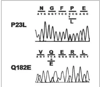
fi ed and analyzed by direct sequencing using Big Dye Terminator v1.1 (Applied Biosystems) on a 310 ABI PRISM genetic an-alyzer. The direct sequence analysis showed a heterozy-gous C > T mutation at position 68 (c.68 C>T) of the GCH1 gene, leading to an amino acid change from nonpolar pro-line (P) to nonpolar leucine (L) at position 23 (P23L) and a new heterozygous C > G transition at nucleotide posi-tion 544 (c.544 C>G) of the GCH1 gene, producing substitu-tion of the non-charged amino acid glutamine (Q) by nega-tively charged glutamic acid (E) at position 182 (Q182E) (Fig 1). The mendelian inheritance was confi rmed in the par-ents, with the mother being a P23L carrier and the father a Q182E carrier (Fig 2). Our patient is therefore a compound heterozygote for two mutant alleles in the GCH1 gene: the P23L mutation, of maternal origin, and the Q182E mutation, of paternal origin (Fig 2). The new Q182E mutation was not found in 100 normal chromosomes.
Table. Plasma levels of phenylalanine and tyrosine after oral loading with phenylalanine.
Time (hours)
Phenylalanine (µmol/L)
Tyrosine (µmol/L)
Phenylalanine/ Tyrosine
0 73 87 0.8
1 320 93 3.4
2 651 105 6.2
4 836 96 8.7
6 692 95 7.3
Fig 1. Direct-sequencing results for the GCH1 gene in an affect-ed patient showing two heterozygous mutations: the previous-ly described C > T mutation at nucleotide position 68 leading to an amino acid change from proline to leucine at position 23 (P23L); and a new C > G mutation at nucleotide position 544 leading to an amino acid change from glutamine to glutamic acid at position 182 (Q182E).
Arq Neuropsiquiatr 2007;65(4-B)
1226
Dopa-responsive dystonia: missense mutation pattern Scola et al.
After a follow-up of four years, the patient sustained the excellent response to low doses of levodopa.
All studies were done following informed consent of parents.
DISCUSSION
Genetic studies of autosomal dominant DRD fi rstly revealed linkage to the long arm of chromosome 14 and later showed this to be caused by a heterozygous mutation in the GCH1 gene located on 14q22.1-q22.27,8. This gene has a high degree of variability, and to date more than 100 different mutations have been iden-tifi ed in the GCH1 coding region5,9,10. The disease is usually confi rmed by means of biochemical tests, and abnormalities of this gene were only observed in 20 to 87% of patients2,8,9,11,12. GCH1 is responsible for cata-lyzing the formation of tetrahydrobiopterin (BH4), an essential cofactor of tyrosine hydroxylase, which is the rate-limiting step for dopamine biosynthesis2,13,14. Where as, the decrease of neopterin as well as biop-terin in cerebrospinal fl uid suggested the decrease of GCH1 enzyme as the cause of DRD2,5,13,14.
The main clinical features of dopa-responsive dy-stonia are onset of dydy-stonia at 4-8 years of age, usu-ally in the lower limbs, causing an abnormal gait, di-urnal fl uctuation of signs and symptoms and other associated features such as hyperrefl exia and exten-sor plantar refl exes, parkinsonian signs (such as mask faces), or tremor1-5,15,16. Although diurnal variation is a typical feature of DRD, characterized by worsening of symptoms from day to day and improving after sleep, as in our patient, such marked fl uctuation is present only in half patients with DRD1-5,15. DRD patients can have a marked intrafamilial variability of the clini-cal manifestations between affected members of the same family14,16.
Although individuals with DRD never develop hy-perphenylalaninemia, a subclinical defect in phenyla-lanine metabolism caused by partial BH4 defi ciency in the liver can often be detected by the phenylalanine loading test, which analyzes plasma phenylalanine-to-tyrosine ratios following an oral phenylalanine load6.
In our patient, the phenotype included dystonia after a dramatic response to L-dopa trial, and mo-lecular analysis revealed a compound heterozygos-ity for two mutations in the GCH1 gene: a previously described mutation of maternal origin (P23L) and a novel mutation of paternal origin (Q182E).
The P23L mutation in the GCH1 allele was previous-ly described in four different families17,18. It is
particu-larly interesting the fact that this sequence change was previously detected in a patient with DRD com-bined with another mutation, suggesting that P23L could represent a rare polymorphism in the popula-tion or, alternatively, that this mutapopula-tion could only be pathogenic in association with another mutation, as in our case18.
The new Q182E mutation that was found in our patient led to the substitution of a neutrally charged amino acid by a negatively charged one, a net charge alteration that can adversely affect the structure and function of the GCH1 gene at critical positions of the amino acid sequence. Another mutation (Q182X) that caused DRD was previously reported in this same ami-no acid position, highlighting the important func-tional role of this part of the protein10. The Q182E mutation in the paternal allele might be suffi cient to reduce GCH1 activity below a critical threshold, although neither mutation alone was suffi cient to cause disease in either of the carrier parents18. These observations agree with the hypothesis that the new mutation, in combination with the other previously described, is responsible for the phenotype in our case.
DRD should be considered in any child who pres-ents with paroxysmal or progressive hypertonia of unknown etiology, and its dramatic response to L-dopa has to be emphasized. Fluctuations in response, increased dose requirements, or long-term adverse effects such as the “on-off” phenomenon have not been described4, 12, 19-21. The detection of the speci
fi c mutation is important to confi rm the diagnosis, and to provide accurate treatment and genetic counseling to the patients and their family. Treatment of these patients is one of the most satisfying experiences in neurology4, 19.
Acknowledgment – The authors are grateful to Drs
Car-lo Marrone, MD and Ehrenfried Wittig, MD for their col-laboration in the case.
REFERENCES
1. Segawa M. Hereditary progressive dystonia with marked diurnal fl uc-tutation. Brain Dev 2000; 22 (Suppl 1): S65-S80.
2. Segawa M, Nomura Y, Nishiyama N. Autosomal dominant guanosine triphosphate cyclohydrolase I defi ciency (Segawa disease). Ann Neurol 2003;54:S32-S45.
3. Tarsy D, Simon D. Current concepts: dystonia. N Engl J Med 2006;355: 818-829.
4. Araujo AQC, Miranda SBM. Doença de Segawa: distonia progressive sensível à L-dopa. Arq Neuropsiquiatr 1993; 51: 532-536.
5. Venna N, Sims KB, Grant PE. Case 26-2006: a 19-year-old woman with diffi culty walking. N Engl J Med 2006; 355: 831-839.
6. Hyland K, Fryburg JS, Wilson WG, et al. Oral phenylalanine loading in dopa-responsive dystonia: a possible diagnostic test. Neurology 1997; 48: 1290-1297.
Arq Neuropsiquiatr 2007;65(4-B)
1227
Dopa-responsive dystonia: missense mutation pattern Scola et al.
8. Ichinose H, Ohye T, Takahashi E, et al. Hereditary progressive dystonia with marked diurnal fuctuation caused by mutations in the GTP cyclo-hydrolase I gene. Nat Genet 1994; 8: 236-242.
9. Furukawa Y. Genetics and biochemistry of dopa-responsive dystonia: signifi cance of striatal tyrosine hydroxylase protein loss. Adv Neurol 2003; 91: 401-410.
10. BIOMD database. Available at: ht p://www.bh4.org/BH4_databases_ biomdb2.asp (accessed August 25, 2007).
11. Furukawa Y, Kish SJ. Dopa-responsive dystonia: recent advances and remaining issues to be addressed. Mov Disord 1999;14:709-715. 12. Steinberger D, Korinthenberg R, Topka H, Berghauser M, Wedde R,
Muller U. Dopa-responsive dystonia: mutation analysis of GCH1 and analysis of therapeutic doses of L-dopa. Neurology 2000;55:1735-1737. 13. Cheng WW, Kong CK. Girl with dopa-responsive dystonia. J Pediatric
Child Health 2001;37:300-305.
14. Hahn H, Trant MR, Brownstein MJ, Harper RA, Milstien S, Butler IJ . Neurologic and psychiatric manifestations in a family with a mutation in exon 2 of the guanosine triphosphate-cyclohydrolase gene. Arch Neu-rol 2001;58:749-755.
15. Segawa M, Hosana A, Miyagawa F, Nomura Y, Imai H. Hereditary pro-gressive dystonia with marked diurnal fl uctuation. Adv Neurol 1976;
14:215-233.
16. Lopez-Laso E, Camino R, Mateos ME, et al. Dopa-responsive infantile hypokinetic rigid syndrome due to dominant guanosine triphosphate cyclohydrolase 1 defi ciency. J Neuro Sci 2007;256:90-93.
17. Leuzzi V, Carducci C, Carducci C, Cardona F, Artiola C, Antonozzi I. Autosomal dominant GTP-CH defi ciency presenting as a dopa-respon-sive myoclonus-dystonia syndrome. Neurology 2002;59:1241-1243 18. Jarman PR, Bandmann O, Marsden CD, Wood NW. GTP
cyclohydro-lase I mutations in patients with dystonia responsive to anticholinergic drugs. J Neurol Neurosurg Psychiatry 1997; 63: 304-308.
19. Jan MMS. Misdiagnoses in children with dopa-responsive dystonia. Pe-diatr Neurol 2004;31:298-303.
20. Nygaard TG, Waran SP, Levine RA, Naini AB, Chutorian AM. Dopa-responsive dystonia simulating cerebral palsy. Pediatr Neurol 1994;11: 236-240.