Influence of the borohydride concentration on the composition of the
amorphous Fe–B alloy produced by chemical reduction of synthetic,
nano-sized iron oxide particles
Part II. Goethite
V.G. de Resende
a,b,∗, E. De Grave
a, G.M. da Costa
b, J. Janssens
caDepartment of Subatomic and Radiation Physics, University of Ghent, Proeftuinstraat 86, B-9000 Gent, Belgium bUniversidade Federal de Ouro Preto, Departamento de Qu´ımica, 35400 Ouro Preto, MG, Brazil
cDepartment of Chemistry, University of Antwerpen, B-2020 Antwerpen, Belgium
Received 23 November 2006; accepted 22 December 2006 Available online 7 January 2007
Abstract
A systematic study of the effect of the NaBH4 concentration upon the composites produced by chemical reduction of nano-sized goethite
(␣-FeOOH) particles suspended in water is presented. Amorphous Fe–B alloys with boron contents between 2 and 8 at.% have been produced. The
obtained samples were characterized by X-ray diffraction, wet chemical analyses, thermal analyses, scanning electron microscopy, transmission M¨ossbauer spectroscopy (TMS) and integral low-energy electron M¨ossbauer spectroscopy (ILEEMS). It was observed that the boron content of this amorphous composite sensitively depends on the borohydride concentration. The TMS analyses have shown that the contribution of the Fe1−xBx
phase, which is present as a superposition of a broad sextet and a doublet, accounts for about 38% of the total spectral area. ILEEMS has further revealed that the reduction process mainly affects the surface layers of the goethite grains, without causing any changes in both particle shape and sizes.
© 2007 Elsevier B.V. All rights reserved.
Keywords: Goethite; Amorphous Fe–B; Chemical reduction; M¨ossbauer spectroscopy; ILEEMS
1. Introduction
A few methods are available for the preparation of amorphous transition-metal boron (TM-B) alloy particles, e.g., vapour depo-sition, splat cooling, melt spinning, and sputtering [1]. These methods differ in nature and complexity, and the appearance and properties of the resulting alloys are therefore dependent on the employed method[2].
Several authors have stressed the importance of improving existing methods for preparing amorphous TM-B alloys and, more specifically, of devising alternative methods to prepare these alloys in the form of particles rather than films or foils. A novel synthesis route involving the formation of fine metal
∗Corresponding author at: Department of Subatomic and Radiation Physics,
University of Ghent, Proeftuinstraat 86, B-9000 Gent, Belgium. Fax: +32 9 264 6697.
E-mail address:[email protected](V.G. de Resende).
and metal-boride particles via sodium borohydride (NaBH4)
reduction of metal ions in aqueous solution has been
success-fully applied [3]. This development has opened a wealth of
new possibilities for various applications of amorphous (TM-B) alloys such as ferrofluids, catalysts and recording media[4–7]. Furthermore, it has been found that the reduction process can produce amorphous alloys with high boron content, which has been demonstrated in the case of Fe–Co–B and Fe–Ni–B[3,8,9]. It was learned from earlier experiments that the composi-tion of the amorphous TM-B alloys obtained by reduccomposi-tion of metal ions in solution using sodium borohydride depends on several parameters such as the reducing agent concentration, pH of the aqueous medium, stirring speed of the solution, kind
and amount of the complex-forming agent[4,10–13]. All these
reaction parameters have been investigated, and all of them have been found to have an effect on the global composition of the TM-B alloy and its compositional homogeneity, on the particle size, and on thermal stability of the powder.
An earlier study by some of the present authors was focused on a new route for synthesis of amorphous (Fe–B) alloy particles
[14]. That route consisted of the chemical reduction by sodium borohydride of well-crystalline hematite (␣-Fe2O3) powder in
aqueous suspension. The final product is a highly magnetic black powder, which showed to be stable at room temperature. Under a given condition the NaBH4treatment converts about half of the
hematite to an amorphous Fe–B alloy and to a small fraction of submicron-sized, amorphous metallic-Fe nodules. Heating the treated sample at 400◦C resulted in the crystallization and/or
oxidation of more than half of the amorphous Fe–B phase to␣
-Fe, Fe3O4 and B2O3. Furthermore, after treatment at 800◦C
the metallic iron and the amorphous Fe–B have completely vanished, and the resulting product consists of hematite and FeBO3.
More recently, the present authors reported their results of treating synthetic nano-sized and natural micron-sized hematite
with various sodium borohydride concentrations [15]. They
showed that the particle size of the parent oxide, as well as the borohydride concentration play an important role in determining the composition of the final products: the amount of amorphous Fe1−xBxalloy formed in the reduction process strongly increases
with increasing NaBH4concentration and, for a given
concen-tration, with decreasing particle size.
It was tempting to examine how other Fe oxides respond to borohydride reduction and goethite, being the second most abundant iron oxide in the environment, was a logical choice in that respect. In this contribution, the authors report on the results of the reduction of synthetic small-particle goethite by sodium borohydride (SBH).
2. Experimental
Compounds of the type Fe1−xBx(0.01 <x< 0.09) were synthesized by
reac-tion of goethite particles with SBH. The precursor considered in this study was a synthetic goethite with a mean particle size of about 21 nm. This goethite was prepared using the method described by Schwertmann and Cornell[16].
As the nature and overall properties of the materials obtained were expected to be grossly determined by the reaction conditions (pH, amounts of reactants, reaction time, order of addition of the reactants, etc.), these conditions were carefully controlled and monitored.
Three mixtures consisting of different weight proportions of iron oxide and SBH powders were used (1:1, 1:4 and 1:8). The suspensions were obtained by addition of 1 g of goethite to 50 ml of distilled water. To each suspension, 100 ml of a 0.2 M HCl solution was drop-wise added. After the initial reactions, amounts of SBH powder, equal to the respective amounts present in the initial mixtures, were added to the obtained suspensions. This was done in four consecutive steps, each one involving one-fourth of the total amount of powder to be added. Hence, the oxide:borohydride weight proportions in the final products were 1:2, 1:8, and 1:16, respectively. These samples are henceforward denoted as BGS1, BGS2 and BGS3, respectively. All reactions were initiated at room temperature and occurred in a time span of 30 min. After thorough washing with distilled water, the products were dried at 70◦C.
X-ray diffractograms (XRD) were recorded in the range of 20–85◦(2θ) with
goniometer speed of 1◦/min by means of an XRD-6000 Shimadzu
diffractrom-eter equipped with Fe K␣radiation and Mn filter. Counts were registered every
0.02◦(2θ). Standard silicon was added in order to correct the positions of the
diffraction lines. Cell parameters were calculated by least-square refinement after subtracting the background and the K␣2contribution and using intensity
and angular weighting of the most intense peaks. Particle sizes were evaluated from the widths at half-maximum of the diffraction peaks using the well-known Scherrer formula.
The iron and boron contents in the final products were determined by wet chemical and ICP (inductively coupled plasma) analysis, respectively.
DSC measurements in the range 25–550◦C, heating rate of 5◦C/min and
under an atmosphere of synthetic air (50 ml/min), were carried out with the powders contained in open aluminium pans.
M¨ossbauer spectra (MS) at a number of different temperatures were collected with spectrometers operating at constant acceleration mode with triangular ref-erence signals.57Co(Rh) sources were used. At room temperature (RT), both conventional transmission M¨ossbauer spectroscopy (TMS spectra) and inte-gral low-energy electron M¨ossbauer spectroscopy (ILEEMS spectra) have been applied. All MS were computer-analyzed in terms of model-independent dis-tributions of hyperfine-parameter values and numerical data quoted hereafter refer to maximum probability values. Isomer shifts are referenced with respect to␣-Fe at RT.
Finally, SEM (scanning electron microscopy) images of the powder samples dispersed in acetone were collected with a Jeol apparatus.
3. Results and discussion
The reaction of the goethite with SBH resulted in products with different colours and magnetism, depending on the amount of borohydride used in the reaction. However, the particle size of the treated samples is approximately the same as for the original goethite. When the amount of SBH increased, the colour of the particles in suspension changed from yellowish-green to dark-green. These colours persisted even after washing and drying the samples. The grains of sample BGS3 are the most magnetic, as experienced by using a hand magnet.
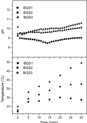
Fig. 1(top) shows that, except for the lowest SBH concen-tration (sample BGS1), the pH of the reaction medium initially increases during the chemical-reduction process. The temper-ature of the medium (Fig. 1, bottom) increases for all three
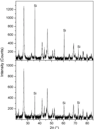
Fig. 2. X-ray diffraction patterns of the synthetic goethite (top) and treated sample BGS3 (bottom). Si corresponds to standard silicon which was added to the sample.
samples. Forster et al.[12]reported that the reaction with boro-hydride proceeds via iron-hydroborate intermediates, which presumably involve exothermal reactions, thus increasing pH and temperature of the reaction medium. These intermediates have not yet been isolated due to the reaction being extremely fast.
Although macroscopic changes were observed in the treated samples, the XRD patterns of all samples merely show the diffraction lines of goethite (Fig. 2). The presence of any other crystalline phase is not observed, implying that the phase respon-sible for the magnetism and colour of the samples is amorphous, most likely amorphous Fe–B alloy as reported by de Resende et al.[14].
The total iron and boron contents of the treated samples, as well as the global compositions of the amorphous Fe–B alloys are listed in Table 1. The bulk compositions were cal-culated assuming that no other phases, in addition to goethite and Fe1−xBx, alloy are present. The results show that the boron
content increases as the SBH concentration increases, which is
Table 1
Boron and iron content (wt.%), and the global composition of the amorphous Fe–B alloy
Goethite/NaBH4(g) Sample Boron Fetotal Fe1−xBx
0 GS 0.00 62.2±0.3 –
1:2 BGS1 0.4±0.1 61.3±0.1 Fe0.98B0.02 1:8 BGS2 1.5±0.1 62.7±0.6 Fe0.93B0.07 1:16 BGS3 1.7±0.1 62.4±0.4 Fe0.92B0.08
in line with the results reported for Fe1−xBxsystems prepared
by the reduction of Fe ions using borohydride in aqueous solu-tions[4]. As mentioned by these authors, the SBH concentration is the most important reaction parameter, which may determine the boron content in the bulk composition.
Differential scanning calorimetry (DSC) was carried out to characterize the crystallization temperature and thermal sta-bility of the amorphous Fe–B alloys. It is well known that
goethite undergoes a thermal decomposition at∼260◦C,
giv-ing rise to the formation of hematite. The DSC curve for the synthetic goethite (sample GS) is shown inFig. 3(top). The pres-ence of several endothermic processes in the temperature range
between 170 and 265◦C is evident. During thermal
decom-position, goethite continuously looses mass due to removal of crystalline water molecules and/or remaining hydroxyl groups from the hematite structure. The transformation of goethite to hematite proceeds without the formation of any other intermedi-ary phase. In DSC curves only one peak should thus be obvious, but in many cases one observes two or more shoulders besides
the main peak (Fig. 3). Schwertmann[17] proposed that the
additional peaks are due to the transition of two phases of well-crystallized goethite. Another explanation for this multi-peak
appearance of the DSC curve was given by Goss[18]who
pro-posed that the satellite DSC peaks are due to non-structural water in the goethite.
DSC curves for all three reduced samples show a single
endothermic peak and one or two exothermic peaks (Fig. 3).
The endothermic peak, centered at approximately 270◦C, is
attributed to the thermal decomposition of␣-FeOOH as
men-tioned in the previous paragraph. If the multi-peak DSC signal of
sample GS is indeed due to the presence of non-structural water on the surface of the␣-FeOOH particles, then the results for the
treated samples suggest that this non-structural water is removed from the surface of the goethite particles during the reaction with SBH. The DSC curve of sample BGS1 shows, in addition to the endothermic process associated to the decomposition of goethite, the presence of a weak exothermic peak centered at
about 480◦C. On the other hand, the DSC curves of samples
BGS2 and BGS3 reflect the occurrence of two exothermic
pro-cesses in the range between 415 and 507◦C. These exothermic
processes are believed to be associated to the crystallization and subsequently oxidation of the amorphous Fe–B phases[19].
The MS at RT of well-crystallized goethite and without iso-morphic substitution is composed of an asymmetric sextet with magnetic hyperfine field (Hhf) equal to 380 kOe. TheHhfvalue
is approximately 500 kOe at 77 K[20,21]. For the GS sample,
theHhf at RT was found to be 378 kOe whereas at 16 K it is
505 kOe. These values are in good agreement with the above mentioned data.
TMS spectra at RT and at 16 K for the treated samples are shown inFig. 4. All spectra consist of a predominant sextet due to non-reacted goethite, superimposed to a quadrupole doublet labeled Fe0or to a broad sextet or to both. The relative areas RA of the doublet and of the broad sextet increase with increasing SBH concentration and on lowering the temperature for a given sample RA of the doublet decreases while that of the broad sextet increases.
Several trial fits were attempted to obtain an adequate model to describe the experimental TMS spectra, leading to parameter
Fig. 4. Temperature dependence of the M¨ossbauer spectra at RT (left) and at 16 K (right) of goethite samples treated with sodium borohydride.
Table 2
M¨ossbauer results of the treated samples
T(K) Broad sextet Fe0doublet
Hhf (kOe)
RA (%) δ(mm/s) EQ (mm/s)
RA (%) δ(mm/s)
BGS1
16 245a 7 0.48a – – –
40 245 7 0.47a – – –
80 245 6 0.44a – – –
RT – – – 0.85a 3 0.36a
BGS2
16 240 30 0.48a – – –
40 242 27 0.47a – – –
80 234 25 0.41 0.86 3 0.41
RT 224 22 0.36 0.82 7 0.36
BGS3
16 234a 38 0.48a – – –
40 239 33 0.47a 0.85a 4 0.47a
80 238 30 0.42 0.85a 7 0.42
RT 225 30 0.35 0.85 8 0.35
The values of isomer shifts are with reference to metallic iron at room tempera-ture.Note:Hhf= maximum probability hyperfine fields; 2εQ= quadrupole shifts; EQ= quadrupole splitting;δ= isomer shift;T= temperature and RA = relative areas.
aFixed parameter.
values that consistently vary with temperature and SBH con-centration. Some constraints that were used in these trial fits are:
• the range of hyperfine-field value of the broad sextet was
chosen to be the same for all temperatures;
• the area ratios of outer lines to middle lines to inner lines for all elemental magnetic components were forced to be equal to 3:2:1;
• for the broad sextet component, the quadrupole shift was taken to be zero;
• the Fe0doublet and broad sextet were constrained to exhibit the same, but adjustable centre-shift values at any giving tem-perature.
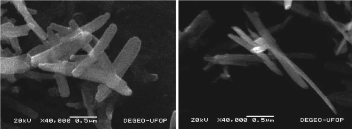
Fig. 6. Scanning electron microscopy images of samples GS (left) and BGS3 (right).
The numerical results of the fittings are listed in Table 2. These results, combined with the X-ray diffraction, thermal and chemical analyses results have prompted the authors to suggest that the Fe0 doublet and the broad sextet are both due to the
same amorphous Fe1−xBx alloy phase which exhibits a more
or less broad distribution for the compositional parameter x. As suggested in the companion paper concerning the chemical reduction of hematite[15]the idea is that at a given tempera-ture, Fe1−xBxgrains withxexceeding a certain threshold value
(depending on temperature) are (super)paramagnetic, giving rise to the Fe0doublet; whereas other grains with lower B content produce the sextet.
To examine the composition of the surface layers of the pow-der, an ILEEMS measurement at RT for the BGS3 has been performed (Fig. 5). ILEEMS is a variant of conventional
trans-mission M¨ossbauer spectroscopy[22]by which the low-energy
electrons are counted. These electrons, with energy of∼10 eV, are produced by after effects following the decay of the probe nuclei in the absorber. As a consequence of this low energy, only an extremely thin surface layer of the material is probed. In com-parison with the transmission spectrum (Fig. 4), the emission spectrum shows an additional very weak sextet component. This sextet is attributed to a layer of hematite present at the surface of the as-prepared goethite grains, as reported by De Grave et al.[22]. Additionally, it is observed that the goethite phase has a significantly weaker contribution to the total ILEEMS spectrum as compared to its contribution to the TMS, i.e., 46% instead of 62%, respectively. This observation is a clear evidence that the surface layers (2–3 nm) of BGS3 are enriched with Fe–B grains. For completeness, it should be mentioned that the
hyper-fine parameters of the␣-FeOOH and Fe–B phases obtained from
the ILEEMS are within the error limits identical to the values fitted to the TMS spectra.
SEM images of the synthetic goethite and with the highest amount of SBH are reproduced inFig. 6. These images show that the particles appear to be non-porous and that the reaction with SBH did not significantly affect their morphology, in contrast to what was observed earlier for hematite particles that had been subjected to the same treatment.
In summary, goethite appears to be less reactive with the boro-hydride as compared to synthetic hematite with similar particle
size. The amount of reduced goethite increases with increasing SBH concentration, but even for the highest amount of added
NaBH4(16 g), only 38% of the␣-FeOOH is reduced to form
the amorphous Fe–B alloy, while for hematite the reduction was
found to be 63% [15]. Also the boron content of the
amor-phous Fe–B alloy is on average lower in the case of the goethite reduction.
Acknowledgements
This work was partially funded by CNPq and Fapemig (Brazil), and by the Fund for Scientific Research, Flanders, Bel-gium. The authors gratefully acknowledge the Program Alban (the European Union Programme of High Level Scholarships for Latin America) for a grant (E04M034189BR) received by V.G. de Resende to finalize her MSc studies in the Department of Subatomic and Radiation Physics.
References
[1] S. Linderoth, S. Mørup, A. Meagher, J. Larsen, M.D. Bentzon, B.S. Clausen, C.J.W. Koch, S. Wells, S.W. Charles, J. Magn. Magn. Mater. 81 (1989) 138.
[2] S. Linderoth, C.A. Oxborrow, O.V. Nielsen, Nucl. Instrum. Meth. B 76 (1993) 64.
[3] S. Wells, S.W. Charles, S. Mørup, S. Linderoth, J. van Wonterghem, J. Larsen, M.B. Madsen, J. Phys.: Condens. Matter 1 (1989) 8199. [4] S. Linderoth, S. Mørup, J. Appl. Phys. 69 (8) (1991) 5256.
[5] Z. Hu, Y. Hsia, J. Zheng, J. Shen, Q. Yan, L. Dai, J. Appl. Phys. 70 (1991) 436.
[6] J. Shen, Z. Hu, Q. Zhang, L. Zhang, Y. Chen, J. Appl. Phys. 71 (1992) 5217.
[7] G. Principi, A. Maddalena, A. Gupta, G. Bottoni, D. Candolfo, A. Cecchetti, A.R. Corradi, Nucl. Instrum. Meth. B 76 (1993) 143.
[8] J. Saida, M. Ghafari, Y. Nakamura, A. Inoue, T. Masumoto, Nucl. Instrum. Meth. B 76 (1993) 223.
[9] S. Morup, S.A. Sethi, S. Linderoth, C. BenderKoch, M.D. Bentzon, J. Mater. Sci. 27 (1992) 3010.
[10] S. Linderoth, S. Mørup, J. Appl. Phys. 67 (9) (1990) 4472.
[11] J. Rivas, M.A.L. Quintela, M.G. Bonome, R.J. Duro, J.M. Greneche, J. Magn. Magn. Mater. 122 (1993) 1.
[12] G.D. Forster, L.F. Barqu´ın, R.L. Bilsborrow, Q.A. Pankhurst, I.P. Parkin, W.A. Steer, J. Mater. Chem. 9 (1999) 2537.
[14] V.G. de Resende, G.M. da Costa, E. De Grave, L. Datas, J. Mater. Sci. 41 (2006) 6843.
[15] V.G. de Resende, E. De Grave, G.M. da Costa, J. Janssens, J. Alloy Compd. 440 (2007) 236.
[16] U. Schwertmann, R.M. Cornell, Iron Oxides in the Laboratory— Preparation and Characterization, Wiley-VCH, 2000.
[17] U. Schwertmann, Thermochim. Acta 78 (1984) 39.
[18] E.J. Goss, Mineral. Mag. 51 (1987) 437.
[19] G.D. Foster, L.F. Barqu´ın, N.S. Cohen, Q.A. Pankhurst, I.P. Parkin, J. Mater. Proc. Technol. 92/93 (1999) 525.
[20] E. Murad, U. Schwertmann, Clay Miner. 18 (1983) 301.
[21] E. De Grave, R.E. Vandenberghe, Hyperfine Interact. 28 (1986) 643. [22] E. De Grave, R.E. Vandenberghe, C. Dauwe, Hyperfine Interact. 161 (2005)