Epidemiology of enteroaggregative
Escherichia
coli
infections and associated outcomes in the
MAL-ED birth cohort
Elizabeth T. Rogawski1,2*, Richard L. Guerrant2, Alexandre Havt3, Ila F. N. Lima3, Pedro H. Q. S. Medeiros3, Jessica C. Seidman4, Benjamin J. J. McCormick4, Sudhir Babji5,
Dinesh Hariraju5, Ladaporn Bodhidatta6, Jasmin Shrestha7, Japhat Anania8, Athanasia Maro8, Amidou Samie9, Pablo Peñataro Yori10, Shahida Qureshi11,
Mustafa Mahfuz12, Pascal O. Bessong9, Margaret N. Kosek10,13, Tahmeed Ahmed12, Zulfiqar A. Bhutta11, Dennis R. Lang14, Michael Gottlieb14, Eric R. Houpt2, Aldo A.
M. Lima3, the MAL-ED Network Investigators¶
1Department of Public Health Sciences, University of Virginia, Charlottesville, Virginia, United States of America,2Division of Infectious Diseases and International Health, University of Virginia, Charlottesville, Virginia, United States of America,3Clinical Research Unit and Institute of Biomedicine, Federal University of Ceara, Fortaleza, Brazil,4Fogarty International Center, National Institutes of Health, Bethesda, Maryland, United States of America,5Division of Gastrointestinal Sciences, Christian Medical College, Vellore, India,
6Department of Enteric Diseases, Armed Forces Research Institute of Medical Sciences, Bangkok, Thailand,7Walter Reed-AFRIMS Research Unit, Nepal, Kathmandu, Nepal,8Haydom Global Health Research Center, Haydom Lutheran Hospital, Haydom, Tanzania,9Department of Microbiology, University of Venda, Thohoyandou, South Africa,10 Asociacio´n Bene´fica PRISMA, Iquitos, Peru,11Department of Paediatrics and Child Health, Aga Khan University, Karachi, Pakistan,12 Nutrition and Clinical Services Division, International Centre for Diarrhoeal Disease Research, Dhaka, Bangladesh,13Department of International Health, Bloomberg School of Public Health, Johns Hopkins University, Baltimore, Maryland, United States of America,14Foundation for the National Institutes of Health, Bethesda, Maryland, United States of America
¶ Membership of the MAL-ED Network Investigators is provided in the Acknowledgments. *etr5m@virginia.edu
Abstract
Background
EnteroaggregativeE.coli(EAEC) have been associated with mildly inflammatory diarrhea in outbreaks and in travelers and have been increasingly recognized as enteric pathogens in young children with and without overt diarrhea. We examined the risk factors for EAEC infections and their associations with environmental enteropathy biomarkers and growth outcomes over the first two years of life in eight low-resource settings of the MAL-ED study.
Methods
EAEC infections were detected by PCR gene probes foraatAandaaiCvirulence traits in 27,094 non-diarrheal surveillance stools and 7,692 diarrheal stools from 2,092 children in the MAL-ED birth cohort. We identified risk factors for EAEC and estimated the associations of EAEC with diarrhea, enteropathy biomarker concentrations, and both short-term (one to three months) and long-term (to two years of age) growth.
a1111111111 a1111111111 a1111111111 a1111111111 a1111111111 OPEN ACCESS
Citation:Rogawski ET, Guerrant RL, Havt A, Lima IFN, Medeiros PHQS, Seidman JC, et al. (2017) Epidemiology of enteroaggregativeEscherichia coli
infections and associated outcomes in the MAL-ED birth cohort. PLoS Negl Trop Dis 11(7): e0005798.
https://doi.org/10.1371/journal.pntd.0005798
Editor:Stephen Baker, Oxford University Clinical Research Unit, VIET NAM
Received:April 20, 2017
Accepted:July 11, 2017
Published:July 24, 2017
Copyright:This is an open access article, free of all copyright, and may be freely reproduced, distributed, transmitted, modified, built upon, or otherwise used by anyone for any lawful purpose. The work is made available under theCreative Commons CC0public domain dedication.
Data Availability Statement:Public availability of individual participant data would compromise participant privacy. While data are not publicly available due to ethical restrictions, project data for this study are available upon request to others in the scientific community. For access, please contact Stacey Knobler (knoblersl@mail.nih.gov; MAL-ED project management).
Results
Overall, 9,581 samples (27.5%) were positive for EAEC, and almost all children had at least one detection (94.8%) by two years of age. Exclusive breastfeeding, higher enrollment weight, and macrolide use within the preceding 15 days were protective. Although not asso-ciated with diarrhea, EAEC infections were weakly assoasso-ciated with biomarkers of intestinal inflammation and more strongly with reduced length at two years of age (LAZ difference associated with high frequency of EAEC detections: -0.30, 95% CI: -0.44, -0.16).
Conclusions
Asymptomatic EAEC infections were common early in life and were associated with linear growth shortfalls. Associations with intestinal inflammation were small in magnitude, but suggest a pathway for the growth impact. Increasing the duration of exclusive breastfeeding may help prevent these potentially inflammatory infections and reduce the long-term impact of early exposure to EAEC.
Author summary
EnteroaggregativeE.coli(EAEC) are pathogens that infect the intestine and can cause diarrhea. They are also commonly identified among young children in low-resource set-tings, who can carry the pathogen without symptomatic diarrhea. We examined the risk factors for EAEC infections and their associations with child health outcomes over the first two years of life in eight low-resource settings of the MAL-ED study. EAEC infections were detected using molecular methods in more than 30,000 stools collected from 2,092 children in the MAL-ED study. We identified risk factors for EAEC and estimated the associations of EAEC with diarrhea, markers of intestinal health, and child growth. Almost all children were infected with EAEC at least once by two years of age. Exclusive breastfeeding, higher enrollment weight, and recent macrolide antibiotic use were protec-tive against these infections. Although not associated with diarrhea in these children, EAEC infections were associated with intestinal inflammation and reduced length at two years of age. EAEC may impact child development, even in the absence of diarrhea, by causing intestinal inflammation and impairing child growth.
Introduction
EnteroaggregativeEscherichia coli(EAEC) infections have been increasingly recognized as important enteropathogens since their initial discovery by patterns of adherence to HEp-2 cells inE.coliisolates from Chilean children with diarrhea [1]. EAEC have since been associ-ated with foodborne outbreaks of diarrhea [2], traveler’s diarrhea [3–5], diarrhea in adults with HIV infection [6], endemic diarrhea in cities in the US [7], and variably in healthy adult human volunteers [8,9]. A meta-analysis of 41 studies found EAEC to be significantly associ-ated with acute diarrheal illness among both children and adults in developing regions [10]. However, because EAEC are also a highly common infection among children without overt diarrhea in low-resource settings, they have not been found to be a major cause of diarrhea in some endemic settings [11,12]. Regardless, EAEC, independent of diarrheal symptoms, have
Development Project (MAL-ED) is carried out as a collaborative project supported by the Bill & Melinda Gates Foundation, the Foundation for the NIH and the National Institutes of Health/Fogarty International Center. This work was supported by the Fogarty International Center, National Institutes of Health (D43-TW009359 to ETR). The funders had no role in study design, data collection and analysis, decision to publish, or preparation of the manuscript.
been associated with other poor health outcomes in children, such as growth failure [13] and mild to moderate intestinal inflammation [5,13,14].
The genetic determinants and biological mechanism for the virulence of EAEC have been described by a complex array of interacting traits that reside on both the chromosome and plasmid in the organism [15,16]. As presently defined, EAEC are heterogeneous with respect to virulence gene content. TheaggRtrait on the plasmid is a common and well-characterized EAEC gene [17] that regulates many virulence traits, including chromosomalaaiC, which is in the gene clusteraaiA-Ythat encodes the type VI secretion system, as well as plasmid-borne
aatA, which encodes an ABC transporter. In addition, the flagellin of EAEC strain 042 has been shown to trigger inflammation via TLR5 signaling [18,19]. Murine models have helped determine the impact of these virulence genes by providing evidence that EAEC can cause inflammation, enteropathy, and growth shortfalls among mice with dietary protein deficiency [20,21], and even diarrhea among mice with dietary zinc deficiency [22].
Increasing evidence suggests that enteric infections, especially common pathogens like EAEC, may play an important role in morbidity due to enteric disease, beyond symptomatic diarrhea [23]. While mortality from diarrheal diseases has been dramatically reduced to less than half a million deaths per year [24], more than a quarter of the world’s children are moder-ately or severely stunted [25]. Because improved feeding does not eliminate growth shortfalls in low-resource areas where inadequate water and sanitation and heavy burdens of enteric infections are common [26,27], enteric infections and sub-clinical environmental enteropathy likely also contribute to poor child growth outcomes [28,29].
We characterized the epidemiology and impact of EAEC infections among children in the first two years of life in eight low-resource settings of the Etiology, Risk Factors, and Interac-tions of Enteric InfecInterac-tions and Malnutrition and the Consequences for Child Health and Development Project (MAL-ED) study. With twice-weekly active surveillance from near birth to two years of age, the MAL-ED study provides a unique opportunity to assess the impact of both clinical and subclinical enteric infections on early-life growth and development. We examined risk factors for EAEC infections and their associations with diarrhea, environmental enteropathy biomarkers, and growth outcomes over the first two years of life.
Methods
The study design and methods of the MAL-ED study have been extensively described [30]. Briefly, children were enrolled within 17 days of birth and followed until two years of age at eight sites: Dhaka, Bangladesh (BGD), Vellore, India (INV), Bhaktapur, Nepal (NEB), Naush-ahro Feroze, Pakistan (PKN), Fortaleza, Brazil (BRF), Loreto, Peru (PEL), Venda, South Africa (SAV), and Haydom, Tanzania (TZH). Non-diarrheal surveillance stool samples were col-lected monthly and diarrheal stool samples were colcol-lected during 94% of diarrhea episodes identified by active surveillance at twice weekly home visits. Diarrhea was defined as maternal report of three or more loose stools in 24 hours or one stool with visible blood [31]. Monthly surveillance stool samples in the first year of life, quarterly stool samples in the second year of life, and all diarrheal stool samples were tested for more than 50 enteropathogens [32] and stool biomarkers of environmental enteropathy:α-1-antitrypsin (ALA), myeloperoxidase (MPO), and neopterin (NEO) [33]. For EAEC specifically, we picked and pooled five lactose-fermenting colonies resemblingE.coli, and characterized them for virulence genes using a multiplex polymerase chain reaction (PCR) assay. Presence of the enteroaggregativeE.coli
We included all stool samples that were tested for EAEC in this analysis even if they were not tested for the full suite of other pathogens.
Fieldworkers also collected information on other illnesses, medicines, and feeding practices at home visits. Sociodemographic information was collected by questionnaire biannually and summarized using a construct of access to improved water and sanitation (as defined by WHO guidelines [34]), wealth measured by eight assets, years of maternal education, and average monthly household income (Water, Assets, Maternal education, and Income, WAMI) [35]. Plasmaα-1-acid glycoprotein (AGP), a marker of systemic inflammation, was measured at 7, 15, and 24 months. Urinary lactulose:mannitol excretion ratios were measured at 3, 6, 9 and 15 months and converted into a sample-based z-score (LMZ) using the Fortaleza, Brazil cohort as the internal reference population [36]. Weight and length were measured monthly and con-verted into weight-for-age (WAZ) and length-for-age (LAZ) z-scores using the 2006 WHO child growth standards [37]. Length measurements from Pakistan were excluded due to mea-surement quality concerns.
Ethics statement
The study was approved by the Institutional Review Board for Health Sciences Research, Uni-versity of Virginia, USA as well as the respective governmental, local institutional, and collabo-rating institutional ethical review boards at each site: Ethical Review Committee, ICDDR,B (BGD); Committee for Ethics in Research, Universidade Federal do Ceara; National Ethical Research Committee, Health Ministry, Council of National Health (BRF); Institutional Review Board, Christian Medical College, Vellore; Health Ministry Screening Committee, Indian Council of Medical Research (INV); Institutional Review Board, Institute of Medicine, Tribhu-van University; Ethical Review Board, Nepal Health Research Council; Institutional Review Board, Walter Reed Army Institute of Research (NEB); Institutional Review Board, Johns Hopkins University; PRISMA Ethics Committee; Health Ministry, Loreto (PEL); Ethical Review Committee, Aga Khan University (PKN); Health, Safety and Research Ethics Commit-tee, University of Venda; Department of Health and Social Development, Limpopo Provincial Government (SAV); Medical Research Coordinating Committee, National Institute for Medi-cal Research; Chief MediMedi-cal Officer, Ministry of Health and Social Welfare (TZH). Informed written consent was obtained from the parent or guardian of each participating child on their behalf.
Data analysis
We identified risk factors for EAEC detection in surveillance stools using log-binomial regres-sion with general estimating equations (GEE) and robust variance to account for correlation between stools within children, adjusting for site and a restricted quadratic spline [38] for age. Variables were assessed individually in this model and were included in the multivariable model if statistically significant (p<0.05). We estimated the association between EAEC and
diarrheal versus non-diarrheal stools using Poisson regression with the robust variance estima-tor to estimate risk ratios [39] since log-binomial models did not converge, adjusting for the age spline, site, the interaction between age and site, and antibiotic use within the preceding 15 days.
BecauseCampylobacterwas the most common pathogen detected in stools and has been previ-ously shown to be associated with intestinal inflammation in the MAL-ED cohort [40,41], we assessed potential interactions between the effects of EAEC andCampylobacteron MPO by including an interaction term between presence of EAEC andCampylobacter. All estimates were adjusted for site, the age spline, sex, WAMI, percent exclusive breastfeeding in previous month, contemporary presence ofCampylobacterin the stool sample, and a qualitative description of stool consistency (for stool biomarkers only).
Finally, we estimated the association between EAEC detection and short-term and long-term growth using multivariable linear regression. Short-long-term growth was defined by the change in WAZ and LAZ over both the one and three months following each monthly stool collection. We compared differences in short-term growth velocity between children who had surveillance stools with and without EAEC detection, using GEE and adjusting for site, age, sex, WAMI, percent exclusive breastfeeding in the exposure month, and detection of Campylo-bacterin the stool.
We further assessed the interaction between MPO levels and EAEC positivity to explore the role of intestinal inflammation in the potential effect of EAEC on short-term growth impairment. In the adjusted short-term growth models examining WAZ and LAZ velocity over the one and three months following EAEC testing, we estimated the additive interaction effect of EAEC detection and high MPO concentration in the same stool using an interaction term. High MPO was defined as an MPO concentration in the highest quartile on the logarith-mic scale among all non-diarrheal stools collected at that child’s site and 3-month age period. Values defining high MPO (range: 2,515–33,190 ng/mL) were higher than previous reports from non-tropical settings (<2,000 ng/mL) [42].
Effects on long-term growth were then estimated as the total difference in size at two years of age as a function of the percent surveillance stools positive for EAEC. The long-term model was adjusted for the WAZ and LAZ measurements at enrollment (within 17 days of birth), site, sex, WAMI, the age at which exclusive breastfeeding first stopped, and the percent surveillance stools positive forCampylobacterin the first 2 years of life. Adjusting for the same covariates, we assessed the potential synergistic interaction between the effects of EAEC andCampylobacter
on growth at 2 years given that both have been associated with gut inflammation, by including an interaction term between an indicator for a high frequency of detection (at least 50% surveil-lance stools positive) of EAEC and an indicator for a high frequency of detection of Campylo-bacter. We also repeated the model described above, but focused on EAEC detections in specific age periods (1–6, 7–12, and 15–24 months) and growth outcomes at 2 years to assess if there were specific age periods of susceptibility.
Results
We included 27,094 non-diarrheal surveillance stools and 7,692 diarrheal stools that were tested for EAEC from 2,092 children who each contributed at least one stool sample in the MAL-ED birth cohort. 1,736 (83.0%) of these children were followed to two years of age. Over-all, 9,581 samples (27.5%) were positive for EAEC;aatAwas detected in 41.6% (n = 3,982) of EAEC-positive stool samples,aaiCwas detected in 31.4% (n = 3,007), and both genes together were detected in 27.1% (n = 2,592).
child when including surveillance and diarrheal stools. The median number of detections in surveillance stools among children who completed two years of follow-up was 2 in Peru, 3 in South Africa, and 4 or 5 at all other sites.
Risk factors
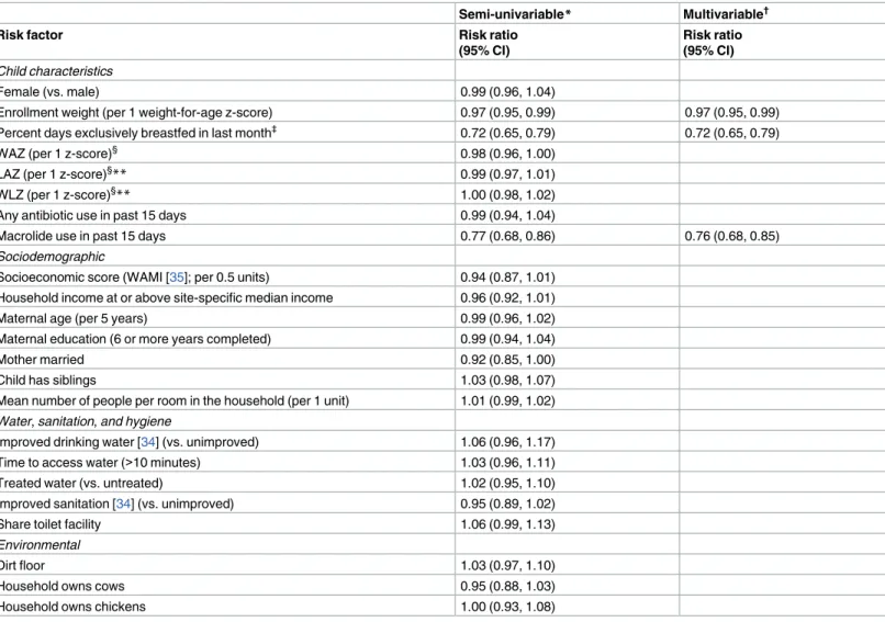
Because of the near ubiquity of EAEC detection in these study sites, few factors were identified that were associated with EAEC detection in surveillance stools. Enrollment weight, exclusive breastfeeding, and recent macrolide use were the only protective factors in the multivariable analysis, and only the associations with the latter two had a substantial magnitude of effect (Table 1). Socioeconomic status (WAMI) was weakly protective, but the association was not statistically significant. Macrolide use in the past 15 days, but not cephalosporin use nor any other antibiotic use, was associated with a reduction in EAEC detection. However, macrolide use in the past 16–30 days was not protective (RR: 0.94, 95% CI: 0.85, 1.05). This short-term only effect of macrolide use was consistent across all sites and ages.
EAEC and diarrhea
Adjusting for age, site, and their interaction, EAEC was not associated with diarrhea and was found significantly more often in surveillance stools compared to diarrheal stools (RR: 0.86, 95% CI: 0.82, 0.90). This association remained when adjusting for recent antibiotic use and specifically macrolide use, as well as if restricted to only those children with no antibiotic use in the past 30 days. Similarly, presence of EAEC in stools was not associated with persistent diarrhea (duration of 14 days or more; RR: 0.93, 95% CI: 0.73, 1.18) compared to non-diar-rheal stools.
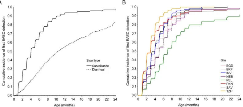
Fig 1. Incidence of EAEC.Cumulative incidence of first EAEC detection in A) surveillance and diarrheal stools at all sites and B) surveillance stools by site among 2,092 children with at least one stool sample in the MAL-ED birth cohort. BGD–Dhaka, Bangladesh; BRF–Fortaleza, Brazil; INV–Vellore, India; NEB–Bhaktapur, Nepal; PEL–Loreto, Peru; PKN–Naushahro Feroze, Pakistan; SAV–Venda, South Africa; TZH–Haydom, Tanzania.
Association with markers of environmental enteropathy
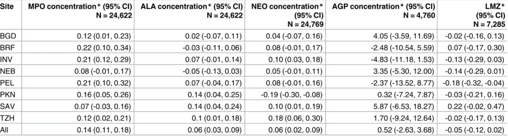
EAEC detection was associated with higher contemporary concentrations of MPO (MPO 0.14 ln(ng/mL), 95% CI: 0.11, 0.18 higher in the presence of EAEC), a marker of intestinal inflam-mation, at all sites (Table 2). It was also associated with higher levels of ALA (permeability) and NEO (intestinal inflammation) overall, with some variation across sites. However, the magnitudes of these associations were very small (1.15 ng/mL difference in MPO) compared to the range of observed concentrations in the study (MPO interquartile range: 2,050–12,920 ng/mL). In addition, EAEC was not associated with AGP, a marker of systemic inflammation, nor the lactulose-mannitol ratio, a marker of intestinal permeability, measured during the same month as the stool collection.
Table 1. Risk factors for EAEC detection in monthly surveillance stools among 2,091 children in the MAL-ED cohort with at least one surveillance stool.
Semi-univariable* Multivariable†
Risk factor Risk ratio
(95% CI)
Risk ratio (95% CI)
Child characteristics
Female (vs. male) 0.99 (0.96, 1.04)
Enrollment weight (per 1 weight-for-age z-score) 0.97 (0.95, 0.99) 0.97 (0.95, 0.99) Percent days exclusively breastfed in last month‡ 0.72 (0.65, 0.79) 0.72 (0.65, 0.79)
WAZ (per 1 z-score)§ 0.98 (0.96, 1.00)
LAZ (per 1 z-score)§
** 0.99 (0.97, 1.01)
WLZ (per 1 z-score)§** 1.00 (0.98, 1.02)
Any antibiotic use in past 15 days 0.99 (0.94, 1.04)
Macrolide use in past 15 days 0.77 (0.68, 0.86) 0.76 (0.68, 0.85)
Sociodemographic
Socioeconomic score (WAMI [35]; per 0.5 units) 0.94 (0.87, 1.01) Household income at or above site-specific median income 0.96 (0.92, 1.01)
Maternal age (per 5 years) 0.99 (0.96, 1.02)
Maternal education (6 or more years completed) 0.99 (0.94, 1.04)
Mother married 0.92 (0.85, 1.00)
Child has siblings 1.03 (0.98, 1.07)
Mean number of people per room in the household (per 1 unit) 1.01 (0.99, 1.02)
Water,sanitation,and hygiene
Improved drinking water [34] (vs. unimproved) 1.06 (0.96, 1.17) Time to access water (>10 minutes) 1.03 (0.96, 1.11)
Treated water (vs. untreated) 1.02 (0.95, 1.10)
Improved sanitation [34] (vs. unimproved) 0.95 (0.89, 1.02)
Share toilet facility 1.06 (0.99, 1.13)
Environmental
Dirt floor 1.03 (0.97, 1.10)
Household owns cows 0.95 (0.88, 1.03)
Household owns chickens 1.00 (0.93, 1.08)
*Adjusted for site and age only (using restricted quadratic splines) †Adjusted for site, age, and all other variables with estimates in this column
‡ Included as a continuous variable; risk ratio is scaled for the comparison of exclusive breastfeeding on all days in previous month to exclusive breastfeeding on no days in the previous month
§At most recent measurement prior to stool collection. WAZ–weight-for-age z-score; LAZ–length-for-age z-score; WLZ–weight-for-length z-score. **Excluding Pakistan.
EAEC was associated with elevated MPO independently ofCampylobacter, but their com-bined effect on MPO was less than additive when both pathogens were present. Detection of EAEC alone was associated with an adjusted 0.17 (95% CI: 0.13, 0.21) higher ln(MPO) concen-tration,Campylobacteralone was associated with an adjusted 0.19 (95% CI: 0.15, 0.24) higher concentration, and the detection of both pathogens was associated with an adjusted 0.27 (95% CI: 0.21, 0.34) higher concentration.
Effects of EAEC infection on growth
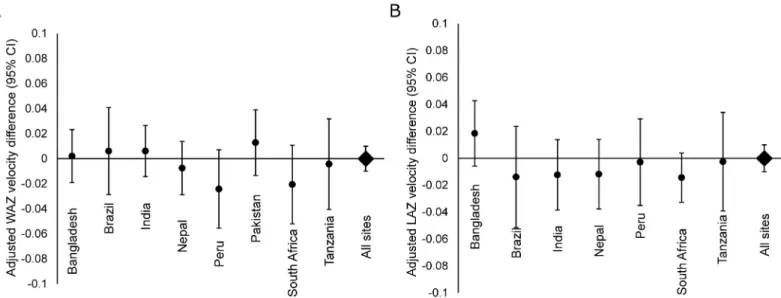
Detection of EAEC was not associated with short term differences in growth velocity in both the one and three months following each monthly stool collection overall or at any site (Fig 2). Furthermore, there was no evidence of an interaction between EAEC detection and MPO in the same stool (pfor interaction: 0.9 and 0.5 for 1-month WAZ and LAZ velocity, respec-tively); concurrent detection of EAEC and a high level of MPO were also not associated with short-term WAZ and LAZ velocity.
Over the course of the first two years of life, there was no difference at 2 years in WAZ (overall difference: -0.05, 95% CI: -0.18, 0.08) associated with a linear increase in EAEC stool positivity (Fig 3). In contrast, more detections of EAEC were associated with significant decre-ments in LAZ (Fig 3). The difference in LAZ at 2 years of age between a child at the 90th
Table 2. Associations between EAEC detection and markers of inflammation and gut permeability in surveillance and diarrheal stools among 2,076 children in the MAL-ED cohort with at least one biomarker measurement.
Site MPO concentration*(95% CI) N = 24,622
ALA concentration*(95% CI) N = 24,622
NEO concentration* (95% CI) N = 24,769
AGP concentration*(95% CI) N = 4,760
LMZ* (95% CI) N = 7,285
BGD 0.12 (0.01, 0.23) 0.02 (-0.07, 0.11) 0.04 (-0.07, 0.16) 4.05 (-3.59, 11.69) -0.02 (-0.16, 0.13) BRF 0.22 (0.10, 0.34) -0.03 (-0.11, 0.06) 0.08 (-0.01, 0.17) -2.48 (-10.54, 5.59) 0.07 (-0.17, 0.30) INV 0.21 (0.12, 0.29) 0.07 (-0.01, 0.14) 0.10 (0.03, 0.18) -4.83 (-11.18, 1.53) -0.13 (-0.29, 0.03) NEB 0.08 (-0.01, 0.17) -0.05 (-0.13, 0.03) 0.05 (-0.01, 0.11) 3.35 (-5.30, 12.00) -0.14 (-0.29, 0.01) PEL 0.21 (0.10, 0.32) 0.07 (-0.04, 0.17) 0.08 (-0.01, 0.16) -2.37 (-13.52, 8.77) -0.18 (-0.32, -0.04) PKN 0.16 (0.05, 0.26) 0.14 (0.04, 0.25) -0.19 (-0.30, -0.08) 0.32 (-7.24, 7.87) -0.03 (-0.21, 0.16) SAV 0.07 (-0.03, 0.16) 0.14 (0.04, 0.24) 0.10 (0.01, 0.19) 5.87 (-6.53, 18.27) 0.22 (-0.02, 0.47) TZH 0.12 (0.02, 0.21) 0.1 (0.01, 0.18) 0.18 (0.06, 0.30) 1.70 (-9.24, 12.64) -0.02 (-0.17, 0.13) All 0.14 (0.11, 0.18) 0.06 (0.03, 0.09) 0.06 (0.02, 0.09) 0.52 (-2.63, 3.68) -0.05 (-0.12, 0.02)
*Difference in concentration comparing stools with and without EAEC detection, adjusted for site, age, sex, WAMI, percent exclusive breastfeeding, presence ofCampylobacterin stool sample, and stool consistency (MPO, ALA, NEO models only).
LMZ: Urinary lactulose:mannitol excretion ratio z-score measured at 3, 6, 9, and 15 months using the BRF cohort as the internal reference population ALA:α-1-antitrypsin (ln(mg/g))
MPO: myeloperoxidase (ln(ng/mL)) NEO: neopterin (ln(nmol/L))
AGP:α-1-acid glycoprotein (mg/dL) measured at 7, 15, and 24 months. BGD–Dhaka, Bangladesh
BRF–Fortaleza, Brazil INV–Vellore, India NEB–Bhaktapur, Nepal PEL–Loreto, Peru PKN–Naushahro Feroze Pakistan
SAV–Venda, South Africa TZH–Haydom, Tanzania
percentile of EAEC stool positivity from 0–2 years (50% stools positive) compared to a child at the 10thpercentile for EAEC stool positivity (11% stools positive) was -0.30 LAZ (95% CI: -0.44, -0.16). Among site-specific estimates, this association was greatest in Brazil (LAZ differ-ence at 2 years: -0.89, 95% CI: -1.24, -0.54) and South Africa (LAZ differdiffer-ence at 2 years: -0.70, 95% CI: -1.09, -0.31).
There was evidence for an antagonistic interaction between high frequency of EAEC detec-tion (at least 50% of stools positive) and high frequency ofCampylobacterdetection on the adjusted LAZ difference at two years, such that high detection of both pathogens was
Fig 2. Short-term growth.Adjusted site-specific associations between EAEC detection in monthly surveillance stools and A) weight-for-age z-score (WAZ) velocity and B) length-for-age z-score (LAZ) velocity over the subsequent month among 2,050 children in the MAL-ED cohort with at least one surveillance stool and at least one month of complete anthropometric measurements and testing for EAEC andCampylobacter.
https://doi.org/10.1371/journal.pntd.0005798.g002
Fig 3. Long-term growth.Adjusted site-specific association between EAEC detection in monthly surveillance stools and A: weight-for-age z-score (WAZ) and B: length-for-age z-score (LAZ) at two years of age among 1,727 children in the MAL-ED cohort who had anthropometric measurements at two years. Estimates are the z-score difference associated with a high frequency of EAEC detection compared to a low frequency of EAEC detection. Definitions for high and low frequency are based on the 10thand 90thpercentiles of stool positivity in the cohort. Low:11% of surveillance stools positive for EAEC; high: 50% of surveillance stools positive for EAEC.
associated with a similar decrement in LAZ (-0.29, 95% CI: -0.74, 0.15) as that for high detec-tion of either pathogen alone (EAEC: -0.38, 95% CI: -0.54, -0.22;Campylobacter: -0.29, 95% CI: -0.43, -0.14).
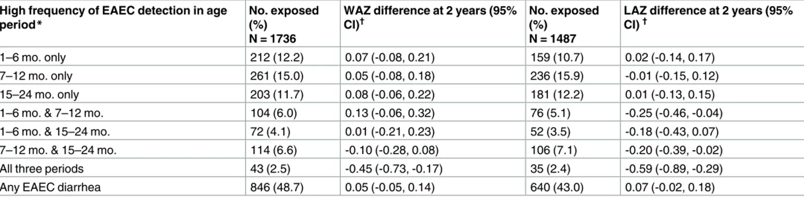
A high frequency of EAEC detection during only one of the periods 1–6 months, 7–12 months, and 15–24 months was not associated with LAZ decrements, whereas high frequency of detection in any two of the three time periods was associated with small non-significant length decrements, and high frequency of detection in all three time periods was associated with the largest length decrements (Table 3). There were no additional differences in growth between children who had at least one detection of EAEC in a diarrheal stool compared to children who did not after accounting for EAEC detection in surveillance stools (Table 3).
Discussion
We identified widespread acquisition of EAEC within the first few months of life across diverse settings in South Asia, South America, and Africa. In all sites except Peru, EAEC was detected at least once by two years of age in more than 90% of enrolled children. Slightly lower detec-tion of EAEC in Peru may be due to the relatively high rates of macrolide use observed at this site in MAL-ED [43]. A high prevalence of EAEC in children with and without diarrhea was also found in the seven-site Global Enteric Multicenter Study, a prospective matched case-con-trol study of moderate-to-severe diarrhea [11]. There was no evidence in either study that EAEC was a major cause of diarrhea of any duration.
Few risk factors for EAEC were identified in this analysis, and surprisingly, components of socioeconomic status and our index, the WAMI, were not consistently protective. Only exclu-sive breastfeeding, enrollment weight, and recent macrolide use were associated with reduced EAEC detections. Exclusive breastfeeding is protective against enteric infections through mul-tiple pathways, including limits on environmental exposure through contaminated food and water and directly through antimicrobial factors like lactoferrin and antibodies present in breastmilk [44]. The percent days of exclusive breastfeeding accounts for temporary cessation and return to exclusivity, and the protective association of this construct emphasizes that the age of first stopping exclusivity may be less important than the practice of exclusive breastfeed-ing itself, which may occur in multiple episodes [45]. The association of EAEC infections with
Table 3. Effects of EAEC detection in monthly surveillance stools on weight (WAZ) and length (LAZ) attainment at 2 years of age among 1,727 chil-dren in the MAL-ED cohort with anthropometric measurements at 2 years.
High frequency of EAEC detection in age
period* No. exposed(%)
N = 1736
WAZ difference at 2 years (95% CI)†
No. exposed (%)
N = 1487
LAZ difference at 2 years (95% CI)†
1–6 mo. only 212 (12.2) 0.07 (-0.08, 0.21) 159 (10.7) 0.02 (-0.14, 0.17)
7–12 mo. only 261 (15.0) 0.05 (-0.08, 0.18) 236 (15.9) -0.01 (-0.15, 0.12)
15–24 mo. only 203 (11.7) 0.08 (-0.06, 0.22) 181 (12.2) 0.01 (-0.13, 0.15)
1–6 mo. & 7–12 mo. 104 (6.0) 0.13 (-0.06, 0.32) 76 (5.1) -0.25 (-0.46, -0.04) 1–6 mo. & 15–24 mo. 72 (4.1) 0.01 (-0.21, 0.23) 52 (3.5) -0.18 (-0.43, 0.07) 7–12 mo. & 15–24 mo. 114 (6.6) -0.10 (-0.28, 0.08) 106 (7.1) -0.20 (-0.39, -0.02)
All three periods 43 (2.5) -0.45 (-0.73, -0.17) 35 (2.4) -0.59 (-0.89, -0.29)
Any EAEC diarrhea 846 (48.7) 0.05 (-0.05, 0.14) 640 (43.0) 0.07 (-0.02, 0.18)
*At least 50% of surveillance stools in the period were positive for EAEC.
†Adjusted for site, anthropometric measurement at enrollment, sex, WAMI, age at stopping exclusive breastfeeding, percent surveillance stools positive for
Campylobacterin the first 2 years of life. All LAZ estimates exclude Pakistan
lower enrollment weight is consistent with the increased susceptibility of malnourished mice to EAEC infection compared to well-nourished mice [20].
Antimicrobial resistance is a common feature of EAEC [46–48], and at least one EAEC-spe-cific resistance island has been characterized [49]. This island does not contain resistance genes for macrolides, which may explain the protective association with macrolide use (unlike either cephalosporin or any class of antibiotic use). The specificity of protection by macrolides may provide EAEC with a competitive advantage over other enteropathogens since non-macrolide antibiotic use was highly frequent at many of the MAL-ED sites [43]. Further char-acterization of the antimicrobial resistance of these isolates will be necessary to confirm this hypothesis.
Because only recent macrolide use was protective against EAEC infections, clearance of EAEC may be incomplete or more likely, reinfection with EAEC occurs quickly. In addition, alterations of the microbiota by macrolides could increase susceptibility to later EAEC infec-tions, as is evident in murine infections [21]. Therefore, antibiotic use to clear EAEC infections is likely not justified; however, increasing the duration of exclusive breastfeeding (even if in separated episodes) may delay the acquisition of these common, potentially inflammatory infections.
EAEC detection was associated with markers of intestinal inflammation, most strongly with increased fecal MPO. While the magnitudes of the associations were small relative to the range of observed concentrations, the increase in average levels of fecal MPO associated with EAEC [0.17 ln(ng/ml)] was comparable to that seen withCampylobacterinfections [0.19 ln (ng/ml)], which is a recognized cause of inflammatory enteritis [50,51]. EAEC has been previ-ously associated with markers of inflammation, specifically with lactoferrin [52] and the proin-flammatory cytokines interleukin (IL)-1b [14] and IL-8 [13,14,53]. The relevance of elevated intestinal inflammation to potential systemic inflammation associated with EAEC is not clear; there was no evidence that EAEC was associated with elevated AGP, a marker of systemic inflammation, though we note AGP was tested less frequently in this study and could have captured highly acute responses that may not have been temporarily coincident with stool sampling.
The association between EAEC and intestinal inflammation suggests a potential mecha-nism for the observed association between EAEC and growth. Intestinal inflammation [54] and specifically higher levels of fecal MPO [55–57], have been associated with poor linear growth among children in Brazil, Bangladesh, and the Gambia. However, because the magni-tudes of association with inflammatory biomarkers were very small, this pathway may not be a major contributor to the overall growth impact, or equally, the biomarkers measured may be suboptimal markers.
This analysis provides a comprehensive longitudinal assessment of EAEC infections in early life across diverse low-resource settings, drawing on a large number of stool collections, biomarker assessments, and repeated anthropometric measurements. The study was limited by the potentially suboptimal assessment of pathogenic EAEC since the virulence genes for EAEC are not well understood [58], and there may have been differences in strain variability across sites. Our gene probes,aatAandaaiC, were chosen as characteristic plasmid and chro-mosomal traits of EAEC, respectively [59], and may not be perfectly discriminating for patho-genic EAEC. Genetic probes generally associate with laboratory phenotypes, not necessarily clinical disease [49,60]. In a study of children in Mali,aatAandaaiCwere not associated with diarrhea when considering presence of either gene alone or in combination [61]. Furthermore, EAEC is able to acquire additional virulence genes that could increase its pathogenicity, such as the acquisition of Stx2 phage (a characteristic of enterohemorrhagicE.coli) in a German outbreak of EAEC-associated gastroenteritis [62]. The potential inability to distinguish patho-genic versus non-pathopatho-genic EAEC may contribute to the weak associations observed between EAEC, inflammatory biomarkers, and short-term growth velocity.
In conclusion, we found that EAEC infections were very common in the eight MAL-ED sites over the first two years of life. While often acutely subclinical, repeated EAEC detections were associated with longer-term linear growth deficits. Further work is needed better quantify the contribution of intestinal inflammation caused by EAEC to impaired growth. Refining our understanding of virulence traits may further help elucidate mechanisms of pathogenesis as well as the potential for vaccine-mediated or other approaches to control these increasingly recognized enteric pathogens. Because these infections may cause lasting consequences in terms of environmental enteropathy and relate to child growth deficits, a better understanding of the mechanisms involved and relevant biomarkers are critical to developing targeted inter-ventions to prevent these consequences for the world’s poorest children.
Acknowledgments
We thank the staff and participants of the MAL-ED Network for their important contributions.
MAL-ED network investigators
Sunder Shrestha10, Rita Shrestha10, Manjeswori Ulak10, Aubrey Bauck11, Robert Black11, Laura E Caulfield11, William Checkley11,6, Margaret N Kosek11, Gwenyth Lee11, Kerry Schulze11, Pablo Peñataro Yori11, Laura E. Murray-Kolb12, A Catharine Ross12, Barbara Schaefer12,6, Suzanne Simons12, Laura Pendergast13, Cla´udia B Abreu14, Hilda Costa14, Alessandra Di Moura14, Jose´ Quirino Filho14,6, Alexandre Havt14, A´ lvaro M Leite14, Aldo AM Lima14, Noe´lia L Lima14, Ila F Lima14, Bruna LL Maciel14, Pedro HQS Medeiros14, Milena Moraes14, Fran-cisco S Mota14, Reinaldo B Oria´14,Josiane Quetz14, Alberto M Soares14,Rosa MS Mota14, Crys-tal L Patil16, Pascal Bessong17, Cloupas Mahopo17, Angelina Maphula17, Emanuel Nyathi17, Amidou Samie17, Leah Barrett18, Rebecca Dillingham18, Jean Gratz18, Richard L Guerrant18, Eric Houpt18, William A Petri, Jr18, James Platts-Mills18, Rebecca Scharf18, Elizabeth T. Rogawski18, Binob Shrestha19, Sanjaya Kumar Shrestha19, Tor Strand19,15, Erling Svensen20,8
1A.B. PRISMA, Iquitos, Peru,2Aga Khan University, Karachi, Pakistan,3Armed Forces
Research Institute of Medical Sciences, Bangkok, Thailand,4Christian Medical College, Vel-lore, India,5Duke University, Durham, NC, USA,6Fogarty International Center/National Institutes of Health, Bethesda, MD, USA,7Foundation for the NIH, Bethesda, MD, USA,
8Haydom Lutheran Hospital, Haydom, Tanzania,9icddr,b, Dhaka, Bangladesh,10Institute of
Medicine, Tribhuvan University, Kathmandu, Nepal,11Johns Hopkins University, Baltimore, MD, USA,12The Pennsylvania State University, University Park, PA, USA,13Temple Univer-sity, Philadelphia, PA, USA,14Universidade Federal do Ceara, Fortaleza, Brazil,15University of Bergen, Norway,16University of Illinois at Chicago, IL, USA,17University of Venda, Thohoy-andou, South Africa,18University of Virginia, Charlottesville, VA, USA,19Walter Reed/ AFRIMS Research Unit, Kathmandu, Nepal,20Haukeland University Hospital, Bergen, Norway
Author Contributions
Conceptualization:Elizabeth T. Rogawski, Richard L. Guerrant, Aldo A. M. Lima.
Data curation:Jessica C. Seidman, Benjamin J. J. McCormick.
Formal analysis:Elizabeth T. Rogawski.
Funding acquisition:Richard L. Guerrant.
Investigation:Alexandre Havt, Ila F. N. Lima, Pedro H. Q. S. Medeiros, Sudhir Babji, Dinesh Hariraju, Ladaporn Bodhidatta, Jasmin Shrestha, Japhat Anania, Athanasia Maro, Amidou Samie, Pablo Peñataro Yori, Shahida Qureshi, Mustafa Mahfuz, Pascal O. Bessong, Marga-ret N. Kosek, Tahmeed Ahmed, Zulfiqar A. Bhutta, Eric R. Houpt, Aldo A. M. Lima.
Methodology:Elizabeth T. Rogawski, Richard L. Guerrant, Jessica C. Seidman, Benjamin J. J. McCormick, Pascal O. Bessong, Margaret N. Kosek, Tahmeed Ahmed, Zulfiqar A. Bhutta, Eric R. Houpt, Aldo A. M. Lima.
Project administration:Pascal O. Bessong, Margaret N. Kosek, Tahmeed Ahmed, Zulfiqar A. Bhutta, Dennis R. Lang, Michael Gottlieb.
Supervision:Richard L. Guerrant, Ladaporn Bodhidatta, Jasmin Shrestha, Japhat Anania, Athanasia Maro, Amidou Samie, Pablo Peñataro Yori, Shahida Qureshi, Mustafa Mahfuz, Pascal O. Bessong, Margaret N. Kosek, Tahmeed Ahmed, Zulfiqar A. Bhutta, Dennis R. Lang, Michael Gottlieb, Eric R. Houpt, Aldo A. M. Lima.
Visualization:Elizabeth T. Rogawski.
Writing – review & editing:Richard L. Guerrant, Alexandre Havt, Ila F. N. Lima, Pedro H. Q. S. Medeiros, Jessica C. Seidman, Benjamin J. J. McCormick, Sudhir Babji, Dinesh Hariraju, Ladaporn Bodhidatta, Jasmin Shrestha, Japhat Anania, Athanasia Maro, Amidou Samie, Pablo Peñataro Yori, Shahida Qureshi, Mustafa Mahfuz, Pascal O. Bessong, Margaret N. Kosek, Tahmeed Ahmed, Zulfiqar A. Bhutta, Dennis R. Lang, Michael Gottlieb, Eric R. Houpt, Aldo A. M. Lima.
References
1. Nataro JP, Kaper JB, Robins-Browne R, Prado V, Vial P, Levine MM. Patterns of adherence of diarrhea-genic Escherichia coli to HEp-2 cells. Pediatr Infect Dis J. 1987; 6: 829–831. PMID:3313248
2. Scavia G, Staffolani M, Fisichella S, Striano G, Colletta S, Ferri G, et al. Enteroaggregative Escherichia coli associated with a foodborne outbreak of gastroenteritis. J Med Microbiol. 2008; 57: 1141–1146.
https://doi.org/10.1099/jmm.0.2008/001362-0PMID:18719185
3. Cohen MB, Hawkins JA, Weckbach LS, Staneck JL, Levine MM, Heck JE. Colonization by enteroaggre-gative Escherichia coli in travelers with and without diarrhea. J Clin Microbiol. 1993; 31: 351–353. PMID:8432822
4. Adachi JA, Jiang ZD, Mathewson JJ, Verenkar MP, Thompson S, Martinez-Sandoval F, et al. Enteroag-gregative Escherichia coli as a major etiologic agent in traveler’s diarrhea in 3 regions of the world. Clin Infect Dis Off Publ Infect Dis Soc Am. 2001; 32: 1706–1709.https://doi.org/10.1086/320756PMID:
11360211
5. Jiang Z-D, Greenberg D, Nataro JP, Steffen R, DuPont HL. Rate of occurrence and pathogenic effect of enteroaggregative Escherichia coli virulence factors in international travelers. J Clin Microbiol. 2002; 40: 4185–4190.https://doi.org/10.1128/JCM.40.11.4185-4190.2002PMID:12409395
6. Wanke CA, Mayer H, Weber R, Zbinden R, Watson DA, Acheson D. Enteroaggregative Escherichia coli as a potential cause of diarrheal disease in adults infected with human immunodeficiency virus. J Infect Dis. 1998; 178: 185–190. PMID:9652439
7. Nataro JP, Mai V, Johnson J, Blackwelder WC, Heimer R, Tirrell S, et al. Diarrheagenic Escherichia coli infection in Baltimore, Maryland, and New Haven, Connecticut. Clin Infect Dis Off Publ Infect Dis Soc Am. 2006; 43: 402–407.https://doi.org/10.1086/505867PMID:16838226
8. Nataro JP, Deng Y, Cookson S, Cravioto A, Savarino SJ, Guers LD, et al. Heterogeneity of enteroag-gregative Escherichia coli virulence demonstrated in volunteers. J Infect Dis. 1995; 171: 465–468. PMID:7844392
9. Mathewson JJ, Johnson PC, DuPont HL, Satterwhite TK, Winsor DK. Pathogenicity of enteroadherent Escherichia coli in adult volunteers. J Infect Dis. 1986; 154: 524–527. PMID:3525699
10. Huang DB, Nataro JP, DuPont HL, Kamat PP, Mhatre AD, Okhuysen PC, et al. Enteroaggregative Escherichia coli is a cause of acute diarrheal illness: a meta-analysis. Clin Infect Dis Off Publ Infect Dis Soc Am. 2006; 43: 556–563.https://doi.org/10.1086/505869PMID:16886146
11. Kotloff KL, Nataro JP, Blackwelder WC, Nasrin D, Farag TH, Panchalingam S, et al. Burden and aetiol-ogy of diarrhoeal disease in infants and young children in developing countries (the Global Enteric Multi-center Study, GEMS): a prospective, case-control study. Lancet Lond Engl. 2013; 382: 209–222.
https://doi.org/10.1016/S0140-6736(13)60844-2
12. Platts-Mills JA, Babji S, Bodhidatta L, Gratz J, Haque R, Havt A, et al. Pathogen-specific burdens of community diarrhoea in developing countries: a multisite birth cohort study (MAL-ED). Lancet Glob Health. 2015; 3: e564–75.https://doi.org/10.1016/S2214-109X(15)00151-5PMID:26202075 13. Steiner TS, Lima AA, Nataro JP, Guerrant RL. Enteroaggregative Escherichia coli produce intestinal
inflammation and growth impairment and cause interleukin-8 release from intestinal epithelial cells. J Infect Dis. 1998; 177: 88–96. PMID:9419174
14. Greenberg DE, Jiang Z- D, Steffen R, Verenker MP, DuPont HL. Markers of inflammation in bacterial diarrhea among travelers, with a focus on enteroaggregative Escherichia coli pathogenicity. J Infect Dis. 2002; 185: 944–949.https://doi.org/10.1086/339617PMID:11920319
15. Kaper JB, Nataro JP, Mobley HL. Pathogenic Escherichia coli. Nat Rev Microbiol. 2004; 2: 123–140.
https://doi.org/10.1038/nrmicro818PMID:15040260
16. Nataro JP, Kaper JB. Diarrheagenic Escherichia coli. Clin Microbiol Rev. 1998; 11: 142–201. PMID:
9457432
17. Nataro JP, Yikang D, Yingkang D, Walker K. AggR, a transcriptional activator of aggregative adherence fimbria I expression in enteroaggregative Escherichia coli. J Bacteriol. 1994; 176: 4691–4699. PMID:
18. Steiner TS, Nataro JP, Poteet-Smith CE, Smith JA, Guerrant RL. Enteroaggregative Escherichia coli expresses a novel flagellin that causes IL-8 release from intestinal epithelial cells. J Clin Invest. 2000; 105: 1769–1777.https://doi.org/10.1172/JCI8892PMID:10862792
19. Harrington SM, Strauman MC, Abe CM, Nataro JP. Aggregative adherence fimbriae contribute to the inflammatory response of epithelial cells infected with enteroaggregative Escherichia coli. Cell Micro-biol. 2005; 7: 1565–1578.https://doi.org/10.1111/j.1462-5822.2005.00588.xPMID:16207244 20. Roche JK, Cabel A, Sevilleja J, Nataro J, Guerrant RL. Enteroaggregative Escherichia coli (EAEC)
Impairs Growth while Malnutrition Worsens EAEC Infection: A Novel Murine Model of the Infection Mal-nutrition Cycle. J Infect Dis. 2010; 202: 506–514.https://doi.org/10.1086/654894PMID:20594107 21. Bolick DT, Roche JK, Hontecillas R, Bassaganya-Riera J, Nataro JP, Guerrant RL. Enteroaggregative
Escherichia coli strain in a novel weaned mouse model: exacerbation by malnutrition, biofilm as a viru-lence factor and treatment by nitazoxanide. J Med Microbiol. 2013; 62: 896–905.https://doi.org/10. 1099/jmm.0.046300-0PMID:23475903
22. Bolick DT, Kolling GL, Moore JH, de Oliveira LA, Tung K, Philipson C, et al. Zinc deficiency alters host response and pathogen virulence in a mouse model of enteroaggregative Escherichia coli-induced diar-rhea. Gut Microbes. 2014; 5: 618–627.https://doi.org/10.4161/19490976.2014.969642PMID:
25483331
23. Rogawski ET, Guerrant RL. The burden of enteropathy and “subclinical” infections. Pediatr Clin North Am. 2017;In press.
24. GBD 2015 Mortality and Causes of Death Collaborators. Global, regional, and national life expectancy, all-cause mortality, and cause-specific mortality for 249 causes of death, 1980–2015: a systematic anal-ysis for the Global Burden of Disease Study 2015. Lancet Lond Engl. 2016; 388: 1459–1544.https:// doi.org/10.1016/S0140-6736(16)31012-1PMID:27733281
25. de Onis M, Blo¨ssner M, Borghi E. Prevalence and trends of stunting among pre-school children, 1990– 2020. Public Health Nutr. 2012; 15: 142–148.https://doi.org/10.1017/S1368980011001315PMID:
21752311
26. Dewey KG, Adu-Afarwuah S. Systematic review of the efficacy and effectiveness of complementary feeding interventions in developing countries. Matern Child Nutr. 2008; 4 Suppl 1: 24–85.https://doi. org/10.1111/j.1740-8709.2007.00124.xPMID:18289157
27. Nabwera HM, Fulford AJ, Moore SE, Prentice AM. Growth faltering in rural Gambian children after four decades of interventions: a retrospective cohort study. Lancet Glob Health. 2017; 5: e208–e216.
https://doi.org/10.1016/S2214-109X(16)30355-2PMID:28104187
28. Crane RJ, Berkley JA. Progress on growth faltering. Lancet Glob Health. 2017; 5: e125–e126.https:// doi.org/10.1016/S2214-109X(16)30357-6PMID:28104171
29. Cumming O, Cairncross S. Can water, sanitation and hygiene help eliminate stunting? Current evi-dence and policy implications. Matern Child Nutr. 2016; 12 Suppl 1: 91–105.https://doi.org/10.1111/ mcn.12258PMID:27187910
30. MAL-ED Network Investigators. The MAL-ED study: a multinational and multidisciplinary approach to understand the relationship between enteric pathogens, malnutrition, gut physiology, physical growth, cognitive development, and immune responses in infants and children up to 2 years of age in resource-poor environments. Clin Infect Dis Off Publ Infect Dis Soc Am. 2014; 59 Suppl 4: S193–206.https://doi. org/10.1093/cid/ciu653PMID:25305287
31. Richard SA, Barrett LJ, Guerrant RL, Checkley W, Miller MA, MAL-ED Network Investigators. Disease surveillance methods used in the 8-site MAL-ED cohort study. Clin Infect Dis Off Publ Infect Dis Soc Am. 2014; 59 Suppl 4: S220–224.https://doi.org/10.1093/cid/ciu435PMID:25305290
32. Houpt E, Gratz J, Kosek M, Zaidi AKM, Qureshi S, Kang G, et al. Microbiologic methods utilized in the MAL-ED cohort study. Clin Infect Dis Off Publ Infect Dis Soc Am. 2014; 59 Suppl 4: S225–232.https:// doi.org/10.1093/cid/ciu413PMID:25305291
33. Kosek M, Guerrant RL, Kang G, Bhutta Z, Yori PP, Gratz J, et al. Assessment of environmental enterop-athy in the MAL-ED cohort study: theoretical and analytic framework. Clin Infect Dis Off Publ Infect Dis Soc Am. 2014; 59 Suppl 4: S239–247.https://doi.org/10.1093/cid/ciu457PMID:25305293
34. World Health Organization and United Nations Children’s Fund Joint Monitoring Programme for Water Supply and Sanitation, (JMP). Progress on Drinking Water and Sanitation: Special Focus on Sanitation [Internet]. UNICEF, New York and WHO, Geneva; 2008. Available:http://www.wssinfo.org/fileadmin/ user_upload/resources/1251794333-JMP_08_en.pdf
36. Kosek MN, Lee GO, Guerrant RL, Haque R, Kang G, Ahmed T, et al. The MAL-ED study: Age and sex normalization of intestinal permeability for the assessment of enteropathy in infancy. J Pediatr Gastro-enterol Nutr. 2017;In press.
37. World Health Organization. WHO Child Growth Standards: Length/height-for-age, weight-for-age, weight-for-length, weight-for-height and body mass index-for-age, Methods and development [Internet]. 2006. Available:http://www.who.int/childgrowth/standards/Technical_report.pdf?ua=1
38. Howe CJ, Cole SR, Westreich DJ, Greenland S, Napravnik S, Eron JJ. Splines for Trend Analysis and Continuous Confounder Control. Epidemiology. 2011; 22: 874–875.https://doi.org/10.1097/EDE. 0b013e31823029ddPMID:21968779
39. Zou G. A modified poisson regression approach to prospective studies with binary data. Am J Epide-miol. 2004; 159: 702–706. PMID:15033648
40. Amour C, Gratz J, Mduma E, Svensen E, Rogawski ET, McGrath M, et al. Epidemiology and impact of Campylobacter infection in children in eight low-resource settings: results from the MAL-ED study. Clin Infect Dis Off Publ Infect Dis Soc Am. 2016;https://doi.org/10.1093/cid/ciw542PMID:27501842 41. Kosek M, Ahmed T, Bhutta Z, Caulfield L, Guerrant R, Houpt E, et al. Causal Pathways from
Entero-pathogens to Environmental Enteropathy: Findings from the MAL-ED Birth Cohort Study. EBioMedi-cine.https://doi.org/10.1016/j.ebiom.2017.02.024PMID:28396264
42. Saiki T. Myeloperoxidase concentrations in the stool as a new parameter of inflammatory bowel dis-ease. Kurume Med J. 1998; 45: 69–73. PMID:9658754
43. Rogawski ET, Platts-Mills JA, Seidman JC, John S, Mahfuz M, Ulak M, et al. Use of antibiotics in chil-dren younger than two years in eight countries: a prospective cohort study. Bull World Health Organ. 2017; 95: 49–61.https://doi.org/10.2471/BLT.16.176123PMID:28053364
44. Mittal SK. How protective is breast-feeding in diarrhoeal diseases? In: Walker-Smith JA, McNeish AS, editors. Diarrhoea and Malnutrition in Childhood. Boston: Butterworth & Co.; 1986. pp. 214–220.
45. Ambikapathi R, Kosek MN, Lee GO, Mahopo C, Patil CL, Maciel BL, et al. How multiple episodes of exclusive breastfeeding impact estimates of exclusive breastfeeding duration: report from the eight-site MAL-ED birth cohort study. Matern Child Nutr. 2016; 12: 740–756.https://doi.org/10.1111/mcn.12352
PMID:27500709
46. Yamamoto T, Echeverria P, Yokota T. Drug resistance and adherence to human intestines of enteroag-gregative Escherichia coli. J Infect Dis. 1992; 165: 744–749. PMID:1552205
47. Gassama A, Aïdara-Kane A, Chainier D, Denis F, Ploy M-C. Integron-associated antibiotic resistance in enteroaggregative and enteroinvasive Escherichia coli. Microb Drug Resist Larchmt N. 2004; 10: 27– 30.https://doi.org/10.1089/107662904323047763PMID:15140390
48. Mendez Arancibia E, Pitart C, Ruiz J, Marco F, Gasco´n J, Vila J. Evolution of antimicrobial resistance in enteroaggregative Escherichia coli and enterotoxigenic Escherichia coli causing traveller’s diarrhoea. J Antimicrob Chemother. 2009; 64: 343–347.https://doi.org/10.1093/jac/dkp178PMID:19474067 49. Okeke IN, Wallace-Gadsden F, Simons HR, Matthews N, Labar AS, Hwang J, et al. Multi-Locus
Sequence Typing of Enteroaggregative Escherichia coli Isolates from Nigerian Children Uncovers Multi-ple Lineages. PLOS ONE. 2010; 5: e14093.https://doi.org/10.1371/journal.pone.0014093PMID:
21124856
50. Riddle MS, Gutierrez RL, Verdu EF, Porter CK. The chronic gastrointestinal consequences associated with Campylobacter. Curr Gastroenterol Rep. 2012; 14: 395–405. https://doi.org/10.1007/s11894-012-0278-0PMID:22864805
51. Young KT, Davis LM, Dirita VJ. Campylobacter jejuni: molecular biology and pathogenesis. Nat Rev Microbiol. 2007; 5: 665–679.https://doi.org/10.1038/nrmicro1718PMID:17703225
52. Bouckenooghe AR, Dupont HL, Jiang ZD, Adachi J, Mathewson JJ, Verenkar MP, et al. Markers of enteric inflammation in enteroaggregative Escherichia coli diarrhea in travelers. Am J Trop Med Hyg. 2000; 62: 711–713. PMID:11304060
53. Jiang Z-D, Okhuysen PC, Guo D-C, He R, King TM, DuPont HL, et al. Genetic susceptibility to enter-oaggregative Escherichia coli diarrhea: polymorphism in the interleukin-8 promotor region. J Infect Dis. 2003; 188: 506–511.https://doi.org/10.1086/377102PMID:12898436
54. Campbell DI, McPhail G, Lunn PG, Elia M, Jeffries DJ. Intestinal inflammation measured by fecal neop-terin in Gambian children with enteropathy: association with growth failure, Giardia lamblia, and intesti-nal permeability. J Pediatr Gastroenterol Nutr. 2004; 39: 153–157. PMID:15269619
56. DeBoer MD, Scharf RJ, Leite AM, Fe´rrer A, Havt A, Pinkerton R, et al. Systemic inflammation, growth factors, and linear growth in the setting of infection and malnutrition. Nutr Burbank Los Angel Cty Calif. 2017; 33: 248–253.https://doi.org/10.1016/j.nut.2016.06.013PMID:27712965
57. Platts-Mills JA, Taniuchi M, Uddin J, Uddin Sobuz S, Mahfuz M, Gaffar SMA, et al. Association between enteropathogens and malnutrition in children aged 6–23 months in Bangladesh: a case-control study. Am J Clin Nutr. 2017;In press.
58. Jensen BH, Olsen KEP, Struve C, Krogfelt KA, Petersen AM. Epidemiology and Clinical Manifestations of Enteroaggregative Escherichia coli. Clin Microbiol Rev. 2014; 27: 614–630.https://doi.org/10.1128/ CMR.00112-13PMID:24982324
59. Taniuchi M, Walters CC, Gratz J, Maro A, Kumburu H, Serichantalergs O, et al. Development of a multi-plex polymerase chain reaction assay for diarrheagenic Escherichia coli and Shigella spp. and its evalu-ation on colonies, culture broths, and stool. Diagn Microbiol Infect Dis. 2012; 73: 121–128.https://doi. org/10.1016/j.diagmicrobio.2012.03.008PMID:22541788
60. Chattaway MA, Harris R, Jenkins C, Tam C, Coia JE, Gray J, et al. Investigating the link between the presence of enteroaggregative Escherichia coli and infectious intestinal disease in the United Kingdom, 1993 to 1996 and 2008 to 2009. Euro Surveill Bull Eur Sur Mal Transm Eur Commun Dis Bull. 2013; 18.
61. Boisen N, Scheutz F, Rasko DA, Redman JC, Persson S, Simon J, et al. Genomic Characterization of Enteroaggregative Escherichia coli From Children in Mali. J Infect Dis. 2012; 205: 431–444.https://doi. org/10.1093/infdis/jir757PMID:22184729