CXCL8 Chemokines in the Skin Associated with Parasite
Density in Canine Visceral Leishmaniasis
Daniel Menezes-Souza1,2,3, Renata Guerra-Sa´3,4, Cla´udia Martins Carneiro1,2,5, Juliana Vitoriano-Souza1, Rodolfo Cordeiro Giunchetti1,2,4, Andre´a Teixeira-Carvalho6, Denise Silveira-Lemos7, Guilherme
Correˆa Oliveira8, Rodrigo Correˆa-Oliveira2, Alexandre Barbosa Reis1,2,5*
1Laborato´rio de Imunopatologia, Nu´cleo de Pesquisas em Cieˆncias Biolo´gicas, Universidade Federal de Ouro Preto, Ouro Preto, Minas Gerais, Brasil,2Laborato´rio de Imunologia Celular e Molecular, Centro de Pesquisas Rene´ Rachou, Fundac¸a˜o Oswaldo Cruz, Belo Horizonte, Minas Gerais, Brasil,3Laborato´rio de Bioquı´mica e Biologia Molecular, Nu´cleo de Pesquisas em Cieˆncias Biolo´gicas, Universidade Federal de Ouro Preto, Ouro Preto, Minas Gerais, Brasil,4Departamento de Cieˆncias Biolo´gicas, Instituto de Cieˆncias Exatas e Biolo´gicas, Universidade Federal de Ouro Preto, Ouro Preto, Minas Gerais, Brasil,5Departamento de Ana´lises Clı´nicas, Escola de Farma´cia, Universidade Federal de Ouro Preto, Ouro Preto, Minas Gerais, Brasil,6Laborato´rio de Biomarcadores de Diagno´stico e Monitorac¸a˜o, Centro de Pesquisas Rene´ Rachou, Fundac¸a˜o Oswaldo Cruz, Belo Horizonte, Minas Gerais, Brasil,7Laborato´rio de Imunoparasitologia, Nu´cleo de Pesquisas em Cieˆncias Biolo´gicas, Universidade Federal de Ouro Preto, Ouro Preto, Minas Gerais, Brasil,8Laborato´rio de Parasitologia Celular e Molecular, Centro de Pesquisas Rene´ Rachou, Fundac¸a˜o Oswaldo Cruz, Belo Horizonte, Minas Gerais, Brasil
Abstract
Background:The immune response in the skin of dogs infected withLeishmania infantumis poorly understood, and limited studies have described the immunopathological profile with regard to distinct levels of tissue parasitism and the clinical progression of canine visceral leishmaniasis (CVL).
Methodology/Principal Findings: A detailed analysis of inflammatory cells (neutrophils, eosinophils, mast cells, lymphocytes, and macrophages) as well as the expression of chemokines (CCL2, CCL4, CCL5, CCL13, CCL17, CCL21, CCL24, and CXCL8) was carried out in dermis skin samples from 35 dogs that were naturally infected withL. infantum. The analysis was based on real-time polymerase chain reaction (PCR) in the context of skin parasitism and the clinical status of CVL. We demonstrated increased inflammatory infiltrate composed mainly of mononuclear cells in the skin of animals with severe forms of CVL and high parasite density. Analysis of the inflammatory cell profile of the skin revealed an increase in the number of macrophages and reductions in lymphocytes, eosinophils, and mast cells that correlated with clinical progression of the disease. Additionally, enhanced parasite density was correlated with an increase in macrophages and decreases in eosinophils and mast cells. The chemokine mRNA expression demonstrated that enhanced parasite density was positively correlated with the expression of CCL2, CCL4, CCL5, CCL21, and CXCL8. In contrast, there was a negative correlation between parasite density and CCL24 expression.
Conclusions/Significance:These findings represent an advance in the knowledge about skin inflammatory infiltrates in CVL and the systemic consequences. Additionally, the findings may contribute to the design of new and more efficient prophylactic tools and immunological therapies against CVL.
Citation:Menezes-Souza D, Guerra-Sa´ R, Carneiro CM, Vitoriano-Souza J, Giunchetti RC, et al. (2012) Higher Expression of CCL2, CCL4, CCL5, CCL21, and CXCL8 Chemokines in the Skin Associated with Parasite Density in Canine Visceral Leishmaniasis. PLoS Negl Trop Dis 6(4): e1566. doi:10.1371/journal.pntd.0001566 Editor:Hechmi Louzir, Institut Pasteur de Tunis, Tunisia
ReceivedJuly 18, 2011;AcceptedJanuary 29, 2012;PublishedApril 10, 2012
Copyright:ß2012 Menezes-Souza et al. This is an open-access article distributed under the terms of the Creative Commons Attribution License, which permits unrestricted use, distribution, and reproduction in any medium, provided the original author and source are credited.
Funding:This work was supported by Conselho Nacional de Desenvolvimento Cientifico e Tecnologico - CNPq, Fundacao de Amparo a Pesquisa do Estado de Minas Gerais, Rede Mineira de Bioterismo - FAPEMIG. The funders had no role in study design, data collection and analysis, decision to publish, or preparation of the manuscript.
Competing Interests:The authors have declared that no competing interests exist. * E-mail: [email protected]
Introduction
Visceral leishmaniasis (VL), caused by Leishmania (Leishmania) infantum[syn.Leishmania(Leishmania)chagasi], is endemic in over 88 countries in Europe and Latin America and is transmitted by the bite of the female sand fly (phlebotomine) [1]. The skin is considered a key reservoir compartment for amastigotes in both asymptomatic and symptomaticLeishmania-infected dogs, and the important role of dogs in VL transmission in urban areas is supported by the high parasite loads found in the skin of infected animals and their shared
habitat with humans [2–4]. Previous investigations have revealed that symptomaticLeishmania-infected dogs exhibit an intense diffuse dermal inflammatory infiltrate and a high parasitic burden in comparison with their asymptomatic counterparts [4]. On this basis it was proposed that the immunopathological changes in the skin and the levels of cutaneous parasitism are directly related to the clinical severity of the disease.
histopathological finding in tissues is a granulomatous inflamma-tory reaction associated with the presence ofLeishmaniaamastigotes within macrophages [18]. In the skin of Leishmania infantum– infected dogs, the histopathological alterations consist of variable degrees of focal or diffuse inflammatory infiltrate in the dermis and variable numbers of plasma cells, macrophages, lymphocytes, and isolated neutrophils [19,20].
Furthermore, it has recently been demonstrated that parasite density in the skin, bone marrow, and spleen compartments increases according to the severity of the clinical manifestation of CVL [6,14,16]. Calabrese et al. [21] evaluated histopathological aspects of the skin in naturally infected dogs and showed that low parasite load is associated with an intense inflammatory reaction driven mainly by mast cells, indicating that these cells exert a role in innate immunity and in the resistance against canineLeishmania infection.
Recently, different aspects of the immune response in Leishmania-infected dogs have been studied, particularly the profile of cytokines in distinct compartments [5,9,12,17,22–24]. However, the role of chemokines in disease progression or parasite burdens of the visceralising species represents an important approach for understanding immunopathology in CVL.
Chemokines are chemotactic factors that coordinate recruit-ment of leukocytes that are involved in homeostasis as well as innate and adaptive immune responses. In the context of experimental or natural infection in CVL, an up-regulation of the chemokines in the spleen has been described, although only CXCL10 and CCL5 were markedly elevated in oligosymptomatic dogs [24]. In addition, the augmented levels of chemokines suggested an accumulation of infiltrating monocytes attracted by CCL3 and CCL2. CD4+
Th1 and CD8+
cells also accumulated and may have been recruited by CXCL10, with further expression induced through IFN-csecretion [24].
Considering the importance of chemokines on the pattern of CVL and the lack of studies on this topic, understanding the chemokine profile during ongoingL. infantuminfection in dogs is a prerequisite for identifying the mechanisms for resistance or susceptibility in this experimental model. In the present study, the
immunopathology of CVL was investigated by performing detailed analyses of the RNA expression of different chemokines (CCL2, CCL4, CCL5, CCL13, CCL17, CCL21, CCL24, and CXCL8) and the occurrence of inflammatory cells (neutrophils, eosinophils, mast cells, lymphocytes, and macrophages). We focused on selected chemokines in order to characterize their role in recruiting particular cell types to the inflammatory infiltrate in skin from L. infantum–infected dogs. Thus, we found that the chemokines CCL2, CCL4, CCL5, and CCL21 attract macro-phages; CCL5 and CCL4 attract inflammatory lymphocytes, particularly Th1-type cells; CCL24 attracts eosinophils; and CXCL8 attracts neutrophils, monocytes, and lymphocytes. The chemokine CCL17 helps to establish the inflammatory infiltrate, a characteristic feature of various inflammatory skin conditions, by attracting CCR4-bearing cells, which are especially polarized to Th2-type cells and regulatory T cells [25]. This study was carried out using the skin from 35 dogs that were naturally infected withL. infantum.
Materials and Methods
Study population and clinical evaluation
The study was approved by the Committees of Ethics in Animal Experimentation of the Universidade Federal de Ouro Preto (protocol no. 083/2007) and of the Universidade Federal de Minas Gerais (protocol no. 020/2007) and the City Council of Belo Horizonte (protocol no. 001/2008). All procedures in this study were according to the guidelines set by the Brazilian Animal Experimental Collage (COBEA), Federal Law number 11794. The study population comprised 51 adult dogs (aged between 2 and 6 years) of both sexes that had been captured by the Center of Zoonosis Control in Belo Horizonte (Minas Gerais, Brazil), a region with a high prevalence of CVL and human VL. The animals were maintained under quarantine at the kennels of the Instituto de Cieˆncias Biolo´gicas (Universidade Federal de Minas Gerais) prior to tissue collection for 40 days and treated for intestinal helminthic infections (Endal Plus; Schering-Plough Coopers, Sa˜o Paulo, SP, Brazil). We treated kennels with pyrethroid insecticide monthly during the quarantine and throughout the experiments. Experimental animals were catego-rized on the basis of serological results from an indirect immunofluorescence assay test, the ‘‘gold standard’’ immunolog-ical test in Brazil for the diagnosis of CVL. Sixteen dogs with negative immunofluorescence assay test results from serum samples diluted 1:40 and negative results forLeishmaniain tissue smears (bone marrow, ear skin, spleen, liver, and popliteal lymph node) were considered to be non-infected and were used as the control group (CD, n= 16). Thirty-five animals with positive immunofluorescence assay titers $1:40 were considered CVL positive and comprised the infected animal groups. The infected animal groups were subdivided on the basis of the presence or absence of signs of infection according to Mancianti et al. [25] as follows: an asymptomatic group (AD,n= 10), in which indicative signs of the disease were absent; an oligosymptomatic group (OD, n= 10), in which a maximum of three clinical signs of the disease were present, including opaque bristles, localized alopecia, and/or moderate weight loss; and a symptomatic group (SD,n= 15), in which characteristic clinical signs of the disease were present, including cutaneous lesions, onycogryphosis, opaque bristles, severe loss of weight, apathy, and keratoconjunctivitis.
Sample collection and assessment of skin parasite load Animals were euthanized with sodium thiopental (Abbott Laboratories, Abbott Park, IL, USA; 30 mg/kg body weight) Author Summary
and samples of skin tissue were collected from ear areas without lesions. A skin fragment from each group was used for tissue imprints on microscopic slides coded for blinded analysis. The samples were fixed in methanol, stained with Giemsa, and examined under an optical microscope. Leishmania amastigote stages were counted and parasite densities were expressed as LeishmanDonovan Units (LDU), corresponding to the number of Leishmaniaamastigotes per 1000 nucleated cells per skin imprint as described by Stauber [27], with some modifications according to Reis et al. [7,8]. Parasite densities were categorized statistically into tertiles as absent (LDU = 0; CD group,n= 16), low (LDU = 1– 9; LP group,n= 12), medium (LDU = 10–130; MP group,n= 11), and high (LDU = 131–7246; HP group,n= 12).
Extraction of total RNA and synthesis of first strand cDNAs
A second fragment of ear skin was stored at 280uC until required for RNA analysis. Total RNA was extracted by homogenizing approximately 20 mg of skin tissue with 1 mL of TRIzol reagent (Invitrogen Brasil, Sa˜o Paulo, SP, Brazil) in a rotor stator. The lysate was incubated at room temperature for 10 min, mixed with chloroform (200mL) by tube inversion, and
centrifuged at 12,0006gfor 10 min at 4uC. The aqueous phase
was collected, and RNA extraction was done by using the SV Total RNA Isolation System (Promega, Madison, WI, USA) according to the manufacturer’s recommendations, which included a DNase treatment step. The efficiency of DNAse treatment was evaluated by PCR amplification of the cDNA reaction mix without the addition of the ThermoScript enzyme. Finally, each quantitative PCR (q-PCR) run was performed with two internal controls assessing both potential genomic DNA contamination (no reverse transcriptase added) and purity of the reagents used (no cDNA added). Strand cDNAs were synthesized from 1.0mg of total RNA using the ThermoScript RT-PCR System (Invitrogen Brasil) with oligo-dT primers according to the manufacturer’s instructions.
Design of primers for gene evaluation
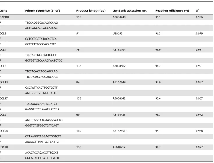
Primers were designed with the aid of Gene Runner version 3.05 (Hasting Software Inc. 2004) using specific canine sequences obtained from GenBank (http://www.ncbi.nlm.nih.gov/genbank/). The se-quences of the primers are listed in Table 1. The primers were synthesized by Eurogentec (Southampton, UK) and reconstituted in nuclease-free water.
Table 1.Sequences of primers used for quantification of mRNA expression by real-time PCRa.
Gene Primer sequence (59–39) Product length (bp) GenBank accession no. Reaction efficiency (%) R2
GAPDH 115 AB038240 99.1 0.996
F TTCCACGGCACAGTCAAG R ACTCAGCACCAGCATCAC
CCL2 91 U29653 96.3 0.979
F CCTGCTGCTATACACTCA R GCTTCTTTGGGACACTTG
CCL4 76 AB183194 95.9 0.981
F TCCTACTGCCTGCTGCTT R GCTGGTCTCAAAGTAATCTGC
CCL5 136 AB098562 98.7 0.991
F TTCTACACCAGCAGCAAG R TTCTACACCAGCAGCAAG
CCL13 84 AB162849 97.6 0.987
F CCCTATTCACTTGCTGCTT R AGTGGCTGCTGGTGATTC
CCL17 128 AB054642 95.4 0.967
F TCCAAGGCAAGTCCATCT R GAGGTCTCCAAATGATCCA
CCL21 60 AB164433 96.7 0.972
F AGTCTGGCAAGAAGGGAAAG R GGGTCTGTGGCTGTTCAGT
CCL24 149 AB162851.1 95.3 0.968
F CCTAAGGCAGGAGTGGTCTT R AGGGCTTTGGTGCTCATTG
CXCL8 116 AF048717 98.7 0.977
F ACACTCCACACCTTTCCAT R GGCACACCTCATTTCCATTG
aF: Forward primer, R: Reverse primer. GenBank accession number of the sequence used to design primers and their product length are shown, as well as each PCR
efficiency andR2
.
Real-time PCR and cloning and sequencing of amplicons q-PCR was performed on an ABI Prism 7000 DNA Sequence Detection System using SYBR Green PCR Master Mix (PE Applied Biosystems, Foster City, CA, USA), with 100 mM of each primer and cDNA diluted to 1:5. The samples were incubated at 95uC for 10 min and then submitted to 40 cycles of 95uC for 15 s and 60uC for 1 min, during which time fluorescence data were collected. The efficiency of each pair of primers was evaluated by serial dilution of cDNA according to the protocol developed by PE Applied Biosystems. In order to evaluate gene expression of the chemokines CCL2, CCL4, CCL5, CCL13, CCL17, CCL21, CCL24, and CXCL8, three replicate analyses were performed, and the amount of target RNA was normalized with respect to the endogenous control (housekeeping) gene GAPDH. Data were expressed according to the 22DDCtmethod using the mean value of the DCt of the control group as the calibrator [26]. After normalization, the expression levels of chemokines in the infected groups were considered up-regulated or down-regulated compared to expression levels in the control group. PCR products were cloned with pGEM-T Easy Vector (Promega) and sequenced to
check specificity by using an ABI 3100 Automated Sequencer (PE Applied Biosystems) and a Dye Terminator Kit.
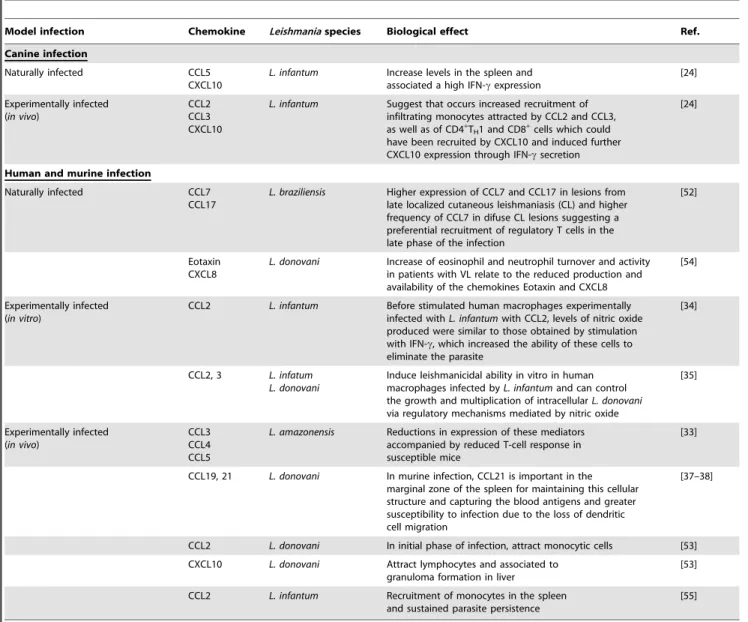
Table 2 presents a summary of the different chemokines and their biological effects duringLeishmania infection in dogs, mice, and humans. These data illustrate how recruitment of specific cells might influence the pathogenesis ofLeishmaniainfection.
Histological analysis
For standard histological examination (morphometric analysis and leukocyte differential counting) sections were coded and stained with hematoxylin and eosin and subsequently underwent blinded analysis under an optical microscope (model CH3RF100, Olympus Optical Co., Tokyo, Japan). The inflammatory cells (neutrophils, eosinophils, macrophages, mast cells, and lympho-cytes) that were recruited to the dermis were counted, and the results are expressed in percentages. Cell types in the cellular infiltrate in the dermis were quantified by using 20 random images (total area = 1.56106mm2) that adequately represented a slide. Thus, the density and predominance of cells in the inflammatory infiltrate and their distribution within the skin layers were assessed
Table 2.Chemokines inLeishmania spp. infection.
Model infection Chemokine Leishmaniaspecies Biological effect Ref.
Canine infection
Naturally infected CCL5 CXCL10
L. infantum Increase levels in the spleen and associated a high IFN-cexpression
[24]
Experimentally infected (in vivo)
CCL2 CCL3 CXCL10
L. infantum Suggest that occurs increased recruitment of infiltrating monocytes attracted by CCL2 and CCL3, as well as of CD4+
TH1 and CD8+cells which could
have been recruited by CXCL10 and induced further CXCL10 expression through IFN-csecretion
[24]
Human and murine infection
Naturally infected CCL7 CCL17
L. braziliensis Higher expression of CCL7 and CCL17 in lesions from late localized cutaneous leishmaniasis (CL) and higher frequency of CCL7 in difuse CL lesions suggesting a preferential recruitment of regulatory T cells in the late phase of the infection
[52]
Eotaxin CXCL8
L. donovani Increase of eosinophil and neutrophil turnover and activity in patients with VL relate to the reduced production and availability of the chemokines Eotaxin and CXCL8
[54]
Experimentally infected (in vitro)
CCL2 L. infantum Before stimulated human macrophages experimentally infected withL. infantumwith CCL2, levels of nitric oxide produced were similar to those obtained by stimulation with IFN-c, which increased the ability of these cells to eliminate the parasite
[34]
CCL2, 3 L. infatum L. donovani
Induce leishmanicidal ability in vitro in human macrophages infected byL. infantumand can control the growth and multiplication of intracellularL. donovani
via regulatory mechanisms mediated by nitric oxide
[35]
Experimentally infected (in vivo)
CCL3 CCL4 CCL5
L. amazonensis Reductions in expression of these mediators accompanied by reduced T-cell response in susceptible mice
[33]
CCL19, 21 L. donovani In murine infection, CCL21 is important in the marginal zone of the spleen for maintaining this cellular structure and capturing the blood antigens and greater susceptibility to infection due to the loss of dendritic cell migration
[37–38]
CCL2 L. donovani In initial phase of infection, attract monocytic cells [53] CXCL10 L. donovani Attract lymphocytes and associated to
granuloma formation in liver
[53]
CCL2 L. infantum Recruitment of monocytes in the spleen and sustained parasite persistence
[55]
and registered quantitatively. The images displayed in the 406
objective were digitized through a Leica DM5000B microscope with a coupled camera using the program Leica Application Suite (version 2.4.0 R1, Leica Microsystems Ltd., Heerbrugg, Switzer-land). For the analysis of images, Leica QWin V3 (Leica Microsystems Ltd.) was used to count all cell nuclei, excluding the pilose follicles, skin annexes, and epidermal cells.
Statistical analysis
Statistical analyses were performed using the GraphPad Prism software package version 5.0 (GraphPad Software, San Diego, CA, USA). Normality of the data was established using the Kolmogorov-Smirnoff test. The Kruskal-Wallis test was used for comparative studies between groups, followed by Dunn’s test for multiple comparisons. Spearman’s rank correlation was also computed in order to investigate relationships between the expression of chemokine mRNAs with clinical forms and skin parasite density as well as cell counts. In all cases, differences were considered significant when the probabilities of equality (pvalues) were#0.05.
Results
Clinical progression in CVL was correlated with increased parasite density and the presence of mononuclear cells in the skin of dogs naturally infected byL. infantum
In order to investigate the relationship between clinical forms of CVL and skin parasite density as well as cellular infiltrate, correlation analyses were conducted with these parameters inL. infantum–infected animals (n= 35) (Fig. 1). The main histopatholog-ical findings are shown in photomicrographs (Fig. 1A). Histopath-ological examination of the skin showed no histHistopath-ological changes within the CD group (Fig. 1A, panels 1 and 2). In the LP and AD groups (Fig. 1A, panels 3 and 4), there was a mild inflammatory infiltrate, composed mainly of mononuclear cells, while in the OD and MP groups, this infiltration was mild to moderate, as shown in Fig. 1A (panels 5 and 6). In panels 7 and 8 of Fig. 1A, which represent sections of ear skin in the SD group, an intense cellular infiltrate composed mainly of mononuclear cells was observed.
The intensity and predominance of cells in the inflammatory infiltrate and their distribution within the skin layers were assessed (Fig. 1B, 1C). Our results demonstrated a positive correlation between cellular infiltrate and clinical status (r= 0.5400, p= 0.0004) (Fig. 1B) and skin parasite density (r= 0.7352, p,0.0001) (Fig. 1C). Significant increases in the inflammatory infiltrate in the skin samples were observed in the AD and SD groups as compared with CD animals (Fig. 1B). The HP group had a significant increase in inflammatory infiltrate compared with the CD and LP groups (Fig. 1C). Moreover, the inflammatory infiltrate within the MP group was significantly increased as compared with the CD group (Fig. 1C).
The results also indicated positive correlation among clinical evolution of CVL and the increase of parasite density in the skin (r= 0.4409, p= 0.0080) (Fig. 1D). Additionally, an increase in parasite density (p,0.05) was detected in the skin of dogs showing the maximum clinical score (SD) when compared with the AD group (Fig. 1D).
Assessment of the inflammatory cell profile in the skin revealed an increase in macrophages and reductions in lymphocytes, eosinophils, and mast cells according to the clinical progression of CVL
The study of skin tissue cellularity included an assessment of the percentage of cell types (neutrophils, eosinophils, mast cells,
lymphocytes, and macrophages) present in the inflammatory infiltrate in the skin of dogs that were naturally infected by L. infantum and categorized by clinical status and dogs that were uninfected (Fig. 2A). In this context, we observed a reduction in the percentage of eosinophils in the SD group compared with the CD group (p,0.05), and a negative correlation between this cell population (r=20.4760,p= 0.0059) and clinical status. Additionally, there was a decrease (p,0.05) in the percentage of mast cells in the OD and SD groups when compared with the CD group. Similarly, a negative correlation was observed in the percentage of mast cells (r=20.6018,p= 0.0002) compared with the clinical form of CVL. For lymphocytes, we observed an increased (p,0.05) percentage in the AD group in comparison with the SD group and control dogs. Furthermore, we also observed an increase (p,0.05) in the OD group as compared with the SD group. The analysis of correlation between lymphocyte counts and clinical status showed a negative correlation between the increase of lymphocytes versus the clinical outcome in CVL (r=20.6283, p,0.0001) (Fig. 2A). Significant increases (p,0.05) were observed in the OD and SD groups in the population of macrophages in comparison to the CD group, and a positive correlation was observed (r= 0.5553,p,0.0010) between the percentage of macrophages and degree of disease.
Enhanced skin parasite density was correlated with an increase of macrophages and decreases of eosinophils and mast cells in the skin of dogs naturally infected byL. infantum
An assessment of cellular infiltrate in the skin of dogs naturally infected by L. infantum and uninfected dogs was performed by categorizing them according to skin parasite density (Fig. 2B). Although neutrophil and lymphocyte subsets did not have significant changes, a shift in the cell profiles related to the innate immune response was observed. The percentage of eosinophils decreased (p,0.05) in the MP and HP groups when compared with the CD group. Associated with these observations, a negative correlation between the percentage of eosinophils and skin parasite density was found (r=20.3885, p= 0.0255). The percentage of mast cells was lower in the LP, MP, and HP groups when compared with the CD group (p,0.05). Accordingly, a significant increase (p,0.05) in the percentage of macrophages in the MP and HP groups in comparison with the CD group was found. Furthermore, a positive correlation between the percentage of macrophages and skin parasite density (r= 0.4163,p= 0.0198) was observed.
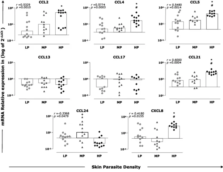
up-regulation occurred in all infected groups when compared to CD, and increased levels were observed in the HP group in comparison with the LP and MP groups (2.1-fold and 1.7-fold, respectively; p,0.05). Moreover, a positive correlation could be established between the expression of CCL5 and skin parasite density (r= 0.5480,p= 0.0014).
CCL13 and CCL17 were down-regulated in all groups in comparison to the CD group; however, significant differences were not found between experimental groups. For the chemokine CCL21, we observed increased expression in all groups compared with the CD group, and high levels were found in the HP group compared with the LP and MP groups (1.4-fold and 1.3-fold,
respectively; p,0.05). In addition, a positive correlation was observed between CCL21 expression and parasite density (r= 0.6000,p= 0.0004).
The expression of CCL24 was up-regulated in the LP and MP groups and down-regulated in the HP group compared to the CD group. In addition, higher CCL24 expression was observed in the MP group when compared with the HP group (0.9-fold;p,0.05). Furthermore, a negative correlation was observed between CCL24 expression and skin parasite density (r=20.3368, p= 0.0479). With regard to CXCL8, we observed an increase in the target transcript levels in the LP and HP groups and down-regulation in the MP group compared with CD. In addition, CXCL8 Figure 1. Histopathological and parasite density analyses of skin of dogs naturally infected withL. infantum.Animals were categorized according to their clinical status into asymptomatic (AD,n= 10), oligosymptomatic (OD,n= 10), or symptomatic (SD,n= 15) or categorized according to skin parasite density into low (LP,n= 12), medium (MP,n= 11), or high (HP,n= 12) parasite density. The control group is represented by CD (n= 16). Photomicrographs of cutaneous cellular infiltrates from dogs naturally inflected byL. infantumstained by hematoxylin and eosin (A). Representative cellular infiltrates of study groups are depicted: Control dogs (1 and 2 ); AD or LP (3 and 4); OD or MP (5 and 6); SD or HP (7 and 8).
Left panels:Slides shown at 106magnification; bar, 100mm.Right panels:Slides shown at 406magnification; bar, 25mm. Correlation between
quantitative analysis of cutaneous cellular infiltrate with clinical status (B) or skin parasite density (C) are presented. Correlation between clinical groups and skin parasite density is also presented (D). The results are expressed as the mean number of cells present in cutaneous cellular infiltrates evaluated at 20 fields plus standard deviation. In (D), the results are expressed as the mean of the log number of skin parasite density plus standard deviation. Significant differences (p,0.05) compared with CD and AD or LP groups are indicated by symbols * and#, respectively. Spearman’s correlation indexes (randpvalues) are shown on the graphs when applicable.
expression was significantly higher in the HP group compared with the LP and MP groups (6.9-fold and 8.3-fold, respectively; p,0.05). Positive correlation was observed between CXCL8 expression and skin parasite density (r= 0.4180,p= 0.0155).
Skin parasite density was most strongly correlated with chemokines that induce macrophage migration during CVL
In order to better identify the association between inflammatory cells present in skin and chemokine levels, we performed additional correlation analyses between distinct cell types and cutaneous
chemokine expression (Fig. 4). Interestingly, our results indicated that macrophages were the cell type that was most likely to be recruited by chemokines CCL2 (r= 0.3514; p= 0.0486), CCL4 (r= 0.3600;p= 0.0396), CCL5 (r= 0.3485;p= 0.0469), and CCL21 (r= 0.3440;p= 0.0499) (Fig. 4). Negative correlation was observed between CCL21 levels and neutrophils (r=20.3562;p= 0.0419).
Discussion
The analysis of chemokine expression in lymphoid compart-ments is crucial for assessing central regulation and pathophysi-ological processes, including traffic homeostasis, inflammation, Figure 2. Cellular profile of the skin from dogs naturally infected byL. infantum.(A) Animals were categorized according to their clinical status into asymptomatic (AD,n= 10), oligosymptomatic (OD,n= 10), or symptomatic (SD,n= 15) or categorized according to skin parasite density into low (LP,n= 12), medium (MP,n= 11), and high (HP,n= 12) parasite density. The control groups are represented by CD (n= 16). Relationship between clinical forms and percentage of cells in the skin of dogs naturally infected withL. infantum(A). Relationship between skin parasite density and percentage of cells in the skin of dogs naturally infected withL. infantum(B). The data are presented as boxplots. The box stretches from the lower hinge (defined as the 25th percentile) to the upper hinge (the 75th percentile) and therefore contains the middle half of the score in the distribution. The median is shown as a line across the box. Therefore, one fourth of the distribution is between this line and the bottom or the top of the box. Significant differences (p,0.05) compared with CD and SD or HP are indicated by symbols * andD, respectively. Spearman’s correlation indexes (randpvalues) are shown on the graphs when applicable.
doi:10.1371/journal.pntd.0001566.g002
Figure 3. Skin parasite density and expression of chemokines in the skin from dogs withL. infantum. Animals were categorized according to their skin parasite density into low (LP = gray circles), medium (MP = gray triangles), and high (HP = dark diamonds) parasite load. The results are expressed as scattering of individual values and median of that group (bars) of log10of the relative copy number of mRNA for CCL2, CCL4,
CCL5, CCL13, CCL17, CCL21, CCL24, and CXCL8. Significant differences (p,0.05) compared with LP, MP, and HP are indicated by the symbols *,#, and
and hematopoiesis [27,28]. In this context, few studies have investigated the levels of chemokines in ongoing CVL. In one of these studies, Strauss-Ayali et al. [24] evaluated the expression of CCL2, CCL4, CCL5, and CXCL10 in the spleen of dogs naturally or experimentally infected byL. infantumand found an increase of CCL2 and CCL5 in the experimentally infected dogs. Herein, increased levels of CCL2, CCL4, and CCL5 in dogs with high parasitism were observed, and these chemokines were positively correlated with parasite density. These results indicate a preferential migration of macrophages into the skin, suggesting a host strategy to control parasitism during ongoing CVL. It has been proposed that in leishmaniasis, chemokines CCL2, CCL4, and CCL5 generally play a role not only as chemotactic factors but also as co-activators of macrophages and consequently have a part in the elimination of parasites [29–32].
After stimulation with CCL2, human macrophages experimen-tally infected with L. infantumproduced levels of nitric oxide that were similar to those obtained by stimulation with IFN-c, which increased the ability of these cells to eliminate the parasite [33]. In addition, CCL2 and CCL3 may induce leishmanicidal abilityin vitro in human macrophages infected byL. infantumand can control the growth and multiplication of intracellularL. donovanivia regulatory mechanisms mediated by nitric oxide [34]. In the present work, we demonstrated an increase in the percentage of macrophages in dogs with clinical signs (OD and SD) or with moderate to high parasitism (MP and HP). There was also a positive correlation between the
percentage of macrophages and expression of CCL2, CCL4, and CCL5. Previous data published by our group demonstrated a decrease in absolute values of circulating monocytes as a hallmark found in the symptomatic group and in the group with the higher parasite load [6]. These data may suggest the recruitment of monocytes to other tissues during active CVL, where they might play an important role in immunological connections throughout antigen presentation and parasite clearance. However, the presence of macrophages in the skin infiltrates does not guarantee their ongoing function since histological analysis of skin during CVL described in this and other studies showed an intense cell infiltrate composed of mononuclear cells in animals with high parasitism that were clinically symptomatic [19]. The finding that expression of macrophage chemoattractants was associated with parasite burden contradicts previous in vitro data demonstrating that these chemokines have a macrophage-activating protective effect. This would suggest that the chemokines are recruiting immature or unresponsive macrophages. Moreover, the levels of CXCL8 observed in HP animals, despite inducing macrophage recruitment, seemed to favor the persistence of the parasite in the skin compartment. In addition, high levels of macrophages in the skin of dogs with active CVL (OD and SD) and in dogs with MP and HP density were demonstrated and highlighted the inability of these cells to control parasitism.
Our study represents the first investigation on the involvement of CCL21 in CVL. We also observed increased levels of CCL21 in Figure 4. Correlation between parasite density (LDU) and cell-types presenting into skin fromL. infantuminfected dogs.The results were expressed on graphs as scattering of individual values. Spearman correlation indexes (r) atp,0.05 are shown on graphs. Connecting lines illustrated positive and negative correlation indexes.
animals with high parasitism, independent of the positive correlation between the chemokine and cutaneous parasitism. It has been reported that CCL21 is an important chemokine involved in recruiting antigen-presenting cells (APCs) to lymphoid organs [35]. In murine infection by L. donovani, Ato et al. [36] demonstrated that CCL21 is important in the marginal zone of the spleen for maintaining the structure of its cellular composition and capturing blood antigens during Leishmania infection. Moreover, mice deficient for the gene encoding CCL21 have greater susceptibility to infection when exposed toL. donovanidue to the loss of dendritic cell migration [37]. In this context, we hypothesize that increased skin parasitism has the potential to stimulate the expression of CCL21, resulting in the recruitment of APCs in the skin from the lymphoid organs. However, it is possible that either these cells, like macrophages, do not present a functional profile favoring a Th1-immune response that would be effective against Leishmaniainfection. Alternately, the increase of CCL21 may lead to retention of APCs in the skin and reduce their migration to the regional lymph node where antigens would be presented to T cells. Several studies have reported the involvement of mast cells in regulating immunity against variousLeishmaniaspecies [38–40]. In the present study, a decrease of this population was observed in the skin of animals presenting severe clinical forms of the disease (OD and SD group) and in all groups categorized according to parasite density (LP, MP, and HP) when compared with the control group. This finding could be related to this cell type being involved in attempts to contain the intense skin parasite density, as described in several studies that evaluated a murine model [41–43]. Calabrese et al. [21] described an intense inflammatory skin reaction formed mainly by mast cells, indicating that these cells might exert a role in innate immunity againstL. infantuminfection. Our data regarding mast cells conflict with this possibility, however. The discrepancy might be explained by L. infantum infection causing a diverse range of clinical and histopathological manifestations. Variations in host resistance may help to explain the variations found in the skin parasite load in dogs. Moreover, when dogs from different regions are compared, additional factors must be considered, such as variations in weather conditions (e.g., Leishmaniainfection seems to occur chiefly in dry seasons).
In the present study, decreases in the eosinophil population and CCL24 expression were observed that were related to the clinical progression and skin parasite density. CCL24 is a specific agonist for CCR3, attracting and activating eosinophils in parasitic diseases [44]. Some authors have described a microbicidal capability of eosinophils againstL. donovaniandL. majorparasites [45,46] and suggested this cell type could play an important role in protection againstLeishmania infection [47]. Moreover, Amusate-gui et al. [48] reported that eosinophil counts were higher in dogs that presented cutaneous signs, and they suggested that this finding was associated with allergenic responses. More studies are
necessary to determine the role of eosinophils in the cutaneous immune response in CVL.
The participation of neutrophils in addressing infection by parasites of the genusLeishmaniahas been studied in recent years to understand the mechanisms related to the innate immune response [49–51]. We observed that higher CXCL8 levels existed in dogs presenting high cutaneous parasitism. This chemokine induces neutrophil chemotaxis, and the initial influx of neutrophils seems to be beneficial for the survival ofLeishmaniain the infected tissue [50]. Interestingly, it has been reported that the parasite itself produces a protein with chemoattractant properties, called Leishmania chemotactic factor, which promotes the migration of neutrophils to the site of infection [49], thereby boosting the phagocytosis of the parasite. Peters et al. [51] evaluated the events that occur in the skin during the initial phase of the transmission of L. majorby sand flies and observed that a decrease in neutrophils at the infection site is associated with the inability of parasites to establish infection. This hypothesis is strongly supported by a recently published study from our group that showed a mixed cytokine profile during active CVL with predominantly higher cutaneous levels of interleukin (IL)-10 and transforming growth factor b1 apart from lower expression of IL-12. These findings might represent a key condition that allows persistence of parasite replication in the skin [17].
Herein, our data highlight the skin as an important organ in CVL and suggest that increased levels of CCL2, CCL4, CCL5, and CCL21 are associated with the immunopathogenesis of CVL. Our data also suggest that the expression of these cytokines in skin could be used as biomarkers for disease progression in dogs naturally infected by L. infantum. Our findings represent an advance in the knowledge of the involvement of skin inflammatory infiltrates in CVL and the systemic consequences and may contribute to developing a rational strategy for the design of new and more efficient prophylactic tools and immunological therapies against CVL.
Acknowledgments
The authors wish to express their appreciation of the hard work carried out by the staff of the Fundac¸a˜o Nacional da Sau´de during the execution of this project. The authors are also grateful for the use of facilities at Centro de Bioterismo (Universidade Federal de Minas Gerais), Universidade Federal de Ouro Preto and Rede Mineira de Bioterismo (FAPEMIG), and for support with the provision of experimental animals.
Author Contributions
Conceived and designed the experiments: RGS CMC RCG GCO ABR. Performed the experiments: DMS RGS JVS DSL. Analyzed the data: DMS CMC RCG ABR. Contributed reagents/materials/analysis tools: RGS CMC ATC RCO ABR. Wrote the paper: DMS RGS RCG DSL ABR.
References
1. Desjeux P (2004) Leishmaniasis: current situation and new perspectives. Comp Immunol Microbiol Infect Dis 27: 305–318.
2. Tesh RB (1995) Control of zoonotic visceral leishmaniasis: is it time to change strategies? Am J Trop Med Hyg 52: 287–292.
3. Abranches P, Silva-Pereira MC, Conceicao-Silva FM, Santos-Gomes GM, Janz JG (1991) Canine leishmaniasis: pathological and ecological factors influencing transmission of infection. J Parasitol 77: 557–561.
4. Giunchetti RC, Mayrink W, Genaro O, Carneiro CM, Correa-Oliveira R, et al. (2006) Relationship between canine visceral leishmaniosis and the Leishmania (Leishmania) chagasi burden in dermal inflammatory foci. J Comp Pathol 135: 100–107.
5. Chamizo C, Moreno J, Alvar J (2005) Semi-quantitative analysis of cytokine expression in asymptomatic canine leishmaniasis. Vet Immunol Immunopathol 103: 67–75.
6. Reis AB, Martins-Filho OA, Teixeira-Carvalho A, Carvalho MG, Mayrink W, et al. (2006) Parasite density and impaired biochemical/hematological status are associated with severe clinical aspects of canine visceral leishmaniasis. Res Vet Sci 81: 68–75.
8. Reis AB, Teixeira-Carvalho A, Vale AM, Marques MJ, Giunchetti RC, et al. (2006) Isotype patterns of immunoglobulins: hallmarks for clinical status and tissue parasite density in Brazilian dogs naturally infected by Leishmania (Leishmania) chagasi. Vet Immunol Immunopathol 112: 102–116.
9. Lage RS, Oliveira GC, Busek SU, Guerra LL, Giunchetti RC, et al. (2007) Analysis of the cytokine profile in spleen cells from dogs naturally infected by Leishmania chagasi. Vet Immunol Immunopathol 115: 135–145.
10. Giunchetti RC, Martins-Filho OA, Carneiro CM, Mayrink W, Marques MJ, et al. (2008) Histopathology, parasite density and cell phenotypes of the popliteal lymph node in canine visceral leishmaniasis. Vet Immunol Immunopathol 121: 23–33.
11. Giunchetti RC, Mayrink W, Carneiro CM, Correa-Oliveira R, Martins-Filho OA, et al. (2008) Histopathological and immunohistochemical investiga-tions of the hepatic compartment associated with parasitism and serum biochemical changes in canine visceral leishmaniasis. Res Vet Sci 84: 269–277. 12. Alves CF, de Amorim IF, Moura EP, Ribeiro RR, Michalick MS, et al. (2009) Expression of IFN-gamma, TNF-alpha, IL-10 and TGF-beta in lymph nodes associates with parasite load and clinical form of disease in dogs naturally infected with Leishmania (Leishmania) chagasi. Vet Immunol Immunopathol 128: 349–358.
13. Carrillo E, Moreno J (2009) Cytokine profiles in canine visceral leishmaniasis. Vet Immunol Immunopathol 128: 67–70.
14. Guerra LL, Teixeira-Carvalho A, Giunchetti RC, Martins-Filho OA, Reis AB, et al. (2009) Evaluation of the influence of tissue parasite density on hematological and phenotypic cellular parameters of circulating leukocytes and splenocytes during ongoing canine visceral leishmaniasis. Parasitol Res 104: 611–622.
15. Manna L, Reale S, Vitale F, Gravino AE (2009) Evidence for a relationship between Leishmania load and clinical manifestations. Res Vet Sci 87: 76–78. 16. Reis AB, Martins-Filho OA, Teixeira-Carvalho A, Giunchetti RC,
Carneiro CM, et al. (2009) Systemic and compartmentalized immune response in canine visceral leishmaniasis. Vet Immunol Immunopathol 128: 87–95. 17. Menezes-Souza D, Correa-Oliveira R, Guerra-Sa R, Giunchetti RC,
Teixeira-Carvalho A, et al. (2011) Cytokine and transcription factor profiles in the skin of dogs naturally infected by Leishmania (Leishmania) chagasi presenting distinct cutaneous parasite density and clinical status. Vet Parasitol 177: 39–49. 18. Baneth G, Koutinas AF, Solano-Gallego L, Bourdeau P, Ferrer L (2008) Canine
leishmaniosis - new concepts and insights on an expanding zoonosis: part one. Trends Parasitol 24: 324–330.
19. dos-Santos WL, David J, Badaro R, de-Freitas LA (2004) Association between skin parasitism and a granulomatous inflammatory pattern in canine visceral leishmaniosis. Parasitol Res 92: 89–94.
20. Solano-Gallego L, Fernandez-Bellon H, Morell P, Fondevila D, Alberola J, et al. (2004) Histological and immunohistochemical study of clinically normal skin of Leishmania infantum-infected dogs. J Comp Pathol 130: 7–12.
21. Calabrese KS, Cortada VM, Dorval ME, Souza Lima MA, Oshiro ET, et al. (2010) Leishmania (Leishmania) infantum/chagasi: Histopathological aspects of the skin in naturally infected dogs in two endemic areas. Exp Parasitol 124: 253–257.
22. Correa AP, Dossi AC, de Oliveira Vasconcelos R, Munari DP, de Lima VM (2007) Evaluation of transformation growth factor beta1, interleukin-10, and interferon-gamma in male symptomatic and asymptomatic dogs naturally infected by Leishmania (Leishmania) chagasi. Vet Parasitol 143: 267–274. 23. Panaro MA, Brandonisio O, Cianciulli A, Cavallo P, Lacasella V, et al. (2009)
Cytokine expression in dogs with natural Leishmania infantum infection. Parasitology 136: 823–831.
24. Strauss-Ayali D, Baneth G, Jaffe CL (2007) Splenic immune responses during canine visceral leishmaniasis. Vet Res 38: 547–564.
25. Mancianti F, Gramiccia M, Gradoni L, Pieri S (1988) Studies on canine leishmaniasis control. 1. Evolution of infection of different clinical forms of canine leishmaniasis following antimonial treatment. Trans R Soc Trop Med Hyg 82: 566–567.
26. Bustin SA, Benes V, Garson JA, Hellemans J, Huggett J, et al. (2009) The MIQE guidelines: minimum information for publication of quantitative real-time PCR experiments. Clin Chem 55: 611–622.
27. Sallusto F, Mackay CR, Lanzavecchia A (2000) The role of chemokine receptors in primary, effector, and memory immune responses. Annu Rev Immunol 18: 593–620.
28. Ono SJ, Nakamura T, Miyazaki D, Ohbayashi M, Dawson M, et al. (2003) Chemokines: roles in leukocyte development, trafficking, and effector function. J Allergy Clin Immunol 111: 1185–1199; quiz 1200.
29. Ritter U, Moll H, Laskay T, Brocker E, Velazco O, et al. (1996) Differential expression of chemokines in patients with localized and diffuse cutaneous American leishmaniasis. J Infect Dis 173: 699–709.
30. Muzio M, Bosisio D, Polentarutti N, D’Amico G, Stoppacciaro A, et al. (2000) Differential expression and regulation of toll-like receptors (TLR) in human leukocytes: selective expression of TLR3 in dendritic cells. J Immunol 164: 5998–6004.
31. Dorner BG, Scheffold A, Rolph MS, Huser MB, Kaufmann SH, et al. (2002) MIP-1alpha, MIP-1beta, RANTES, and ATAC/lymphotactin function together with IFN-gamma as type 1 cytokines. Proc Natl Acad Sci U S A 99: 6181–6186. 32. Ji J, Sun J, Soong L (2003) Impaired expression of inflammatory cytokines and chemokines at early stages of infection with Leishmania amazonensis. Infect Immun 71: 4278–4288.
33. Brandonisio O, Panaro MA, Fumarola I, Sisto M, Leogrande D, et al. (2002) Macrophage chemotactic protein-1 and macrophage inflammatory protein-1 alpha induce nitric oxide release and enhance parasite killing in Leishmania infantum-infected human macrophages. Clin Exp Med 2: 125–129. 34. Bhattacharyya S, Ghosh S, Dasgupta B, Mazumder D, Roy S, et al. (2002)
Chemokine-induced leishmanicidal activity in murine macrophages via the generation of nitric oxide. J Infect Dis 185: 1704–1708.
35. Marsland BJ, Battig P, Bauer M, Ruedl C, Lassing U, et al. (2005) CCL19 and CCL21 induce a potent proinflammatory differentiation program in licensed dendritic cells. Immunity 22: 493–505.
36. Ato M, Nakano H, Kakiuchi T, Kaye PM (2004) Localization of marginal zone macrophages is regulated by C-C chemokine ligands 21/19. J Immunol 173: 4815–4820.
37. Ato M, Maroof A, Zubairi S, Nakano H, Kakiuchi T, et al. (2006) Loss of dendritic cell migration and impaired resistance to Leishmania donovani infection in mice deficient in CCL19 and CCL21. J Immunol 176: 5486–5493. 38. Katakura K, Saito S, Hamada A, Matsuda H, Watanabe N (1993) Cutaneous leishmaniasis in mast cell-deficient W/Wv mice. Infect Immun 61: 2242–2244. 39. Wershil BK, Theodos CM, Galli SJ, Titus RG (1994) Mast cells augment lesion size and persistence during experimental Leishmania major infection in the mouse. J Immunol 152: 4563–4571.
40. Maurer M, Lopez Kostka S, Siebenhaar F, Moelle K, Metz M, et al. (2006) Skin mast cells control T cell-dependent host defense in Leishmania major infections. FASEB J 20: 2460–2467.
41. Gordon JR, Galli SJ (1994) Promotion of mouse fibroblast collagen gene expression by mast cells stimulated via the Fc epsilon RI. Role for mast cell-derived transforming growth factor beta and tumor necrosis factor alpha. J Exp Med 180: 2027–2037.
42. MacDonald AJ, Thornton EM, Newlands GF, Galli SJ, Moqbel R, et al. (1996) Rat bone marrow-derived mast cells co-cultured with 3T3 fibroblasts in the absence of T-cell derived cytokines require stem cell factor for their survival and maintain their mucosal mast cell-like phenotype. Immunology 88: 375–383. 43. Weber A, Knop J, Maurer M (2003) Pattern analysis of human cutaneous mast
cell populations by total body surface mapping. Br J Dermatol 148: 224–228. 44. Petkovic V, Moghini C, Paoletti S, Uguccioni M, Gerber B (2004) Eotaxin-3/
CCL26 is a natural antagonist for CC chemokine receptors 1 and 5. A human chemokine with a regulatory role. J Biol Chem 279: 23357–23363.
45. Pearson RD, Uydess IL, Chapman SW, Steigbigel RT (1987) Interaction of human eosinophils with Leishmania donovani. Ann Trop Med Parasitol 81: 735–739.
46. Oliveira SH, Fonseca SG, Romao PR, Ferreira SH, Cunha FQ (1997) Nitric oxide mediates the microbicidal activity of eosinophils. Mem Inst Oswaldo Cruz 92 Suppl 2: 233–235.
47. Watanabe Y, Hamaguchi-Tsuru E, Morimoto N, Nishio Y, Yagyu K, et al. (2004) IL-5-Induced Eosinophils Suppress the Growth of Leishmania amazo-nensis In Vivo and Kill Promastigotes In Vitro in Response to Either IL-4 or IFN-gamma. DNA Cell Biol 23: 412–418.
48. Amusategui I, Sainz A, Rodriguez F, Tesouro MA (2003) Distribution and relationships between clinical and biopathological parameters in canine leishmaniasis. Eur J Epidemiol 18: 147–156.
49. van Zandbergen G, Hermann N, Laufs H, Solbach W, Laskay T (2002) Leishmania promastigotes release a granulocyte chemotactic factor and induce interleukin-8 release but inhibit gamma interferon-inducible protein 10 production by neutrophil granulocytes. Infect Immun 70: 4177–4184. 50. van Zandbergen G, Klinger M, Mueller A, Dannenberg S, Gebert A, et al.
(2004) Cutting edge: neutrophil granulocyte serves as a vector for Leishmania entry into macrophages. J Immunol 173: 6521–6525.
51. Peters NC, Egen JG, Secundino N, Debrabant A, Kimblin N, et al. (2008) In vivo imaging reveals an essential role for neutrophils in leishmaniasis transmitted by sand flies. Science 321: 970–974.
52. Campanelli AP, Brodskyn CI, Boaventura V, Silva C, Roselino AM, et al. (2010) Chemokines and chemokine receptors coordinate the inflammatory immune response in human cutaneous leishmaniasis. Human Immunology 71: 1220–1227.
53. Cotterell SE (1999) Leishmania donovani infection initiates T cell-independent chemokine responses, which are subsequently amplified in a T cell-dependent manner. European Journal of Immunology 29: 203–214.
54. Elshafie AI, A˚ hlin E, Ha˚kansson LD, Elghazali G, Safi SHE, et al. (2011) Activity and turnover of eosinophil and neutrophil granulocytes are altered in visceral leishmaniasis. International Journal for Parasitology 41: 463–469. 55. Rousseau D, Demartino S, Anjuere F, Ferrua B, Fragaki K, et al. (2001)