online | memorias.ioc.fiocruz.br
Leprosy is predominantly a disease of the periph-eral nervous system, although most of the established diagnostic criteria are related to the skin lesions them-selves. Mycobacterium leprae has the unique capacity to invade Schwann cells (Rambukkanna et al. 2000) and this invasion causes nerve damage and sensory impairments that are central to the pathogenesis and clinical manifestations of the disease. A primary sign of leprosy is the loss of sensation in a skin lesion or in an area innervated by an affected peripheral nerve trunk (Saunderson & Groenen 2000).
The presence of M. leprae in the peripheral nerve tis-sue elicits an inflammatory response that is composed of either epithelioid granuloma with lymphocytes or acid fast bacilli (AFB)-loaded macrophages (Shetty & Antia 1997). The inflammatory infiltrate produced also affects both the cutaneous nerve endings that surround adnexal structures and the microvasculature, as well as the intra-epidermal nerve fibres (Antunes et al. 1997).
Understand the mechanisms that enable peripheral neurons to regenerate is needed to identify methods of improving this regeneration. The literature contains few studies of nerve regeneration in leprosy. Most of these studies are restricted to the peripheral nerve trunks, with
little reference to the regeneration of cutaneous nerves (Job & Desikan 1968, Dastur et al. 1970, Junqueira et al. 1980, Jacobs et al. 1993). Nerve fibres regenerate slowly in leprosy and the process is inefficient because of endoneurial fibrosis. Although nerve regeneration has been observed in the nerve trunks of the lower limbs of leprosy patients (LPs), this regenerative process appears to be functionally ineffective, as the sensory impairment persists after treatment (Miko et al. 1993).
Cutaneous nerve fibre expression of the protein gene product (PGP) 9.5 and the nerve growth factor receptors (NGFr) are used as markers for small nerve fibre neuropa-thy and to diagnose and stage the peripheral neuropaneuropa-thy (Lauria et al. 2005). The use of PGP 9.5 immunolabelling provides a well-characterised and reliable method of iden-tifying most of, if not all, the nerve fibres in the human skin (Kelly et al. 2005). The p75 pan-neurotrophin recep-tor (the low-affinity form of NGFr) immunoreactivity in the Schwann cell membrane and on the axonal membrane of the cutaneous nerve fibres suggests an active role in the control and maintenance of normal sensory innervation (Liang & Johansson 1998).
The aim of the present study was to ascertain the sensory function recovery in skin lesions by comparing the sensory status before and after multidrug therapy (MDT) and by correlating improvements in sensory function with the cutaneous nerve branch damage index (CNDI) assigned during the histopathological examina-tion of cutaneous biopsy specimens. In addiexamina-tion, the PGP 9.5 and NGFr-immunolabelled nerve fibres were quanti-fied in cryosections of skin biopsies to assess the nerve regeneration in the leprosy skin lesions after treatment.
Financial support: FIOCRUZ, CNPq + Corresponding author: anemixir@gmail.com Received 31 March 2012
Accepted 14 August 2012
Cutaneous lesions sensory impairment recovery
and nerve regeneration in leprosy patients
Ximena Illarramendi/+, Emanuel Rangel,Alice Machado Miranda,
Ana Claudia Ribeiro de Castro, Giselle de Oliveira Magalhães, Sérgio Luiz Gomes Antunes
Laboratório de Hanseníase, Instituto Oswaldo Cruz-Fiocruz, Rio de Janeiro, RJ, Brasil
It is important to understand the mechanisms that enable peripheral neurons to regenerate after nerve injury in order to identify methods of improving this regeneration. Therefore, we studied nerve regeneration and sensory impairment recovery in the cutaneous lesions of leprosy patients (LPs) before and after treatment with multidrug therapy (MDT). The skin lesion sensory test results were compared to the histopathological and immunohistochemi-cal protein gene product (PGP) 9.5 and the p75 nerve growth factor receptors (NGFr) findings. The cutaneous neural occupation ratio (CNOR) was evaluated for both neural markers. Thermal and pain sensations were the most frequently affected functions at the first visit and the most frequently recovered functions after MDT. The presence of a high cutaneous nerve damage index did not prevent the recovery of any type of sensory function. The CNOR was calculated for each biopsy, according to the presence of PGP and NGFr-immunostained fibres and it was not sig-nificantly different before or after the MDT. We observed a variable influence of MDT in the recovery from sensory impairment in the cutaneous lesions of LPs. Nociception and cold thermosensation were the most recovered sensa-tions. The recovery of sensation in the skin lesions appeared to be associated with subsiding inflammation rather than with the regenerative activity of nerve fibres.
SUBJECTS, MATERIALS AND METHODS
Forty-five untreated LPs aged 12-78 years (mean = 41.8 ± 17.43 years, 52% male) from the Souza Araújo Outpatient Clinic, Oswaldo Cruz Institute, Oswaldo Cruz Foundation (Fiocruz), Rio de Janeiro, were includ-ed in the study. The patients who had leprosy reactions at diagnosis were excluded from the study.
The numbers and morphology of the cutaneous lesions were recorded. Sensory evaluations of the skin lesions were performed by a well-trained physiotherapist using the pinprick test for pain sensation assessment, testing ether for cold thermal sensation [which measures sensa-tion to 20ºC (NIOSH 2010)] and the Semmes-Weinstein monofilament test (MFT) for assessing tactile sensation. Thermal and pain sensation were compared to the normal skin surrounding the lesion or to the opposite side of the body and were classified as abolished, diminished or pre-served. Tactile sensations were classified according to the monofilament force felt by the patient (0 = no sensation, 1 = 300 g, 2 = 4 g, 3 = 2 g, 4 = 0.2 g and 5 = 0.05 g). The lesion with the maximal degree of sensory impairment was selected. Later recovery of a sensory function was designated when evidence of improvement from “abol-ished” to “partial” sensation or from “partial” to “nor-mal” sensation was observed during the final evaluation. In addition, a 2-point increase in the final MFT was also considered to mark the recovery of tactile sensation.
Slit-skin smears were obtained from all of patients to evaluate the bacterial index (BI). A skin biopsy was performed using a 6-mm punch from the most actively affected area of the lesion. The patients were classified according to the Ridley and Jopling criteria (1966) and were treated following Ministry of Health (MS 2004) guidelines with either paucibacillary (PB) or multibacil-lary (MB) regimens according to their BI result. A com-plete evaluation was performed at the end of the MDT (after the skin changes were recorded).
Conventional histopathological evaluation with haematoxylin-eosin (H&E) and Wade staining was per-formed and evaluated by trained pathologists. The pres-ence, type and localisation of the inflammatory infiltrate were registered and the morphological aspects of the cu-taneous nerve branches were studied under light micros-copy using 10X, 20X and 40X objective lenses. A CNDI was obtained in each biopsy from the ratio between the number of cutaneous nerve branches affected by inflam-mation (surrounded and/or invaded by the inflammatory infiltrate) and the total number of visible cutaneous nerve branches in the whole biopsy section. The CNDI was cat-egorised into the following groups based on the values obtained: not available, no cutaneous branches were ob-served, low, the index was between 0.1-0.5, and high, the CNDI was between 0.6-1. The type of inflammatory infil-trates (epithelioid granuloma, AFB-loaded macrophages or mononuclear cell infiltrate) that were associated with the cutaneous nerve branches was also recorded.
Immunohistochemical study - A subgroup of 13 pa-tients with no leprosy reactions during the course of the MDT also had skin biopsies taken at the discharge visit after completing the MDT.
Immunohistochemical labelling for two neural mark-ers, PGP 9.5 and p75 NGFr was performed in a portion of the skin biopsy according to Antunes et al. (1997). The samples were fixed in Lana’s solution, snap-frozen and stored in liquid nitrogen until processing. The biop-sy specimens were cut in a Leica cryostat; the sections were thawed on silane-coated glass slides, allowed to dry and incubated with phosphate buffered saline 0.01M and blocked with normal swine serum for 10 min. The pri-mary antibodies (rabbit anti-PGP 9.5, 1:400 diluted, Ul-traclone, Cambridge, England; anti-NGFr 1:50, DAKO, Carpenteria, USA) were applied and left overnight at 4ºC in a humid chamber. Swine anti-rabbit immunoglobulin-fluorescein isothiocyanate and swine anti-mouse-tetram-ethylrhodamine isothiocyanate were used as conjugates for PGP 9.5 and NGFr antibodies, respectively.
Computer-assisted image analyses quantification of the immunostained nerve fibres in the fields captured by a digital camera were performed using the ImagePro® software (Media Cybernetics, Rockville, Maryland, USA). Four fields (2 comprising the superior dermis and 2 covering the inferior dermis) were examined un-der 20X objective lenses to detect and calculate the PGP and NGFr-immunoreactive areas. The cutaneous neural occupation ratio (CNOR) of each captured image was calculated as the ratio between the area of immunoreac-tive fibres and the whole evaluated area. Thus, CNOR is a quantitative indicator of the cutaneous innervation of each biopsy. To compare CNOR to other variables, it was arbitrarily classified into high or low if it was higher or lower than the median, respectively.
To maintain objectivity, the technician and patholo-gist who performed the morphometry and the quantifi-cation of the immunostained fibres were blinded to the clinical evaluation results.
Data analyses were performed using Open Source Epidemiologic Statistics for Public Health v. 2.3.1 (ope-nepi.com/OE2.3/Menu/OpenEpiMenu.htm) and STA-TA/SE 8.0 for Windows (StataCorp, Texas, USA). The distribution frequencies were calculated for the different variables. Associations between variables were assessed
using Mantel-Haenszel chi-squared (MH χ2) test and the
two-tailed Fisher’s exact test. Comparisons between the variables before and after the MDT were made using the Wilcoxon signed-rank test and a 1-tailed Fisher’s exact test. The CNDI was compared with the degree of cu-taneous sensory impairment, with the type of infiltrate observed in the lesions and with the CNOR.
Ethics - All of the procedures were in accordance with the ethical standards of the Fiocruz Research Ethi-cal Committee and the Helsinki Declaration of 1975, as revised in 1983. All of the subjects provided written in-formed consent.
RESULTS
Only four patients had leprosy reactions during the MDT; they were treated with oral prednisone.
On average, the patients had five lesions (1 to > 20 range). Most of the patients (64%) had erythematous plaques. The selected lesions were located mainly in the upper and lower extremities (84%), followed by the trunk and the face.
At the diagnosis, all of the patients had thermal and pain sensory impairments in the selected lesions, but nine had normal tactile function (Table I). Cold ther-mosensation was abolished in most of the patients (94%), pain sensation was abolished in 81% and tactile sensa-tion was abolished in 11%.
Although the patients did not recover normal thermal and pain sensations in the skin lesions at the follow-up evaluation, 14 (41%) patients recovered some cold ther-mosensation and 13 (38%) patients partially recovered pain sensation (Table I). All of the patients with thermal (n = 2) or pain (n = 6) hypoesthesia at diagnosis remained the same after completing the MDT. Only two patients recovered normal tactile sensation in the skin lesion and half of the patients with initially-abolished tactile sen-sation (2/4) reported partial recovery. However, of the 19 patients who still suffered from tactile hypoesthesia, five (26%) recovered more than two points in the MFT. Of the four patients who developed reactions during the MDT, two patients recovered their abolished sensations, one partially recovered pain sensation and one recovered some tactile sensation. No significant differences were observed in the recovery of thermal, pain or tactile sen-sations between the patients with or without reaction (p = 0.095, p = 0.481 and p = 0.635, respectively).
Inflammatory infiltrate was observed in 34/45 (76%) samples under light microscopy. No differences were observed between the type of lesion and the presence
of inflammatory infiltrate (MH χ2= 0.36, Fisher’s exact test = 0.795). Mononuclear infiltrates (lymphocytes and macrophages) were observed in 10 samples. Although
epithelioid granulomas were more frequently observed in the samples obtained from plaques (n = 17, 50%) than in those obtained from skin patches (n = 7, 21%), these
dif-ferences were not statistically significant (MH χ2 = 1.302, Fisher exact p = 0.4416). Following the Wade staining, AFB was observed in eight biopsy samples, ranging from 1+ to 5+ (mean = 3.375).
Cutaneous nerve branches were observed in 38 (84%) samples (Fig. 1A, B). A mean of 2.5 (minimum = 1, max-imum = 11) dermal nerve branches were observed per sample; most were affected by inflammatory infiltrate. Only five (11%) samples were not invaded or surrounded by inflammatory cells. However, a significantly higher index was observed in the branches with inflammation
(MH χ2 = 7.48, Fisher exact = 0.02) that were obtained from the plaque biopsies (22/38, 58%) than in those ob-tained from the maculae biopsies (9/38, 24%). In
addi-tion, significantly higher (MH χ2 = 7.665, Fisher exact = 0.040) CNDIs were observed in the biopsies with epithe-lioid granulomas (24/24, 100%) than in the biopsies with mononuclear infiltrate (7/10, 70%).
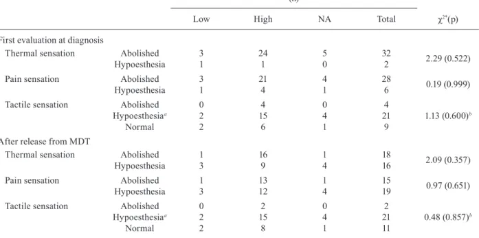
Comparisons between the sensory status of the skin lesion and the index of the affected nerves demonstrated no significant differences in the CNDI by degree of loss of thermal, pain or tactile sensation at either evaluation (Table II). Nonetheless, the highest proportions of high CNDI were observed in the patients who had abolished thermal (24/32, 75%), pain (21/28, 75%) and tactile (4/4, 100%) sensations. In addition, the presence of a high CNDI did not prevent any type of functional recovery. Eight of the 25 patients who had high CNDIs at diag-nosis recovered some thermal and pain sensation (32%), but only four (16%) patients recovered tactile sensation. A higher proportion of the patients with low CNDI at diagnosis (50%, 2/4) recovered their thermal and pain sensations, but none recovered tactile sensation.
Recovery of the nerve fibres was observed in five (39%) of the 13 samples that were taken from patients
TABLE I
Sensory function evaluation of the selected skin lesion in 34 leprosy patients
at diagnosis and after release from multidrug therapy (MDT) according to type of sensory function
At diagnosis
Sensation status after MDT
Total n Abolished
n (%)
Hypoesthesia n (%)
Normal n (%)
Thermal sensation Abolished 18 (56) 14 (44) 0 (0) 32
Hypoesthesia 0 (0) 2 (100) 0 (0) 2
Normal 0 (0) 0 (0) 0 (0) 0
Pain sensation Abolished 15 (55) 13 (45) 0 (0) 28
Hypoesthesia 0 (0) 6 (100) 0 (0) 6
Normal 0 (0) 0 (0) 0 (0) 0
Tactile sensation Abolished 2 (50) 2 (50) 0 (0) 4
Hypoesthesiaa 0 (0) 19 (91) 2 (9) 21
Normal 0 (0) 0 (0) 9 (100) 9
after completing the MDT. However, no nerve fibres were observed in three (23%) biopsy specimens from le-sions in which the nerve fibres were observed in the first sample. A total of 6/9 (67%) lesions that had inflamma-tory infiltrate at the first evaluation recovered or main-tained the same number of nerve fibres. No significant differences were observed in the nerve fibres of the four samples with no inflammatory infiltrate, two of which had no visible nerve fibres at both time points.
On average, 11% of the dermal area of the biopsy sec-tions was immunostained against PGP 9.5 (Fig. 1C) and 26% against p75 NGFr (Fig. 1E). All of the PGP 9.5 im-munostained samples with low CNDI (n = 2) also had low CNOR, but 29% of the samples with high CNDI also had low CNOR. Interestingly, 50% of the NGFr immu-nostained samples with high CNDI had low CNOR and 67% of the samples with low CNDI had high CNOR.
The PGP 9.5 immunostained area was slightly re-duced in six patients in the follow-up biopsy specimen (mean = 9% of positive area) as a result of a decrease in the immunoreactive area. However, in p75 NGFr-stained biopsies, the mean CNOR increased (33%). However,
no significant differences were found in the CNOR be-tween the two biopsies (Fig. 2), as shown with the PGP 9.5 immunostaining (Z = -1.38, p = 0.097) or with the NGFr immunostaining (Z = -0.53, p = 0.319).
DISCUSSION
Variable recovery of sensory function in the skin le-sion after completing the MDT was seen in the present sample and the results differed by sensory functions. No worsening of sensation status was reported after the MDT, even in those patients who had experienced a leprosy reaction. Although the reliability of clinical diagnosis based on pain and thermal testing of skin le-sions has been found to be low (Jain et al. 1986), the use of Semmes-Weinstein monofilaments for the evaluation of tactile sensory function is highly reliable (Birke et al. 2000). Sensory testing in the present study was per-formed by the same physiotherapist following the same technique each time, thus the observed recovery of sen-sory functions was considered consistent.
The pattern of sensory loss observed in the skin le-sions of the patients in the present study is consistent with knowledge of the nerve damage produced by lep-rosy. M. leprae is harboured in both non-myelinating and myelinating Schwann cells, but because it is an ob-ligate intracellular bacterium, it needs to invade unmy-elinating cells to survive within the peripheral nervous system (Rambukkana et al. 2002). As a result, small nerve fibers are easily affected. This is observed clini-cally by the early loss of sensation to thermal or nox-ious stimuli.
During the progression of the infection, M. leprae induces demyelination and axonal damage in order to provide a sufficient intracellular niche for bacterial sur-vival (Rambukkana et al. 2002). Tactile sensation, which is conducted by large myelinated axons, is affected later in the disease, which explains the reduced damage to this sensory modality - relative to nociception and ther-mosensation - that was observed in this sample.
While sensory function impairment was verified clinically, it could not be significantly associated with the histopathology findings. The highest CNDIs were observed in those sections of the lesion with abolished thermal and pain sensations. Other authors have found that higher proportions of nerve bundles are affected by the leprosy inflammatory infiltrate (Paksoy 1987). This difference may be caused by the higher proportion of MB patients selected in the latter study.
Morphological alteration of the cutaneous nerve branches on account of the inflammatory infiltrate is an important diagnostic marker for leprosy in the skin biopsy. However, recognition of the small nerve fibres in H&E-stained sections is difficult, especially when there is appreciable inflammation (Ridley & Jopling 1966). This finding reflects the need for special tech-niques, such as immunohistochemistry, to identify the fibres in the deep and superficial plexuses and to iden-tify their remnants in granulomas (Antunes et al. 1997, Jain et al. 2000).
After the MDT, recovery of the damage at the histo-pathological level is variable, but is present in most cases
when assessed with the resolution of granuloma (Mathew et al. 2004, Prasad et al. 2005). Follow-up biopsies from 26 patients with skin patches that were taken after six-eight months of treatment showed healing of the lesion or reduction in the infiltrate in most cases (Agarwal et al. 1990). However, of 17 patients with borderline tuber-culoid leprosy that showed no evidence of Schwann cells or of axons under S-100 staining at diagnosis, Jain et al. (2000) only observed Schwann cells in two biopsies that were taken after the MDT, suggesting that once nerve damage is established, it is refractory to treatment.
The lack of significant differences between the CNORs registered in both sequential biopsies suggests that sensory function recovery in leprosy may not all be caused by neural regenerative activity. Sensory func-tion could be reestablished because of remission of the leprosy inflammatory process during treatment, which improves the sensory function of the trunk and cutane-ous nerve fibres. Previcutane-ous studies have demonstrated the effectiveness of MDT for resolving the inflammatory infiltrate in the cutaneous nerves, particularly if an anti-inflammatory drug, such as clofazimine, is added to the scheme (Prasad et al. 2005).
The regenerative capacity of unmyelinated fibres is higher than those of myelinated fibres. In addition, re-generation of the large myelinated fibres takes longer on account of the additional need for remyelination (Chen et al. 2007). This need accounts for the higher
frequen-cy of recovery of thermal and pain sensations that we observed compared to the recovery of tactile function. However, M. leprae-induced demyelination and axonal damage resemble peripheral nerve injuries in Wallerian degeneration, in which Schwann cells rapidly proliferate and promote the regeneration of injured nerves (Ram-bukkana et al. 2002).
TABLE II
Comparison between sensory impairment and the cutaneous nerve damage index at diagnosis and after release from multidrug therapy (MDT)
Cutaneous nerve damage index at leprosy diagnosis (n)
χ2*(p)
Low High NA Total
First evaluation at diagnosis
Thermal sensation Abolished 3 24 5 32
2.29 (0.522)
Hypoesthesia 1 1 0 2
Pain sensation Abolished 3 21 4 28
0.19 (0.999)
Hypoesthesia 1 4 1 6
Tactile sensation Abolished 0 4 0 4
Hypoesthesiaa 2 15 4 21 1.13 (0.600)b
Normal 2 6 1 9
After release from MDT
Thermal sensation Abolished 1 16 1 18
2.09 (0.357)
Hypoesthesia 3 9 4 16
Pain sensation Abolished 1 13 1 15
0.97 (0.651)
Hypoesthesia 3 12 4 19
Tactile sensation Abolished 0 2 0 2
Hypoesthesiaa 2 15 4 21 0.48 (0.857)b
Normal 2 8 1 11
a: sensation of monofilaments 1-4 (300-0.2g) was considered as hypoesthesia; b: calculated by grouping abolished and hy-poesthesic into impaired for comparison with normal tactile sensation due to 0 values; NA: not available (no nerves were observed in the sections).
As shown in the present study, MDT has a variable influence on the recovery of sensory impairment in the cutaneous lesions of LPs. Nociception and cold ther-mosensation were the most impaired and the most com-monly recovered sensory functions, but tactile sensation was the only modality that recovered completely. This sensation recovery appears to be more associated with the subsiding inflammatory process.
Open-ended perineurial sleeves of simple sensory nerve formations and branching ending in connective tissue, as well as the naked nerve endings in the skin, provide distal continuity of the endoneurial microenvi-ronment with the surrounding extracellular tissue space. The interactions of Schwann cells, axons, macrophages and inflammatory cells via cell-cell and cell-matrix sig-nalling regulate the permeability of the blood-nerve in-terface in the tissue. Inflammation induces vasodilation and increased vascular permeability in the peripheral nerves and alters the cell population and matrix compo-nents. The homeostasis of the environment is modified, in turn and consequently, the impairment of the impulse conduction is impaired (Schepers & Ringkamp 2010). MDT is known to kill the bacilli, which leads to a focal remission of the inflammatory infiltrate. Therefore, in contrast to the expected nerve regeneration, remission of the inflammation seems to be the most prominent factor in restoring cold thermosensation and sensation follow-ing mechanical noxious stimuli.
The quality of sensory outcome following nerve re-pair is difficult to objectively quantify. Further research is required to quantify the functional sensation of skin lesions and to establish the reliability of the evaluation of the different components of sensory function (thermal, pain and tactile functions) in relation to the pathologic features of biopsies.
ACKNOWLEDGEMENTS
To Dr José Augusto Costa Nery, for the clinical evaluation of the patients.
REFERENCES
Agarwal US, Handa AK, Mathur D, Mehta RD, Mittal A, Dhar N, Mathur NK 1990. Hypopigmented lesions in early leprosy - a clinical and histological study. Indian J Lepr 62: 416-421.
Antunes SL, Sarno EN, Holmqvist G, Johansson O 1997. A compari-son of the expression of NGFr with PGP 9.5 and NSE in the cuta-neous lesions of early leprosy patients using immunohistochem-istry. Int J Lepr Other Mycobact Dis65: 357-365.
Birke JA, Brandsma JW, Schreuders TA, Piefer 2000. A sensory test-ing with monofilaments in Hansen’s disease and normal control subjects. Int J Lepr Other Mycobact Dis68: 291-298.
Chen ZL, Yu WM, Strickland S 2007. Peripheral regeneration. Ann Rev Neurosci30: 209-233.
Dastur DK, Pandya SS Antia NH 1970. Nerves in the arm in leprosy II. Pathology, pathogenesis and clinical correlation. Int J Lepr Other Mycobact Dis38: 30-48.
Jacobs JM, Shetty VP, Antia NH 1993. A morphological study of nerve biopsies from cases of multibacillary leprosy given multi-drug therapy. Acta Neuropathol(Berl) 85: 533-541.
Jain GL, Pasricha JS, Guha SK 1986. Objective grading of the loss of pain and touch sensations in leprosy patients. Int J Lepr Other Mycobact Dis54: 525-529.
Jain M, Singh N, Bhatia A, Arora VK 2000. Histological assessment of dermal nerve damage occurring during multidrug therapy for leprosy. Int J Lepr Other MycobactDis68: 167-171.
Job CK, Desikan KV 1968. Pathologic changes and their distribution in peripheral nerves in lepromatous leprosy. Int J Lepr Other My-cobact Dis 36: 257-270.
Junqueira LCU, Montes CS, Neto EA, Barros C, Tedescu-Marques AJ 1980. The collagen of permanently damaged nerves in human leprosy. Int J Lepr Other Mycobact Dis48: 291-297.
Kelly EJ, Terenghi G, Hazari A, Wiberg M 2005. Nerve fibre and sensory end organ density in the epidermis and papillary dermis of the human hand. Br J Plast Surg58: 774-779.
Lauria G, Cornblath DR, Johansson O, McArthur JC, Mellgren SI, Nolano M, Rosenberg N, Sommer C 2005. European Federation of Neurological Societies. EFNS guidelines on the use of skin biopsy in the diagnosis of peripheral neuropathy. Eur J Neurol 12: 747-758.
Liang Y, Johansson O 1998. Light and electron microscopic demonstra-tion of the p75 nerve growth factor receptor in normal human cuta-neous nerve fibers: new vistas. JInvest Dermatol 111: 114-118.
Mathew D, Kishore BN, Shwethadri GK, Sukumar, Shetty NJ 2004. An evaluation of clinical and histopathological status in paucibac-illary leprosy patients after completion of fixed duration therapy. Indian J Lepr76: 11-18.
Miko TL, Gschmeissner SE, Le Maitre C, Kinfu Y, Kazen R, Pereira JH 1993. Regeneration at the predilective damage sites of nerve trunks in treated leprosy. Lepr Rev 64: 330-337.
MS - Ministry of Health 2004. Guide of control of leprosy, 2nd ed., Fundação Nacional de Saúde, Brasília, 89 pp.
NIOSH - National Institute for Occupational Safety and Health 2003. Manual of analytical methods, 4th ed., US Department of Health, Education, and Welfare, NIOSH, Cincinnati, 157 pp.
Paksoy N 1987. Indeterminate leprosy: a clinical and histopathologi-cal evaluation. Indian J Lepr59: 399-404.
Prasad PVS, Babu A, Kaviarasan PK, Viswanathan P, Tippoo R 2005. MDT-MB therapy in paucibacillary leprosy: A clinicopathologi-cal assessment. Indian J Dermatol Venereol Leprol 71: 242-245.
Rambukkana A 2000. How does Mycobacterium leprae target the pe-ripheral nervous system? Trends Microbiol8: 23-28.
Rambukkana A, Zanazzi G, Tapinos N, Salzer JL 2002. Contact-de-pendent demyelination by Mycobacterium leprae in the absence of immune cells. Science296: 927-931.
Ridley DS, Jopling WH 1966. Classification of leprosy according to immunity. A five-group system. Int J Lepr Other Mycobact Dis 34: 255-273.
Saunderson P, Groenen G 2000. Which physical signs help most in the diagnosis of leprosy? A proposal based on experience in the AMFES project, ALERT, Ethiopia. Lepr Rev71: 34-42.
Schepers RJ, Ringkamp M 2010. Thermoreceptors and thermosensi-tive afferents. Neurosci Biobehav Rev 34: 177-184.