ISSN 0100-879X
BIOMEDICAL SCIENCES
AND
CLINICAL INVESTIGATION
www.bjournal.com.br
www.bjournal.com.br
Volume 45 (3) 179-290 March 2012
Braz J Med Biol Res, March 2012, Volume 45(3) 222-229
doi: 10.1590/S0100-879X2012007500024
Association of MICA gene polymorphisms with liver fibrosis in
schistosomiasis patients in the Dongting Lake region
Zheng Gong, Qi-Zhi Luo, Lin Lin, Yu-Ping Su, Hai-Bo Peng, Kun Du, Ping Yu and Shi-Ping Wang
Institutional Sponsors
The Brazilian Journal of Medical and Biological Research is partially financed by
Faculdade de Medicina de Ribeirão Preto Campus
Ribeirão Preto
Explore High - Performance MS Orbitrap Technology In Proteomics & Metabolomics
Association of
MICA
gene polymorphisms with
liver fibrosis in schistosomiasis patients
in the Dongting Lake region
Zheng Gong
1, Qi-Zhi Luo
1, Lin Lin
1, Yu-Ping Su
3, Hai-Bo Peng
3, Kun Du
1,
Ping Yu
1and Shi-Ping Wang
21Department of Immunology, College of Basic Medical Sciences, Central South University, Changsha, Hunan Province, China 2Key Laboratory of Schistosomiasis in Hunan, Department of Parasitology, College of Basic Medical Sciences, Central South University, Changsha, Hunan Province, China
3Central Blood Bank in Yueyang, Yueyang, Hunan Province, China
Abstract
Major histocompatibility complex class I chain-related A (MICA) is a highly polymorphic gene located within the MHC class I
region of the human genome. Expressed as a cell surface glycoprotein, MICA modulates immune surveillance by binding to its
cognate receptor on natural killer cells, NKG2D, and its genetic polymorphisms have been recently associated with susceptibility
to some infectious diseases. We determined whether MICA polymorphisms were associated with the high rate of Schistosoma parasitic worm infection or severity of disease outcome in the Dongting Lake region of Hunan Province, China. Polymerase
chain reaction-sequence specific priming (PCR-SSP) and sequencing-based typing (SBT) were applied for high-resolution allele
typing of schistosomiasis cases (N = 103, age range = 36.2-80.5 years, 64 males and 39 females) and healthy controls (N = 141, age range = 28.6-73.3 years, 73 males and 68 females). Fourteen MICA alleles and five short-tandem repeat (STR) alleles
were identified among the two populations. Three (MICA*012:01/02, MICA*017 and MICA*027) showed a higher frequency in healthy controls than in schistosomiasis patients, but the difference was not significantly correlated with susceptibility to S. japonicum infection (Pc > 0.05). In contrast, higher MICA*A5 allele frequency was significantly correlated with advanced liver
fibrosis (Pc < 0.05). Furthermore, the distribution profile of MICA alleles in this Hunan Han population was significantly different
from those published for Korean, Thai, American-Caucasian, and Afro-American populations (P < 0.01), but similar to other Han populations within China (P > 0.05). This study provides the initial evidence that MICA genetic polymorphisms may underlie the
severity of liver fibrosis occurring in schistosomiasis patients from the Dongting Lake region.
Key words: Schistosoma japonicum; MICA; NKG2D; Gene polymorphism; Liver fibrosis
Introduction
Correspondence: Ping Yu and Shi-Ping Wang, Department of Immunology, College of Basic Medical Sciences, Central South University,
110 Xiangya Road, Changsha, Hunan, China. Fax: +86-073-1448-7301. E-mail: yuping1953@hotmail.com and spwang@126.com
Received August 21, 2011. Accepted February 13, 2012. Available online March 2, 2012. Published March 19, 2012.
Schistosomiasis is the most prevalent water-borne parasitic disease worldwide and remains a major public health problem in many developing countries. Among the
pathogenic Schistosoma species, S. japonicum is
respon-sible for endemic infection in China, Indonesia, and the
Philippines (1). Recorded cases of schistosomiasis in China
date back more than 2100 years (2). Not surprisingly, one of the first and most substantial human health campaigns of the newly founded People’s Republic of China was to control this parasitic disease. By the beginning of the 2000’s, schistosomiasis had been eradicated in five of the twelve previously endemic provinces of China, accompanied by
dramatic reductions in morbidity and mortality (3,4). Currently, the geographic distribution of schistosomiasis
in China is primarily centered around the Yangtze River, with the worst situations in the Dongting Lake and Poyang
Lake regions (3,4). The Dongting Lake, located in the north
of Hunan Province, is the second largest freshwater lake
in China and provides an ideal habitat for Oncomelania
snails, the intermediate host for S. japonicum. Sporocysts
are generated in the snail and produce infective cercariae that are released from the snail into the surrounding
aque-ous environment. These free-swimming parasitic larvae
Polymorphism of the MICA gene and schistosomiasis 223
the human epidermis. Inside the human host, the para-sites mature in the hepatic portal vein and adult paired
sex worms migrate to the mesenteric veins, depositing eggs that are carried by the circulation to the host’s liver, intestine, urinary bladder or other organs. These eggs
trigger the host immune response, including a localized
inflammatory reaction; however, as the simple immune response is unable to clear these eggs, granuloma for
-mation is induced to wall off the foreign substances. The granulomatous reaction is accompanied by fibrosis, which
is particularly devastating to the function of liver tissues
(5). Thus, the pathogenesis of schistosomiasis involves
the dynamic coordination of many tissue- and
process-specific genes. Undoubtedly, the parasite has evolved to exploit genes that vary in expression among different populations to establish infection.
The human major histocompatibility complex (MHC)
class I chain-related (MIC) gene family consists of seven
members, MICA to MICG; however, only MICA and MICB
are functional genes and all other members represent
pseudogenes (6,7). The human MICA gene contains 6
exons encoding a cell-surface glycoprotein with three extracellular domains (encoded by exons 2, 3, and 4, re
-spectively), a transmembrane fragment (encoded by exon 5), and a carboxy-terminal cytoplasmic tail (encoded by exon 6) (6,8). In response to cellular stress, MICA expres -sion is induced in many cell types, including epithelium,
fibroblasts, keratinocytes, endothelial cells, and monocytes (7,9,10), whereby it regulates the autoimmune response through binding to its cognate receptor (5), the natural
killer (NK) cell receptor D (NKG2D) (11-16). The MICA
gene is highly polymorphic, and the exons encoding the extracellular domains present the highest frequency of
polymorphisms (8).
The two different types of alleles of the gene repre
-sent 76 (http://hla.alleles.org/nomenclature/stats.html)
sequence alleles in the mature protein-coding region
(MICA*001 to *064N, http://www.ebi.ac.uk/imgt/hla/
allele.html) and seven microsatellite alleles. Also known
as short tandem repeats (STR), these microsatellite al
-leles (MICA*A4, *A5, *A5.1, *A6, *A7, *A9, and *A10) were formed by insertion/deletion of variable numbers of trinucleotide GCT repeats within exon 5.
MICA has been identified as a candidate disease
gene by several linkage disequilibrium mapping stud -ies of common autoimmune-related diseases, including
Behcet’s disease, insulin-dependent diabetes mellitus, and
Addison’s disease (17). In our ongoing attempts to char-acterize the pathogenesis of schistosomiasis in the Hunan
Han population, we became intrigued by the underlying
mechanistic features involving autoimmune processes and
hypothesized that ethnicity-related MICA polymorphisms
may confer susceptibility to S. japonicum infection and
affect the outcome of severe liver fibrosis. To this end, we performed polymerase chain reaction-sequence specific
priming (PCR-SSP) and sequencing-based typing (SBT)
to analyze the MICA polymorphisms in schistosomiasis
patients from the Dongting Lake region and compared
the distribution of MICA alleles with data for other ethnic groups around the world.
Material and Methods
Subjects
A cohort of 103 patients of Han nationality attending the
Schistosomiasis Outpatient Clinic of the Teaching Hospi -tal, School of Medical Science, Central South University
(Changsha, China) between 2008 and 2010 were recruited
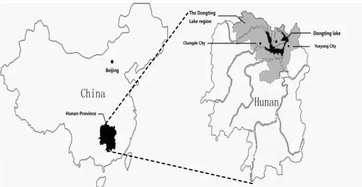
into this study. All patients resided in Changde or Yueyang city in the Dongting Lake region, Hunan Province, China (Figure 1). All patients had a recorded clinical diagnosis
of S. japonicum infection between the ages of 10 and 24
years. Diagnosis of liver fibrosis in these patients was made by B-scan ultrasonography findings fitting World Health
Organization (WHO) criteria. Patients with grade 0 and
grade 1 fibrosis were classified into a mild fibrotic group, and those with grade 2 (moderate) and grade 3 (severe) fibrosis into an advanced fibrotic group. For healthy controls, 141
unrelated Han individuals were recruited from the Central Blood Bank (Yueyang, China) according to the following criteria: residing in Changde or Yueyang city at the time of
blood collection, no previous diagnosis of schistosomiasis,
no family history of schistosomiasis, and a gender ratio
match to the patient cohort. The clinicopathological features of the patients and controls are listed in Table 1. Written informed consent was obtained from each study participant, and the study was approved by the Ethics Review Board
of the Central South University.
High-resolution allele typing of the MICA gene by PCR-SSP and SBT
Genomic DNA was isolated from EDTA-treated pe
-ripheral venous blood using a conventional proteinase K digestion/salting-out extraction method (18). The PCR-SSP method was carried out as previously described with the following minor modifications (19). The human growth
hormone gene was used as an internal control and
ampli-fied (834-bp fragment) by gene-specific primers (sense 5’-GCCTTCCCAACCATTCCCTTA-3’ and anti-sense 5’-GAGAAAGGCCTGGAGGATTC-3’) (19). Ninety-five
primers targeting MICA exons 2-4 were designed ac
-cording to sequences identified in a previous study (19). PCR mixtures contained 100 ng genomic DNA template, 1X PCR buffer, 200 µM each deoxy-nucleoside triphos
-phate, 1.75 mM MgCl2, 0.25 U Taq polymerase (all from
MBI Fermentas, Lithuania), 1.5-2.0 µM allele or group-specific primers, and 0.115 µM internal control primers.
94°C for 4 min; 10 cycles of denaturation at 94°C for 30 s, annealing at 64.5°C for 50 s, and elongation at 72°C for 20 s; 10 cycles of denaturation at 94°C for 30 s, an-nealing at 61.5°C for 50 s, and elongation at 72°C for 30 s; 10 cycles of denaturing at 94°C for 30 s, annealing at
60°C for 50 s, and elongation at 72°C for 40 s, and a final rapid cooling to 4°C. Amplification products (8 µL) were verified by electrophoretic resolution through a 2% agarose gel stained with ethidium bromide and visualized under ultraviolet (UV) illumination. Fifty-five MICA sequence
al-leles, including MICA*001 to MICA*050, were detectable and distinguished by the PCR-SSP analysis employed. Two notable exceptions were that MICA*007:01 could not be distinguished from MICA*026, and MICA*002:01 could not be distinguished from MICA*020.
Since the structure of most MICA-STR alleles can be
obtained from the Anthony Nolan Trust (HLA Informatics Group website: http://www.ebi.ac.uk/imgt/hla/allele.html), the sequences of the alleles genotyped above were used to determine the corresponding STR genotypes. Con
-sidering that there could be some unreported STR from
known sequence alleles and that some rare alleles may
not have been genotyped by the PCR-SSP method, we applied the SBT method as previously described to further
differentiate MICA alleles and validate the results from
PCR-SSP (20,21). A total of 45 samples were genotyped
by PCR-SBT, including some randomly selected samples and all of the MICA*007:01/MICA*026 and MICA*002:01/ MICA*020 alleles.
Statistical analysis
MICA allele distributions were tested for
Hardy-Weinberg equilibrium to assess Mendelian inheritance.
Statistical analysis was performed using the SPSS 11.5 statistical software (SPSS, Inc., USA). Allelic
frequen-cies were calculated by direct counting, and statistical comparisons between groups were performed by the χ2
method, Yates’ correction, or the Fisher exact test. Odds ratios (OR) and 95% confidence intervals (95%CI) were
calculated according to Woolf’s method to determine
disease risk in carriers of specific alleles. Bonferroni’s multiple correction was used for the corrected P (Pc) by multiplying the P value by the number of statistical tests.
Figure 1. Map showing the relative locations of Hunan Province, the Dongting Lake region, Changde City, and Yueyang City in China.
Table 1. Clinicopathological features of schistosomiasis patients and healthy controls.
Characteristics Patients (N = 103) Controls (N = 141)
Gender, N (%)
Male 64 (62.1%) 73 (51.8%) Female 39 (37.9%) 68 (48.2%) Age (years)
Range 36.2-80.5 28.6-73.3
Average 55.6 50.3
Liver fibrosis, N (%)
Polymorphism of the MICA gene and schistosomiasis 225
A two-sided P < 0.05 value was considered to be statisti
-cally significant.
Results
Association between MICA polymorphisms and susceptibility to S. japonicum infection in the Hunan Han population
To determine the correlation between MICA
polymor-phisms and susceptibility to S. japonicum infection, we
genotyped MICA and analyzed the frequencies of distinct
MICA alleles in 103 schistosomiasis patients and 141
healthy controls. As shown in Table 2, a total of 14 MICA
sequence alleles and five STR alleles were identified from the two groups. MICA*018 and MICA*023 were only pres -ent in the pati-ent group and occurred at a low frequency
(0.97 and 0.49%, respectively). MICA*017 and MICA*049
were only present in the healthy controls and occurred at
a low frequency (2.13 and 0.71%, respectively). The 10 remaining alleles were found in both groups. MICA*A5 had the highest frequency in both groups (32.04% for patients and 37.23% for healthy controls). MICA*012:01/02 allele (χ2 = 3.95637, P = 0.04669, OR = 0.501, 95%CI =
0.251-1.001), MICA*017 allele (χ2 = 4.43754, P = 0.03516, OR
= 1.022, 95%CI = 1.004-1.039) and MICA*027 allele (χ2 =
3.94081, P = 0.04713, OR = 0.242, 95%CI = 0.053-1.102)
showed dramatically higher frequencies in healthy controls; however, the difference from the patient groups was not
statistically significant after correcting for multiple tests (Pc > 0.05). Thus, none of the MICA alleles identified by
our analysis correlated with susceptibility to S. japonicum infection in this population.
Association between MICA polymorphisms and
liver fibrosis severity in Hunan Han schistosomiasis
patients
Further analysis was carried out on the 12 MICA
se-quence alleles and 5 STR alleles identified in the patient group to determine the potential association between the
MICA polymorphisms and liver fibrosis severity (Table 3).
Only the MICA*A5 allele showed a significantly higher fre
-quency in schistosomiasis patients with advanced fibrosis compared to patients with mild fibrosis (45.10 vs 26.92%,P =
0.00656, Pc = 0.03279, OR = 2.230, 95%CI = 1.245-3.994). Although the MICA*010 allele also tended to occur more frequently in patients with advanced fibrosis (P = 0.03123),
Table 2.MICA allele frequencies in schistosomiasis patients and healthy controls.
Patients Controls χ2 P Pc
No. of alleles (N = 206)
Allele frequency
(%) No. of alleles (N = 282)
Allele frequency
(%)
MICA alleles
MICA*002:01 35 16.99 36 12.77 1.70867 0.19116 NS
MICA*004 3 1.46 5 1.77 0.07406 0.78551 NS
MICA*007:01/02 2 0.97 2 0.71 0.10025 0.75153 NS
MICA*008:01/02 64 31.07 70 24.82 2.33093 0.12683 NS
MICA*009:01/02 12 5.83 11 3.90 0.98176 0.32176 NS
MICA*010 43 20.87 76 26.95 2.38385 0.12259 NS
MICA*012:01/02 12 5.83 31 10.99 3.95637 0.04669* NS
MICA*017 0 0.00 6 2.13 4.43754 0.03516* NS
MICA*018 2 0.97 0 0.00 2.74913 0.09731 NS
MICA*019 20 9.71 18 6.38 1.83367 0.17569 NS
MICA*023 1 0.49 0 0.00 1.37174 0.24151 NS
MICA*027 2 0.97 11 3.90 3.94081 0.04713* NS
MICA*045 10 4.85 14 4.96 0.00309 0.95567 NS
MICA*049 0 0.00 2 0.71 1.46701 0.22582 NS
STR alleles
MICA*A4 26 12.62 47 16.67 1.53133 0.21591 NS
MICA*A5 66 32.04 105 37.23 1.41152 0.2348 NS
MICA*A5.1 64 31.07 70 24.82 2.33093 0.12683 NS
MICA*A6 18 8.74 18 6.38 0.96613 0.32565 NS
MICA*A9 32 15.53 42 14.89 0.03795 0.84555 NS
this trend was not statistically significant following multiple
testing corrections (Pc > 0.05).
Comparison of MICA distribution between Chinese Han and other ethnic groups
To characterize the differences in MICA gene
poly-morphisms between the distinct ethnic groups, which may modify the susceptibility of different groups to schistoso -miasis or affect associated pathologies, we compared the
distribution of MICA alleles from the healthy controls in this study to those reported for Southern and Northern Han
populations (22,23), as well as for Korean (24), Thai (25),
American-Caucasian (23), and Afro-American populations
(23). As shown in Table 4, no dramatic differences were observed in the MICA allelic distribution between the Han populations from different areas of China (P > 0.05). In
contrast, significant differences were detected between
the Han population from the Dongting Lake region and
all other non-Chinese ethnic groups examined, including Koreans (χ2 = 38.980, P = 0.000), Thais (χ2 = 38.680, P
= 0.001), American-Caucasians (χ2 = 43.161, P = 0.000),
and Afro-Americans (χ2 = 73.575, P = 0.000).
Table 3.MICA allele frequencies in schistosomiasis patients with liver fibrosis.
Advanced fibrotic group Mild fibrotic group χ2 P Pc No. of alleles
(N = 102)
Allele frequency
(%) No. of alleles(N = 104)
Allele frequency
(%)
MICA alleles
MICA*002:01 16 15.69 21 20.19 0.70958 0.39958 NS
MICA*004 0 0.00 3 2.88 2.98579 0.0840 NS
MICA*007:01/02 1 0.98 1 0.96 0.00019 0.98899 NS
MICA*008:01/02 25 24.51 33 31.73 1.3274 0.24927 NS
MICA*009:01/02 5 4.90 7 6.73 0.31395 0.57527 NS
MICA*010 33 32.35 20 19.23 4.64029 0.03123* NS
MICA*012:01/02 4 3.92 7 6.73 0.80397 0.36991 NS
MICA*018 0 0.00 1 0.96 0.98555 0.32083 NS
MICA*019 11 10.78 8 7.69 0.58801 0.44319 NS
MICA*023 0 0.00 1 0.96 0.98555 0.32083 NS
MICA*027 2 1.96 0 0.00 2.05921 0.15129 NS
MICA*045 5 4.90 2 1.92 1.39206 0.23806 NS
STR alleles
MICA*A4 10 9.80 11 10.58 0.03361 0.85454 NS
MICA*A5 46 45.10 28 26.92 7.38996 0.00656* 0.03279*
MICA*A5.1 25 24.51 34 32.69 1.68696 0.1940 NS
MICA*A6 5 4.90 10 9.62 1.69453 0.19301 NS
MICA*A9 16 15.69 21 20.19 0.70958 0.39958 NS
NS = nonsignificant. *P < 0.05 advanced fibrotic group compared with mild fibrotic group (chi-square test).
Table 4. Comparison of the MICA allelic distributions of the Dongting Lake Han healthy population with other ethnic groups.
Dongting Lake
Northern Chinese
Southern Chinese
Korean Thai American-Caucasian Afro-American
χ2 - 22.234 14.367 38.980 38.680 43.161 73.575
Degrees of freedom - 15 12 11 16 16 17
P - 0.102 0.278 0.000* 0.001* 0.000* 0.000*
Polymorphism of the MICA gene and schistosomiasis 227
Discussion
Identification of candidate genes and/or specific al
-leles associated with susceptibility to or the progression of a disease will benefit clinical assessment of disease
predisposition, early diagnosis and preventive therapeutic
intervention. In this study, we report for the first time that
a higher frequency of the MICA allele*A5 correlated sig
-nificantly with the development of advanced liver fibrosis
in schistosomiasis patients from the Dongting Lake region
in Hunan Province, China. In addition, allelic distribution of
MICA in the Chinese Han population is significantly different
from that of many other ethnic groups in the world. Epidemiologic studies on schistosomiasis indicate
that this disease has a genetic basis. Alleles of HLA, in
particular those within the HLA-DR and -DQ loci, have
been correlated with advanced schistosomiasis and severe liver fibrosis outcome (26,27). The human MICA gene is localized within the HLA class I region of chromosome
6, between MICB and HLA-B, and is known to be highly
polymorphic (17). Functionally, MICA binding to NKG2D stimulates the release of interferon-γ (IFN-γ) from NK
cells (28,29). MICA polymorphisms are associated with a
number of NK-involved diseases, such as viral infections
and tumor development (8). In schistosomiasis patients
infected with S. mansoni, IFN-γ has been shown to confer
a protective immune response against liver fibrosis (30). In
addition, non-infected individuals over the age of 70 years
from a Schistosoma endemic area have a significantly
higher number of IFN-γ-producing NK cells than their in
-fected counterparts (31).The importance of NK cells and
IFN-γ in schistosomiasis prompted us to examine whether MICA, with its polymorphic alleles, modifies propensity to
schistosomiasis.
To address this question, we analyzed the MICA al-leles of schistosomiasis patients and compared them with healthy controls of Han nationality from one of the most severe endemic areas in China, the Dongting Lake region.
To our knowledge, this is the first study addressing the
importance of MICA polymorphisms in schistosomiasis.
Our analysis identified a total of 14 sequence alleles and five STR alleles among both populations, but none
of these MICA alleles correlated significantly with the
presence of schistosomiasis. Thus, these specific poly -morphisms do not appear to pre-condition an individual to
Schistosoma infection. However, several alleles, including
MICA*012:01/02, MICA*017, and MICA*027, occurred at higher, yet nonsignificant, frequencies in healthy controls compared to patients. In view of the relatively small number of subjects in this study (103 subjects), it is important to determine whether these findings would be statistically significant if the cohorts were larger.
Although no statistically significant association was ob
-served between the MICA alleles identified in both groups
and susceptibility to schistosomiasis, the MICA*A5 allele did
correlate with infected patients with advanced liver fibrosis. Likewise, the MICA*010 allele also showed a similar trend, but no statistically significant for multiple testing.
Liver fibrosis is a chronic change caused by the granu -lomatous immune response against eggs lodged in the periportal area (5). Both hepatic stellate cells (HSCs) and NK cells play important roles in the development of liver
fibrosis. Upon liver damage, HSCs become “activated”, proliferate and produce excessive extracellular matrix leading to the formation of the fibrotic scar (32). A recent
study has shown that senescent activated HSCs, which
can act to limit fibrosis progression, express significantly
higher levels of immune modulators, such as MICA, than the proliferating activated HSCs; moreover, the senescent
cells are selectively targeted by NK cells, potentially through
interaction of the MICA ligand and NKG2D receptor (33). Several studies have demonstrated that various MICA
family members are elevated in the sera of patients with
autoimmune and cholestatic liver diseases (34-36). In the
3,5-diethoxycarbonyl-1,4-dihydrocollidine (DDC)-induced liver fibrosis model, NK cells ameliorate liver fibrosis by killing
activated HSCs in an NKG2D-dependent manner (37).
The Hunan Han schistosomiasis patients with ad
-vanced liver fibrosis examined in our study had a higher frequency of the MICA*A5 allele. The MICA*A5 allele codes for a valine (V) amino acid at position 129 (IMGT/ HLA Database), while the MICA*A5.1 allele, also identi
-fied in our subjects, may possess either a methionine (M) or V at position 129. It has been shown that MICA M129 confers a higher affinity for the NKG2D receptor, whereas MICA V129 has reduced affinity (38). Therefore, a higher frequency of MICA*A5 may reflect more V129 products.
In this situation, reduced engagement with the NKG2D receptor might lead to less activation of NK cells, and,
subsequently, to less cytotoxicity against activated HSCs and more aggressive development of liver fibrosis. In con
-trast, the MICA*A5.1 allele, producing variant numbers of M129 and V129 products, would not be expected to push the NK-induced cytotoxicity to an extent that would favor either severe or mild liver fibrosis.
So far, few studies have compared MICA polymor-phisms among distinct ethnic groups. We compared our genotyping result with those from other studies of different
ethnic populations and showed that the distribution of MICA alleles does not differ dramatically among Han populations
in different areas of China. However, the distribution is significantly different from that of all non-Chinese ethnic groups examined, implying that a differential susceptibility and progression of MICA-involved diseases exist among
distinct ethnic groups.
Our data show that the MICA gene exhibits great vari
-ability among different ethnic groups. In the schistosomi
References
1. Chitsulo L, Engels D, Montresor A, Savioli L. The global
status of schistosomiasis and its control. Acta Trop 2000; 77: 41-51.
2. Zhou D, Li Y, Yang X. Schistosomiasis control in China. World Health Forum 1994; 15: 387-389.
3. Wang L, Utzinger J, Zhou XN. Schistosomiasis control:
experiences and lessons from China. Lancet 2008; 372: 1793-1795.
4. Zhou XN, Wang LY, Chen MG, Wu XH, Jiang QW, Chen XY,
et al. The public health significance and control of schis -tosomiasis in China - then and now. Acta Trop 2005; 96: 97-105.
5. Wilson MS, Mentink-Kane MM, Pesce JT, Ramalingam TR, Thompson R, Wynn TA. Immunopathology of schistosomia -sis. Immunol Cell Biol 2007; 85: 148-154.
6. Bahram S, Bresnahan M, Geraghty DE, Spies T. A second lineage of mammalian major histocompatibility complex
class I genes. Proc Natl Acad Sci U S A 1994; 91: 6259-6263.
7. Groh V, Bahram S, Bauer S, Herman A, Beauchamp M,
Spies T. Cell stress-regulated human major histocompat
-ibility complex class I gene expressed in gastrointestinal epi -thelium. Proc Natl Acad Sci U S A 1996; 93: 12445-12450.
8. Choy MK, Phipps ME. MICA polymorphism: biology and
importance in immunity and disease. Trends Mol Med 2010; 16: 97-106.
9. Zwirner NW, Dole K, Stastny P. Differential surface expres
-sion of MICA by endothelial cells, fibroblasts, keratinocytes,
and monocytes. Hum Immunol 1999; 60: 323-330. 10. Zwirner NW, Fernandez-Vina MA, Stastny P. MICA, a new
polymorphic HLA-related antigen, is expressed mainly by
keratinocytes, endothelial cells, and monocytes. Immuno-genetics 1998; 47: 139-148.
11. Ostberg JR, Dayanc BE, Yuan M, Oflazoglu E, Repasky EA. Enhancement of natural killer (NK) cell cytotoxicity by
fever-range thermal stress is dependent on NKG2D function and
is associated with plasma membrane NKG2D clustering and increased expression of MICA on target cells. J Leukoc Biol 2007; 82: 1322-1331.
12. Kloss M, Decker P, Baltz KM, Baessler T, Jung G, Ram -mensee HG, et al. Interaction of monocytes with NK cells
upon Toll-like receptor-induced expression of the NKG2D
ligand MICA. J Immunol 2008; 181: 6711-6719.
13. Chung-Ji L, Yann-Jinn L, Hsin-Fu L, Ching-Wen D,
Che-Shoa C, Yi-Shing L, et al. The increase in the frequency of
MICA gene A6 allele in oral squamous cell carcinoma. J Oral Pathol Med 2002; 31: 323-328.
14. Elsner L, Flugge PF, Lozano J, Muppala V, Eiz-Vesper B,
Demiroglu SY, et al. The endogenous danger signals HSP70 and MICA cooperate in the activation of cytotoxic effector
functions of NK cells. J Cell Mol Med 2010; 14: 992-1002. 15. Mei B, Luo Q, Du K, Huo Z, Wang F, Yu P. Association of
MICA gene polymorphisms with Chlamydia trachomatis
in-fection and related tubal pathology in infertile women. Hum Reprod 2009; 24: 3090-3095.
16. Zou Y, Bresnahan W, Taylor RT, Stastny P. Effect of hu
-man cytomegalovirus on expression of MHC class I-related
chains A. J Immunol 2005; 174: 3098-3104.
17. Frigoul A, Lefranc MP. MICA: Standardized IMGT allele nomenclature, polymorphisms and diseases. Trivandrum, Kerala, India: Research Signpost; 2005.
18. Kimura A, Sasazuki T. Eleventh International Histocompat-ibility Workshop reference protocol for the HLA DNA-typing technique. New York: Oxford University Press; 1992.
19. Rees MT, Downing J, Darke C. A typing system for the major histocompatibility complex class I chain related genes A and B using polymerase chain reaction with sequence-specific
primers. Genet Test 2005; 9: 93-110.
20. Zou Y, Han M, Wang Z, Stastny P. MICA allele-level typing
by sequence-based typing with computerized assignment
of polymorphic sites and short tandem repeats within the
transmembrane region. Hum Immunol 2006; 67: 145-151.
21. Quiroga I, Sweeney D, Sutton PM, Ahmad T, Walton R, Barnardo MC, et al. The identification of three novel MICA alleles by sequence-based typing. Tissue Antigens 2006; 67: 321-325.
22. Tian W, Cai JH, Wang F, Li LX. MICA polymorphism in a northern Chinese Han population: The identification of a new MICA allele, MICA*059. Hum Immunol 2010; 71: 423-427.
23. Zhu F, Zhao H, He Y, Zhang W, He J, Xu X, et al. Distribu
-tion of MICA diversity in the Chinese Han popula-tion by polymerase chain reaction sequence-based typing for exons
2-6. Tissue Antigens 2009; 73: 358-363.
24. Pyo CW, Hur SS, Kim YK, Choi HB, Kim TY, Kim TG. Dis
-tribution of MICA alleles and haplotypes associated with
HLA in the Korean population. Hum Immunol 2003; 64: 378-384.
25. Romphruk AV, Naruse TK, Romphruk A, Kawata H, Puapai -roj C, Kulski JK, et al. Diversity of MICA (PERB11.1) and HLA
haplotypes in Northeastern Thais. Tissue Antigens 2001; 58: 83-89.
26. Hirayama K. Immunogenetic analysis of post-schistosomal
liver fibrosis. Parasitol Int 2004; 53: 193-196.
27. McManus DP, Ross AG, Williams GM, Sleigh AC, Wiest P, Erlich H, et al. HLA class II antigens positively and negatively associated with hepatosplenic schistosomiasis in a Chinese
for future in vitro and in vivo studies to elucidate the
un-derlying immunopathological mechanisms associated with
MICA-regulated liver fibrosis in S. japonicum infection.
Acknowledgments
We would like to thank Chief Nurse Gang Yuan for sample collection and all patients and healthy individuals
who generously provided the samples used in this study. We thank the Medjaden Bioscience Limited for assisting with the preparation of this manuscript. Research supported
by grants from the National Natural Science Foundation
of China (Project #30870135) and the Eleventh Five-Year
National Key Technology R&D Program of China (Project
Polymorphism of the MICA gene and schistosomiasis 229
population. Int J Parasitol 2001; 31: 674-680.
28. Czaja MJ, Weiner FR, Takahashi S, Giambrone MA, van der
Meide PH, Schellekens H, et al. Gamma-interferon
treat-ment inhibits collagen deposition in murine schistosomiasis.
Hepatology 1989; 10: 795-800.
29. Baroni GS, D’Ambrosio L, Curto P, Casini A, Mancini R,
Jezequel AM, et al. Interferon gamma decreases hepatic
stellate cell activation and extracellular matrix deposition in rat liver fibrosis. Hepatology 1996; 23: 1189-1199.
30. Henri S, Chevillard C, Mergani A, Paris P, Gaudart J, Camilla
C, et al. Cytokine regulation of periportal fibrosis in humans
infected with Schistosoma mansoni: IFN-gamma is
associ-ated with protection against fibrosis and TNF-alpha with
aggravation of disease. J Immunol 2002; 169: 929-936. 31. Comin F, Speziali E, Martins-Filho OA, Caldas IR, Moura V,
Gazzinelli A, et al. Ageing and Toll-like receptor expression by innate immune cells in chronic human schistosomiasis.
Clin Exp Immunol 2007; 149: 274-284.
32. Bataller R, Brenner DA. Liver fibrosis. J Clin Invest 2005; 115: 209-218.
33. Krizhanovsky V, Yon M, Dickins RA, Hearn S, Simon J, Mi-ething C, et al. Senescence of activated stellate cells limits
liver fibrosis. Cell 2008; 134: 657-667.
34. Mistry AR, O’Callaghan CA. Regulation of ligands for the ac-tivating receptor NKG2D. Immunology 2007; 121: 439-447.
35. Kohga K, Takehara T, Tatsumi T, Ohkawa K, Miyagi T, Hira
-matsu N, et al. Serum levels of soluble major histocompat
-ibility complex (MHC) class I-related chain A in patients with
chronic liver diseases and changes during transcatheter
arterial embolization for hepatocellular carcinoma. Cancer Sci 2008; 99: 1643-1649.
36. Jinushi M, Takehara T, Tatsumi T, Hiramatsu N, Sakamori
R, Yamaguchi S, et al. Impairment of natural killer cell and
dendritic cell functions by the soluble form of MHC class
I-related chain A in advanced human hepatocellular carci-nomas. J Hepatol 2005; 43: 1013-1020.
37. Radaeva S, Sun R, Jaruga B, Nguyen VT, Tian Z, Gao B. Natural killer cells ameliorate liver fibrosis by killing activated
stellate cells in NKG2D-dependent and tumor necrosis factor-related apoptosis-inducing ligand-dependent man-ners. Gastroenterology 2006; 130: 435-452.
38. Kopp R, Glas J, Lau-Werner U, Albert ED, Weiss EH. As