Copyright © 2015 Revista Latino-Americana de Enfermagem This is an Open Access article distributed under the terms of the Creative Commons Attribution Non-Commercial License (CC BY-NC).
This license lets others distribute, remix, tweak, and build upon your work non-commercially, and although their new works must also acknowledge you and be non-commercial, they don’t have to license their derivative works on the same terms.
Corresponding Author:
Valdecy Ferreira de Oliveira Pinheiro Universidade Federal do Rio Grande do Norte Departamento de Enfermagem
Av. Senador Salgado Filho, 3000 Bairro: Lagoa Nova
CEP: 59078-970, Natal, RN, Brasil E-mail: valdecyfopinheiro@gmail.com
1 Paper extracted from doctoral dissertation “Avaliação da eficácia analgésica da associação entre anestesia geral e raquianestesia com morfina combinada à ropivacaína a 0,5% em relação à anestesia geral e analgesia multimodal no pós-operatório de revascularização do miocárdio”, presented to Universidade Federal do Rio Grande do Norte, Natal, RN, Brazil.
2 PhD, Adjunct Professor, Departamento de Enfermagem, Universidade Federal do Rio Grande do Norte, Natal, RN, Brazil. 3 Physician, Hospital Promater, Natal, RN, Brazil.
4 PhD, Adjunct Professor, Departamento de Odontologia, Universidade Federal do Rio Grande do Norte, Natal, RN, Brazil. 5 PhD, Adjunct Professor, Departamento de Farmácia, Universidade Federal do Rio Grande do Norte, Natal, RN, Brazil.
Analgesic efficacy of lidocaine and multimodal analgesia for chest tube
removal: A randomized trial study
1Valdecy Ferreira de Oliveira Pinheiro
2José Madson Vidal da Costa
3Marcelo Matos Cascudo
3Ênio de Oliveira Pinheiro
3Maria Angela Ferreira Fernandes
4Ivonete Batista de Araujo
5Objective: to assess the analgesic efficacy of subcutaneous lidocaine and multimodal analgesia
for chest tube removal following heart surgery. Methods: sixty volunteers were randomly
allocated in two groups; 30 participants in the experimental group were given 1% subcutaneous
lidocaine, and 30 controls were given a multimodal analgesia regime comprising systemic
anti-inflammatory agents and opioids. The intensity and quality of pain and trait and state anxiety
were assessed. The association between independent variables and final outcome was assessed
by means of the Chi-squared test with Yates’ correction and Fisher’s exact test. Results: the
groups did not exhibit significant difference with respect to the intensity of pain upon chest
tube removal (p= 0.47). The most frequent descriptors of pain reported by the participants
were pressing, sharp, pricking, burning and unbearable. Conclusion: the present study suggests
that the analgesic effect of the subcutaneous administration of 1% lidocaine combined with
multimodal analgesia is most efficacious.
Descriptors: Pain Management; Chest Tubes; Analgesia.
Rev. Latino-Am. Enfermagem 2015 Nov.-Dec.;23(6):1000-6 DOI: 10.1590/0104-1169.0498.2642
www.eerp.usp.br/rlae
Introduction
The use of chest tubes is aimed to preserve
hemodynamic stability and cardiopulmonary function
by draining luids, blood and air from the pleural,
pericardial or mediastinal cavities(1). Removing chest
tubes is painful, largely because the visceral pericardium
and pleura are rich in nociceptive ibers(2). The removal
of chest tubes represents a potential stimulus for the
intercostal nerve ibers that innervate the parietal
pleura, chest muscles, and insertions of chest tubes(3).
The adverse effects of this painful procedure have
not yet been duly investigated, and little is known about
the measures applied in intensive care units (ICU) to
control pain related to painful procedures. This lack of
knowledge may contribute to an increase in postoperative
pulmonary complications, such as a decrease in
respiratory muscle strength, pulmonary volumes and
capacity, as well as reducing the effectiveness of cough
and causing an increase in the number of pulmonary
infections. These complications interfere with the clinical
progression of patients and are considered the main
causes of morbidity and mortality in such cases(1) .
The scope of analgesic protocols is quite wide, ranging
from non-pharmacological techniques, relaxation exercise
with opioids, opioids alone(4) and ice packs(5), through to
the use of drugs such as morphine and local anesthetics.
Some approaches combine various drugs to improve
analgesia while reducing their side effects. Systemic
multimodal, or balanced, analgesia consists of intravenous
administration of non-steroidal anti-inlammatory drugs
combined with weak and strong opioids(3) .
Various studies have examined medical treatments
designed to relieve chest tube removel (CTR) pain,
including remifentanyl(6), sulfentanyl(7) fentanyl(8),
intravenous paracetamol(9),cold application in combination
with indomethacin suppository(10),morphine, subcutaneous
bupivacaine and Entonox (nitrous oxide 50% and oxygen
50%), with no statistical difference(11), and morphine and
ketorolac, again with no statistical difference(12).
One of the main analgesic techniques consists of
the subcutaneous administration of lidocaine, which
is used to control pain in several procedures, such
as venous and arterial puncture, venous and arterial
catheter insertion, and chest tube removal, among
others. Nevertheless, patients are often not given
analgesics or any other procedure to control pain(13).
As pain is an expected occurrence when drains are
removed, analgesics should be administered to patients
appropriately before chest tube removal to achieve
satisfactory effects. The objective of the present study
was to analyze the analgesic effect of 1% subcutaneous
lidocaine combined with multimodal analgesia or
an intravenous (IV) analgesic regime by means of
systematic assessment of the intensity of pain during
chest tube removal following heart surgery.
Methods
The present study was a randomized controlled
clinical trial that was approved by the Research Ethics
Committee of the Federal University of Rio Grande do
Norte (Universidade Federal do Rio Grande do Norte -
UFRN), N. 186/05, and registered at the Brazilian Registry
of Clinical Trials, no. RBR 8M444Q, in accordance with the
Declaration of Helsinki. All participants signed an informed
consent form at the preoperative assessment. The study
was carried out at the Promater Hospital, in Natal, in the
Brazilian state of Rio Grande do Norte (RN), in 2013.
The following parameters were used to calculate
the sample size: population size, 354 individuals; type
1 error (α), 0.05; test power (1-ß), 0.80; and 20%
difference between the groups. According to these
criteria, the sample size ought to be 60 participants,
with 30 in each of the two groups. As a function of the
inclusion criteria and losses over the course of the study,
the inal sample was composed by 58 participants, who
were allocated to Group I (GI) – experimental (n= 30)
and GII – control (n= 28), by simple random sampling.
The inclusion criteria were as follows: age 35 to 75
years old; without prior experience of chest drainage; to be
in the postoperative period after heart surgery with chest
tube insertion; to provide surgical access through median
sternotomy; to provide hemodynamic stability with signs
evaluated by bedside monitoring; American Society of
Anesthesiologists (ASA) physical status 2 or 3; and to exhibit
appropriate verbal communication and understanding to
participate in interviews. To assess the latter, the Ramsay
sedation scale was used(14). This scale scores sedation at
six different levels, as follows: 1- anxiety and/or agitation;
2- tranquility, cooperation and orientation; 3- response to
commands only; 4- brisk response to auditory or painful
stimulus; 5- sluggish response to auditory or painful
stimulus; and 6- no response. Only individuals at levels ≤ 3
were included. Individuals who declared a wish to withdraw
from the study were excluded, as were individuals who
developed postoperative complications, including severe
heart and/or respiratory failure and stroke, or who required
reoperation from any cause. Figure 1 depicts the lowchart
At the preoperative visit, after the informed
consent form was signed, all of the participants were
trained in the use of a 10-cm visual analog scale(15)
(VAS) for pain and were advised to describe the quality
of pain using the short-form McGill Pain Questionnaire
(descriptors)(16). On that occasion, the participants were
also instructed on how to behave upon waking up after
surgery at the ICU, as, in order to better assess their
pain, it is better for them to be thoroughly acquainted
with both instruments. Finally, the participants’ levels of
anxiety were assessed using STAI (State-Trait Anxiety
Inventory) and Spielberger’s theoretical framework(17).
The doctors and nurses at the institution where the study
was conducted established that the chest tubes would
be removed 24 hours after surgery. The number, size,
and position of the chest tubes were selected according
to surgical need. Tube sizes 19F and 28F are routinely
used for chest drainage at the institution where the
study was followed.
The individual needs of additional analgesia were
recorded for all experimental and control patients
following the standardization of the ICU post-operative
analgesia.
Before the removal of the chest tubes, the
participants were randomly allocated to the study groups
by the cardiologists using a computer-based database
that had been previously established. In addition to the
standard analgesic regime used at the ICU where the
study was conducted for patients after heart surgery,
the participants in the experimental group were
given four subcutaneous injections of 1% lidocaine at
approximately one cm from the surgical wound margin
using 7 mm in a diamond pattern; the volume of each
dose was 2.0 ml, for a total of 8.0 ml. The participants in
GII were only given one multimodal analgesic(3), which
consisted of the administration of non-steroidal
anti-inlammatory drugs combined with weak and strong
opioids by IV based on the systematic assessment of
the pain intensity (visual analog scale - VAS)(15): pain
< 3 (weak analgesic), dipyrone IV 30 mg/kg every six
hours; pain = 4 to 7, tramadol IV 50 mg/kg every six
hours; and pain = 8 to 10, morphine IV 1 to 2 mg. The
chest tubes were removed by the surgeon. Following the
procedure, nurses were blinded to the composition of
the study groups and assessed the participants when
the surgeon was not present.
In the statistical analysis, for the descriptive
analysis, the categorical data were arranged in tables
of absolute and relative frequencies. As the distribution
of the quantitative data was not normal, the data were Figure 1 - Flow diagram of the study, Natal, RN, Brazil, 2014
Recruited (N=207)
Inclusion criteria
Age 35 to 75 years olde, gender F and M, adequate verbal communication and
understanding. ASA physical status
2 and 3.
No pain or chronic use of analgesics, No heparin for 7
days
Excluded 147
Refused participation (n=53)
Did not meet the criteria (n=79)
Other reasons (n=15)
Randomized at surgery department
n = 60
GI Experimental (n = 28)
Multimodal analgesia + 1% lidocaine, 4 SC injections, at
1 cm from the surgical wond margin, 2.0 ml per
injection, total of 8.0 ml
GII Control (n=30)
Multimodal analgesia
Assessed for anxiety
STAI – Spilberger’s
State-Trait Anxiety
Inventory
Assessed for anxiety STAI – Spilberger’s State-Trait Anxiety
Inventory Assessed for sedation
Ramsay
Pain assessment:
(VNS and VAS) and McGill’s questionnaire
Assessed for sedation Ramsay
Pain assessment:
(VNS and VAS) and McGill’s questionnaire
GI – analyzed (n>= 28)
Excluded from analysis (n = 02)
GII – analyzed (n= 30)
Excluded from analysis (n = 00)
01 sedated Ramsay >4.
expressed as median, minimum, and maximum values.
Data with normal distributions were expressed as the
mean and standard deviation. For the bivariate analysis,
the association among categorical variables relative
to the groups was investigated by means of the
Chi-squared test with Yates’s correction for continuity or
Fisher’s exact test. The Mann-Whitney (U) test was
used to compare the medians or means of continuous
independent variables relative to the groups.
In all of the analyses, standard 0.05 p-values and
95% conidence intervals were applied.
Results
The initial sample was comprised of 60 participants.
However, two individuals in the control group did not
complete the study, with one case requiring reinsertion
of the chest tubes and another refusing to participate
in the assessments. Therefore, 58 participants were
assessed after surgery. Thirty-six (62.1%) participants
were male, and 22 (37.9%) were female; the average
age of the sample was 59.78 (± 8.93) years old.
The groups did not exhibit signiicant difference with
respect to gender, age, surgery time, hospital stay and
ASA physical status. Table 1 describes the distribution
of the results with respect to the assessment of anxiety.
Most of the participants exhibited low-to-average and
mild-to-moderate levels of trait and state anxiety,
respectively. Signiicant association was not observed
between the levels of anxiety and the study groups.
With respect to the assessment of pain as a function
of lidocaine injection, there was no association between
the presence of pain and groups (p= 0.42), as Table 2
shows. In addition, the median intensity of pain with
respect to lidocaine injection did not differ between the
groups (p= 0.27).
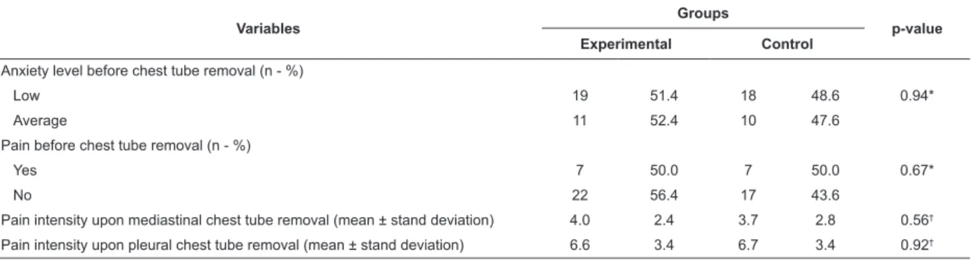
With respect to chest tube removal, no association
was observed between group and anxiety level (p=
0.94) or pain before the procedure (p= 0.67), as shown
in Table 3. The median intensity of pain upon removal
of pleural or mediastinal chest tubes did not differ
signiicantly between the groups.
Table 1 - Data corresponding to the participants’ trait, state anxiety levels and proile per group, Natal, RN, Brazil, 2014
Variables Groups p-value
Experimental Control
Age (years) 60.1 ±8.1 59.7 ±9.5 0.75*
Sex (n - %)
Male 17 47.2 19 52.8
0.23†
Female 13 54.2 9 45.8
Surgery time (hours – mean ± stand deviation) 4.1 0.8 4.3 0.9 0.15*
Hospital stay (days – mean ± stand deviation) 7.1 ±0.9 7.5 ±0.7 0.56*
ASA physical status (n - %)
II 13 44.8 19 61.3 0.20†
III 16 55.2 12 38.7
Trait anxiety (n - %)
Low 8 53.3 7 46.7
Average 17 48.6 18 51.4 0.76†
High 5 62.5 3 37.5
State anxiety (n - %)
Mild 15 51.7 14 48.3
Moderate 13 52.0 12 48.0 0.99†
Intense 2 50.0 2 50.0
* Mann-Whitney (U) test † Chi-Squared test
Table 2 - Sample characterization according to the presence and intensity of pain upon lidocaine injection, Natal, RN,
Brazil, 2014
Variables Groups p-value
Experimental Control
Pain upon lidocaine injection (n - %)
Yes 14 46.7 16 53.3 0.42*
No 16 57.1 12 42.9
Pain intensity upon lidocaine injection (median – minimum – maximum) 0.0 0.0 9.0 3.0 0.0 8.0 0.27†
Table 3 - Sample characterization according to anxiety level and pain intensity upon chest tube removal, Natal-RN,
Brazil, 2014
Variables
Groups
p-value
Experimental Control
Anxiety level before chest tube removal (n - %)
Low 19 51.4 18 48.6 0.94*
Average 11 52.4 10 47.6
Pain before chest tube removal (n - %)
Yes 7 50.0 7 50.0 0.67*
No 22 56.4 17 43.6
Pain intensity upon mediastinal chest tube removal (mean ± stand deviation) 4.0 2.4 3.7 2.8 0.56†
Pain intensity upon pleural chest tube removal (mean ± stand deviation) 6.6 3.4 6.7 3.4 0.92†
* Chi-Squared test † Mann-Whitney (U) testeT
Discussion
There is evidence that adequate pain control is
signiicantly beneicial for patient comfort. Although
multimodal analgesia combined with lidocaine is
considered an option for chest tube removal, this
therapy was not effective in the present study.
Although no association was observed between trait
and state anxiety and group, and despite the orientations
and psychological support provided by the nurses as a
part of the routine care before the procedure, almost
half of the participants exhibited moderate anxiety.
Almost one-third of the participants reported the
presence of pain before chest tube removal. Both groups
localized the majority of their pain to the sternotomy
site, as well as the site of chest tube insertion,
particularly in the case of the pleural drains, followed
by the saphenectomy site. These indings agree with
previous studies, where pain occurred at the sternotomy
site up to postoperative day three(18-19).
There was no signiicant difference in pain associated
with the subcutaneous administration of lidocaine
between the groups. In this regard, it is worth noting
that the intensity of pain reported was mild, which might
be related to the use of multimodal analgesia.According
to the literature(20), discomfort and pain during the
subcutaneous administration of lidocaine is reported by
patients as a whole. These symptoms might be related
to the needle gauge, anesthetic administration speed,
injected solution volume or temperature, patient proile,
or the low pH of the anesthetic.
The groups did not exhibit signiicant differences in
pain associated with the procedure for CTR. However, the
participants in the experimental group (GI) qualiied the
pain upon removal of mediastinal chest tubes as intense,
followed by moderate in almost one-third of the cases.
These indings are supported by several clinical trials
and indicate that patients feel moderate-to-intense pain
even when strong analgesics, such as morphine, and
local anesthetics, including lidocaine, are administered.
Most studies found that patients felt
moderate-to-severe pain during chest tube removal despite the
administration of morphine or local anesthetics(21).
One study compared the eficacy of remifentanyl
0.5 mg/kg versus placebo for alleviating pain due
to chest tube removal. The results revealed that the
patients receiving remifentanyl exhibited signiicantly
less pain than did those receiving a placebo at drain
removal(6). In another study(7) signiicantly lower pain
scores were reported in the groups treated with fentanyl
2 µg/kg IV or sufentanyl 0.2 µg/kg IV, compared with
the patients in the control group who were given 2 ml of
normal saline.
Chest tube removal and pleural drains in
particular are considered a determinant factor for the
development of intense pain after cardiac surgery. It
is also observed that removal of pleural chest tubes
is more painful compared with mediastinal drains(2).
Furthermore, some patients report that the pain or
the discomfort caused by the procedure is one of the
worst experiences in ICU. Insertion of pleural drains is
unavoidable during pleurotomy and causes traumatic
chest injuries due to the perforation of the intercostal
muscles and parietal pleura, which interferes with
the respiratory movements and the position of the
pleural drains(22-23). Another study(24) demonstrated that
patients with subxiphoid pleural drains reported less
pain compared with the ones with intercostal insertion.
Similar indings were reported(25) and described a
technique of subxiphoid pleural drain insertion to
reduce postoperative discomfort due to the chest tube
3. Kelet H, Dahl JB. The value of “multimodal” or
“balanced analgesia” in postoperative pain treatment.
Anesth Analg. 1993;77(5):1048-56.
4. Friesner SA, Miles Curry D, Moddeman GR. Comparison
of two pain-management strategies during chest tube
removal: Relaxation exercise with opioids and opioids
alone. Heart Lung. 2006;35(4):269–76.
5. Chen YR, Hsieh LY. The effectiveness of a cold
application for pain associated with chest tube
removal: a systematic review. Hu Li Za Zhi.
2015;62(1):68-75.
6. Casey E, Lane A, Kuriakose D, McGeary S, Hayes
N, Phelan D, et al. Bolus Remifentanil for chest drain
removal in ICU: a randomized double-blind comparison
of three modes of analgesia in post-cardiac surgical
patients. Intensive Care Med. 2010;36(8):1380-5.
7. Joshi VS, Chauhan S, Kiran U, Bisoi AK, Kapoor
PM. Comparison of analgesic efficacy of fentanyl and
sufentanil for chest tube removal after cardiac surgery.
Anna Card Anaesth. 2007;10(1):42-5.
8. Golmohammadi M, Sane SH. Comparison of fentanyl
with sufentanil for chest tube removal. Iranian Cardiovasc
Res J. 2008;2(1):42-7.
9. Demir Y, Khorshid L. The Effect of Cold Application in
Combination with Standard Analgesic Administration on
Pain and Anxiety during Chest Tube Removal: A
Single-Blinded, Randomized, Double-Controlled Study. Pain
Manag Nurs. 2010;11(3):186-96.
10. Payami MB, Daryei N, Mousavinasab N, Nourizade
E. Effect of cold application in combination with
Indomethacin suppository on chest tube removal pain
in patients undergoing open heart surgery. Iran J Nurs
Midwifery Res. 2014;19(1):77-81.
11. Akrofi M, Miller S, Colfar S, Corry PR, Fabri BM, Pullan
MD, et al. A randomized comparison of three methods of
analgesia for chest drain removal in postcardiac surgical
patients. Anesth Analg 2005;100(1):205–9.
12. Puntillo K, Ley SJ. Appropriately timed analgesics
control pain due to chest tube removal. Am J Crit Care.
2004;13(4):292-302.
13. Chaves LD, Pimenta CAM. Postoperative pain control:
comparison among analgesic methods. Rev. Latino-Am.
Enfermagem. 2003:11(2):215-9.
14. Ramsay MA, Savege TM, Simpson BR, Goodwin R.
Controlled sedation with alphaxalone-alphadolone. Br
Med J. 1974;2(5920):656-59.
15. Jensen MP, Karoly P, Braver R. Postsurgical pain
outcome assessment. Pain 2002;99:101-9.
16. Melzack R. The short-form McGill Pain. Pain.
1987;30:191-7. In our study, the words (descriptors) used by the
participants to describe pain were pressing, sharp,
pricking, and burning, and the most common site of pain
was the drain site (65.0%). Using the same instrument
and type of population, one study(2) reported that
the words used to describe the pain caused by chest
tube removal were fearful (44.8%), sharp and tender
(40.3%), and hot-burning using the same instrument in
the same population.
Regarding the effectiveness of other
non-pharmacological therapies for pain control in the CTR, cold
application, which seemed to be a noninvasive and safe
way to reduce pain, in a systematic review that analyzed
data from 426 patients, 05 trials showed conlicting
results(5). However, the study of Demir and Khorshid(26)
found that cold application reduced patients’ intensity of
pain due to CTR but did not affect anxiety levels or the
type of pain. They nevertheless recommended cold as a
pain-relieving technique during CTR.
As a possible limitation of the present study, we
believe that the fact that the number of participants may
have some kind of inluence on the results. Therefore, the indings cannot be generalized to other patients
who experience CTR. It is recommended that the study
be repeated with more patients who experience CTR
for other reasons. The present study was designed in
two groups; as a result, the possible placebo effect on
the patients’ pain perception was not identiied. It is
recommended that a similar study in three groups be
conducted to exclude the placebo effect. In our study,
patients might have responded differently to pain based
on their physical condition, emotional and cultural states.
Further studies in different settings are suggested.
Conclusion
Thus, the present study suggests that the analgesic
effect of the subcutaneous administration of 1%
lidocaine combined with multimodal analgesia is less
effective and subcutaneous injections are less effective
in relieving pain.
References
1. Charnock Y, Evans D. Nursing management of
chest drains: a systematic review. Aust Crit Care.
2001;14(4):156–60.
2. Puntillo KA, Ley SJ. Appropriately timed analgesics
control pain due to chest tube removal. Am J Crit Care.
17. Spielberger CD, Gorsuch RL, Iushene RE. Inventário
de Ansiedade Traço-Estado - IDATE: Manual de Psicologia
Aplicada [State-Trait Anxiety Inventory – STAI: Manual
of Applied Psychology]. Rio de Janeiro: CEPA; 1979.
18. Mueller XM, Tinguely F, Tevaearai HT, Revelly
JP, Chioléro R, von Segesser LK. Pain location,
distribution, and intensity after cardiac surgery. Chest.
2000;118(2):391-6.
19. Giacomazzi CM, Lagni VB, Monteiro MB. A dor
pós-operatória como contribuinte do prejuízo na função
pulmonar em pacientes submetidos à cirurgia cardíaca
Postoperative pain as a contributor to pulmonary function
impairment in patients submitted to heart surgery. Rev
Bras Cir Cardiovasc. 2006;21(4):386-92.
20. Cepeda MS, Tzortzopoulou A, Thackrey M, Hudcova
J, Arora Gandhi P, Schumann R. Adjusting the pH of
lidocaine for reducing pain on injection. Cochrane
Database Syst Rev. 2010;8(12):CD006581.
21. Bruce EA, Howard RF, Franck LS. Chest drain
removal pain and its management: a literature review. J
Clin Nurs. 2006;15(2):145-54.
22. Jakob H, Kamler M, Hagl S. Doubly angled pleural
drain circumventing the transcostal route relieves
pain after cardiac surgery. Thorac Cardiovasc Surg.
1997;45(5):263-4.
23. Lancey RA, Gaca C, Salm TJV. The use of smaller,
more flexible chest drains following open heart surgery.
Chest. 2001;119(1):19-24.
24. Guizilini S, Bolzan DW, Faresin SM, Ferraz RF,
Tavolaro K, Cancio AA, et al. Pleurotomy with subxyphoid
pleural drain affords similar effects to pleural integrity
in pulmonary function after off-pump coronary artery
bypass graft. J Cardiothorac Surg. 2012;25:7-11.
25. Riebman JB, Yurvati AHO, Laub GW. Improved
technique for pleural drain insertion in cardiovascular
surgery. J Cardiovasc Surg. 1994;35(6):503-5.
26. Demir Y, Khorshid L. The effect of cold application in
combination with standard analgesic administration on
pain and anxiety during chest tube removal: a
single-blinded, randomized, double-controlled study. Pain
Manag Nurs. 2010; 11(3):186-96.
Received: Oct 7h 2014
Accepted: May 3rd 2015
Erratum
Where was written:
“Conclusion: the present study suggests that the analgesic effect of the subcutaneous administration of 1% lidocaine combined with multimodal analgesia is most eficacious.”
Now Read: