INTRODUCTION
The increase in life expectancy in Brazil has generated an increase in the incidence of chronic degenerative pathologies, such as senile osteoporosis in women and also in men. Everyone recognizes the high morbidity of osteoporotic fractures in elderly patients,1,2 but health policy for prevention and treatment of osteoporosis are usually restricted to women after menopause. Although one third of the total femur fractures occur in men, and they have a worse prognosis after fracture than women,3,4 the study of osteoporosis in men is worldwide neglected. This
Densitometric analysis of femoral region in men older
than 50 years old from an ambulatory of urology
Renata Francioni Lopes1, Alexandre Oliveira Marchesi2, Raquel Novaes Fossari3, Michele Chailleaux Cezar4, Cláudia Medina Coeli5, Maria Lucia Fleiuss de Farias6
Received on 11/26/08. Approved on 05/11/09. We declare no conflict of interest.
Institutions (Affiliations): Department of Endocrinology, Hospital Universitário Clementino Fraga Filho (UFRJ), and Unit of Geriatrics and Gerontology of the Air Force Central Hospital.
Institution where the work was done: Air Force Central Hospital (HCA), located at Rua Barão de Itapagipe, 167, Rio de Janeiro, RJ. 1. PhD in Medicine, area of concentration Endocrinology (UFRJ). Physician at the Air Force Central Hospital
2. Radiologist. Head of Radiology at the Air Force Central Hospital 3. Physician. Resident of Internal Medicine at the Air Force Central Hospital 4. Physician. Resident of Internal Medicine at the Air Force Central Hospital
5. PhD in Public Health, area of concentration Epidemiology (UERJ), Post-doctorate in Public Health (Université Montréal, Canada). Associate Professor at Center for Studies in Public Health, (UFRJ)
6. PhD in Medicine, area of concentration Endocrinology (UFRJ). I Associate Professor, of Endocrinology, (UFRJ)
Correspondence to: Renata Francioni Lopes. Rua Passos da Pátria, 105/101, São Domingos, Niterói, RJ, CEP 24210-240. Tel HCA - Unidade de Geriatria e Gerontologia do Hospital Central da Aeronáutica: 55 21 3501-3193 PABX: 55 21 3501-3100. E-mail: [email protected]
ABSTRACT
Introduction: Men osteoporosis remains poorly diagnosed. The objective of this study was to measure bone mineral density (BMD) and the prevalence of osteoporosis in a group of men. Patients and methods: 151 men (ages 50-93 years) in good health, from an outpatient clinic for routine urologic evaluation performed the measurement of bone density of lumbar spine and femoral regions. Results: Age had a negative inluence on femoral neck BMD and T-Score (rs = 0.49 and 0.73, respectively, P ≤ 0.0001) using the Spearman’s rank correlation coeficient. Femoral neck osteoporosis
was detected in 25.16% (n = 38). Most of the osteoporotic patients (81.56%) were over 70 years old, and 47.37% of them were very old (aged 80 years or more). Beside age, hypogonadism induced by GnRH analogues and cyproterone acetate for treatment of prostate cancer, anticoagulants, coronary revascularization history and alcohol were risk factors identiied in about 18% of the osteoporotic group. Conclusion: All men over 70 years old and younger men with risk factors for osteoporosis should be submitted to a bone densitometry.
Keywords: men, hypogonadism, osteoporosis, fractures, bone density.
disease is rarely diagnosed before the fracture, and even after this event rarely an anti-osteoporosis treatment is instituted.
This study measured bone mineral density (BMD) and the prevalence of osteoporosis in a sample of Brazilian men, regularly monitored in the urology clinic.
PATIENTS AND METHODS
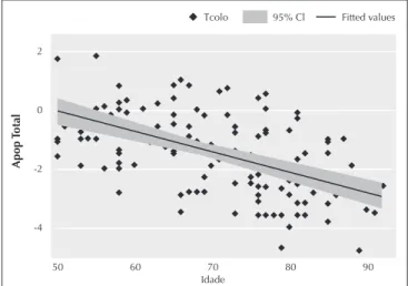
Figure 1. Correlation between age and T-Score in the femur neck in men from 50 to 93 years old (r = 0.73 and P < 0.0001).
Table 1
Bone mineral density in the femur neck (median and interquartiles) by age, in men from 50 from 93 years old
50 to 59 years (n = 39) 0.956 g/cm2 (0.889 to 1.025)
60 to 69 years (n = 37) 0.887g/cm2 (0.830 to 0.988)
70 to 79 years (n = 38) 0.815g/cm2 (0.703 to 0.921)
≥ 80 years (n = 37) 0.717g/cm2 (0.627 to 0.831)
P-valor <0.0001
Apop
Total
2
0
-2
-4
50 60 70
Idade
80 90
Tcolo 95% Cl Fitted values Central da Aeronáutica (Air Force Central Hospital), of age 50
or older and in good clinical conditions were pre-selected. The invitation to participate in the survey was by telephone contact expecting at least forty men by age group. All 163 who agreed to participate in the study were followed in the Geriatrics and Gerontology Unit, where history was collected and directed to diseases and medications that promote osteoporosis, and clinical examination performed. Subsequently, they were referred to a radiographic study of the thoracolumbar spine and pelvis, in addition to the measurement of BMD in lumbar spine and proximal femur on Norland apparatus.
Criteria for the diagnosis of osteoporosis, low bone mass, and normal BMD followed the standard of the World Health Organization (WHO). The international reference of the WHO for the diagnosis of osteoporosis is a T-score of -2.5 standard deviation (SD) or lower at lumbar spine or proximal femur, although the distal radius (peripheral site) can be used for this purpose.
The report of bone densitometry examination (DXA initial report) was based on the oficial positions of the Brazilian Society of Clinical Densitometry, according to minimum requirements.5
Statistical analysis was performed using the Stata program, software version 8.0, 2003. The differences between the median values of bone density by age group were determined by the non-parametric Mann-Whitney U test. The correlations between age and densitometry parameters were evaluated by the correlation coeficient Spearman. P-value less than 0.05 was considered signiicant.
The study was approved by the Research Ethics Committee of the Air Force Central Hospital, without conlict of interest. All patients participating in the study signed a free and informed consent.
RESULTS
From the 163 men initially evaluated, ten did not have bone densitometry and were excluded from the study. Of the remaining 153 men, who completed the form for evaluating the risk of osteoporosis and have done the image testing, 2 were excluded due to polyostotic Paget’s disease.
Thus, 151 men were the object of our densitometric analysis study. The age distribution (mean age and SD) was 37 men at 80 years old or older (83.7 ± 3.7 years), 38 men 70 to 79 years old (75 ± 4.1 years), 37 men 60 to 69 years old (65.4 ± 2.8 years), and 39 men 50 to 59 years old (55.3 ± 2.9 years).
There was a strong correlation between age and BMD in the femur neck (r = -0.49, P = 0.0000), and between age and
T-Score in the same region (r = -0.73, P = 0.0000), as seen in Figure 1.
From 151 men tested, 38 (25.16%) had osteoporosis in the femoral region. The most affected by osteoporosis (81.46%) were men over 70 years old (up to 93 years old) and a signiicant portion (47.37%) was of much older men, i.e., 80 years old or more.
Low BMD in the femoral region, considered osteopenia on densitometry (T-score of <-1.0 to > -2.5 SD compared with young men), was detected in other 47 men, 25 of them were 70 years old or older.
The BMD, median and interquartiles, and the prevalence of osteoporosis and osteopenia in the femoral region by age group are showed in Tables 1 and 2.
Table 2
Prevalence of osteoporosis and osteopenia by densiometric criteria in brazilian men from 50 to 93 years old
50 to 59 years (n = 39)
60 to 69 years (n = 37)
70 to 79 years (n = 38)
≥ 80 years (n = 37)
Femur osteoporosis 2.56% 16.21% 34.21% 48.65% Femur osteopenia 21.51% 37.84% 34.21% 32.43% Normal femur BMD 76.93% 45.95% 31.58% 18.92%
The osteoporosis group, 18.42% (n = 7) were using drugs that potentially cause bone loss: goserelin (n = 4), cyproterone acetate (n = 2) and warfarin sodium (n = 1). The irst two drugs mentioned were used as an antiandrogenic therapy in the treatment of prostate cancer (under control and no evidence of metastases), and anticoagulants in patients with atrial ibrillation. The use of alcohol was observed in two patients with osteoporosis under 70 years old. One 67-year-old patient with a history of myocardial revascularization not receiving anticoagulant presented T-score of -2.8 on femoral neck.
The group with low BMD (osteopenia on densitometry), 10.64% (n = 5) used or were using drugs potentially known to cause bone loss. Two osteopenic patients over 70 years old were using gonadotropin-releasing hormone analog GnRH (goserelin) to treat prostate cancer. A third patient, a 75-year-old former athlete, had a history of chronic use of corticosteroids in the past and T-score of -1.9 on femoral neck. From the osteopenic patients under 70 years old, 2 were chronic users of anticoagulants for atrial ibrillation. Use of alcohol was observed in one patient aged 53 years with T-score of -1.9 on femoral neck. A 66 years old patient with newly diagnosed Parkinson’s disease had T-score of -1.3, and another patient with 58 years, with history of right nephrectomy in youth and serum creatinine of 1.5, showed T-score of -1.4.
Radiographs of thoracolumbar spine and pelvis, revealed no fractures or lytic and/or blastic lesions. As expected, the elderly more often presented spinal osteoarthrosis, which hindered the densitometric analysis of the site. Only in 2 patients (68 and 81 years old with no recognized risk factors for osteoporosis), T-score on lumbar spine was -2.5 SD below the mean, these men also had densitometric patterns for osteoporosis in femoral region. Except for an 80 years old patient with T-score of -2.4 on analysis of femoral region (femoral osteopenia), all other elderly patients classiied as osteopenic at the lumbar spine presented osteoporosis in the femoral region.
It was clear the discrepancy between the values found in the femoral region and the lumbar spine, especially in the elderly. From the 109 densitometry with T-score of lumbar spine within normal limits, the analysis of the femoral region showed osteoporosis in 14, and all these patients were over 60 years old. Furthermore, all bone densitometries classiied as within the normal range, based on the femoral region, were also considered normal in the spine. Therefore, we exclude the lumbar spine site (L2-L4) from the results interpretation and from the discussion below.
DISCUSSION
The prevalence of osteoporosis in the studied male population (25.3%) approximates that of the study performed at the National Institute of Trauma-Orthopedics (INTO) in Rio de Janeiro, which was 19.5%.6 Data are more than twice the prevalence of the disease in the United States, Canada and Europe (approximately 10%).6 When we analyzed the prevalence of the disease in very elderly men, there were even higher values compared to the INTO study (48.7% versus 36.4 %).6
In 2000, Zerbini et al. published the densitometric analysis of 288 Brazilian men aged 50 years or older (mean age 62.5 years), concluding that hip BMD was similar in the population studied, compared to the North American and European, but this study did not include very elderly men (80 years old or more), or presented a percentage of men with osteoporosis, allowing the comparison of prevalences.7
The lack of studies on the prevalence of osteoporosis in men in Brazil and the results found in this study reinforce the importance of health care policies aimed to the male population to prevent and treat osteoporosis, in order to reduce the risk of fractures. Most data from literature comes from foreign populations, and the treatment of male osteoporosis was done with bisphosphonates and teriparatide.8,9
The risk factors related to the appearance of osteoporosis are similar in men and women. Although there is gender difference in bone geometry, the incidence of fractures appears to be similar for the same areal BMD.10 Because men have higher peak bone mass, they usually have fractures of hip, vertebral body or distal radius ten years after women. Hip fractures in men, however, result in a mortality rate, one year after the fracture, of 31% versus 17% in women. About 30% of hip fractures occur in men, and one in eight men over 50 will have an osteoporotic fracture.11
between age and BMD. Virtually the entire group with osteoporosis consisted of men over 70 years old.
Certainly, the increase in life expectancy has contributed to the increased incidence of osteoporosis in the male populations already studied in the last decades. It became more evident that the senile osteoporosis in men aroused public health measures to combat osteoporosis, and researches in the ield of bone metabolism towards the male population.
Tanaka et al. evaluated the risk factors related to osteoporosis in 325 Brazilian men aged 50 years or more, and conirmed that age was a risk factor for osteoporosis. Other risk factors were low BMI, physical inactivity (in the last 12 months), smoking, lack of routine use of thiazide, white ethnicity, and maternal history of osteoporosis after 50 years.12
In a recently published study, which excluded men receiving antiandrogenic therapy, we demonstrated that low BMI and decline of sex steroids explain much of the association between aging, increased bone turnover and osteoporosis.13 The relationship between body composition and BMD was previously described by Zerbini et al.14
The senile osteoporosis tends to be multifactorial, related to age, inactivity, hypogonadism, low intake and/or calcium absorption and hyperparathyroidism secondary to vitamin D deiciency.15,16 Although we are in a tropical country, vitamin D deiciency is expected due to the process of aging in the male population;17 however, the decline in concentrations of 25 OH vitamin D in men takes longer than in women,18 justifying the lower prevalence of osteoporosis in men. Senescence is characterized by the decline of physiological functions and, physiologically, there is a decrease in muscle mass, lentiication and damage in the process of digestion, and reduction of renal and hepatic functions, favoring osteoporosis in both sexes.
The prevention of senile osteoporosis is based on scheduled physical activity, in addition to calcium and vitamin D supplementation.
Florindo et al., studying 326 Brazilian men have shown that physical activity in the last 10 to 20 years, and even in the last 12 months, can contribute to preserve bone mineral density in men aged 50 years or older. The positive correlation between physical activity and bone mineral density in Florindo’s group proved to be independent of age and BMI.19
Saraiva et al., studying 177 institutionalized elderly (52 men) and 243 outpatients (75 men) from São Paulo, found high prevalence of disability and insuficiency of vitamin D in the elderly population studied, more pronounced in the institutionalized group. In this assessment, 71.2% of the institutionalized group and 43.8% of the outpatients have
shown values of 25 OHD lower than the minimum level recommended (50 nmol/L). 20
In our study of vitamin D in Brazilian men, outpatients aged 50 years or older in Rio de Janeiro (n = 152), we observed high prevalence of hypovitaminosis D (49.4%) and deiciency of vitamin D (31. 9%). In the group with osteoporosis, 82.7% had hypovitaminosis D or deiciency of vitamin D. Bone mineral density at femoral neck was associated with levels of 25 OHD (r = 0.317, P = 0.002).21
These works carried out in Brazil demonstrate the importance of vitamin D supplementation during the aging process to prevent and treat male osteoporosis in our country.
The studies on the treatment of osteoporosis in men with bisphosphonates and teriparatide include men of all ages, with younger men suffering from the so called “idiopathic osteoporosis” or secondary osteoporosis.9,22-25 The use of testosterone and/or growth hormone replacement (GH) remains only in cases of deiciency (hypogonadism and GH deiciency). 26,27
In our study, the men came from an urology outpatient clinic and were older. Probably it was the reason for a higher frequency of osteoporosis secondary to drugs for treatment of prostate cancer and other chronic diseases.
We observed cases of osteoporosis and low BMD in men below 70 years old. The chronic consumption of alcohol promotes bone loss, even in young people. It is common with excessive intake, malnutrition and increased risk of falls and fractures, but it seems that alcohol has a direct effect on the bone, inhibiting its formation, reducing the number of osteoblasts, the amount of osteoid matrix and the secretion of osteocalcin.35The real effect of alcohol on BMD of these studied population is dificult to measure due to the small number of drinkers. Attention should be paid to this habit or addiction, so common in our environment, since the amount and frequency of alcohol intake tended to be minimized by men.
In our study, the low incidence of osteoporosis in men below 60 years old suggests that we should not ask for routine bone densitometry in this age group, according to the national and international guidelines for the investigation of osteoporosis in men. The age group between 60 to 70 years old has shown considerable increase in the prevalence of osteoporosis, and in the future they might be routinely assessed. Remains, therefore, the request for routine bone densitometry in men with aged 70 years or older, and in an individualized way in the younger, taking into account the potential risk factors related, especially the family history and the use of drugs potentially deleterious to bone mass.
CONCLUSIONS
Our indings are in accordance with the global data, which show an exponential increase of osteoporosis in the aging male: about 25% of the studied men over 50 years old had femoral neck osteoporosis. Of these, most were over 70 years old. Drugs for prostate cancer, anticoagulant, history of coronary artery bypass surgery, and alcohol use were risk factors found in 18% of the osteoporotic population. These indings lead us to recommend bone densitometry for all men over 70 years old, and for younger men with risk factors.
In Brazil, prevention and treatment of male osteoporosis have become necessary: the attention given to women’s bone health must be shared with men.
REFERENCES
1. Magaziner J, Simonsick EM, Kashner TM, Hebel JR, Kenzora JE. Predictors of functional recovery one year following hospital discharge for hip fracture: a prospective study. J Gerontol 1990;45:101-7.
2. Aharonoff GB, Koval KJ, Skorvon ML, Zucherman JD. Hip fractures in the elderly; predictors of one year mortality. J Orthop Trauma 1997;11:162-5.
3. Melton III LJ, Atkinson EJ, O’Connor MK, O’Fallon WM, Riggs BL. Bone density and fracture risk in men. J Bone Miner Res 1998;13:1915-23.
4. Lois E, Wehren MD. Osteoporosis increases mortality risk in men. Geratric Times 2002; III:4.
5. Zerbini CAF, Pippa MGB, Eis SR, Lazaretti-Castro M, Mota Neto H, Tourinho TF et al. Densitometria Clínica: Posições Oiciais 2006.
Rev Bras Reumatol 2007;47:25-31.
6. Saúde em Foco. Nível de osteoporose entre homens é alto. http:// www.aabnb.com.br/journal/jun-05.asp Acesso em 10/10/2007. 7. Zerbini CAF, Latorre MRQ, Jaime PC, Tanaka T, Pippa MGB. Bone
mineral density in Brazilian men 50 years and older. Braz J Med Biol Res 2000;33:1429-35.
8. Finkelstein JS, Hayes A, Hunzelman JL, Wyland JJ, Lee H, Neer RM. The effects of parathyroid hormone, alendronate, or both in men with osteoporosis. N Engl J Med 2003; 349:1216-26. 9. Saad GK, Shane E, Boonen S, Marín F, Donley DW, Taylor KA et al.
Teriparatide or alendronate in glucocorticoid-induced osteoporosis. N Engl J Med 2007;357:2028-39.
10. Kaufman JM, Johnell O, Abadie E, Adami S, Audran M, Ayouac B
et al. Background for studies on the treatment of male osteoporosis: state of the art. Ann Rheum Dis 2000;59:765-72.
11. Campion JM, Maricic MJ. Osteoporosis in men. Am Fam Physician 2003;67:1521-6.
12. Tanaka T, Latorre MRQ, Jaime PC, Florindo A, Guadalupe MGB, Zerbini CAF. Risk factors for proximal femur osteoporosis in men aged 50 years or older. Osteoporos Int 2001; 12:942-7.
13. Lopes RF, Ferreira SAGJ, Coeli CM, Farias MLF. Low body mass index and declining sex steroids explain most age-related bone loss in Brazilian men. Osteoporos Int 2008. Disponível em http://www. springerlink.com/content/ DOI 10.1007/s00198-008-0796-7/fulltext. pdf. Acesso em 20 de novembro de 2008.
14. Zerbini CAF, Latorre MRQ, Jaime PC, Tanaka T, Pippa MGB. Body composition and bone mineral density in men. J Bone Miner Res 1999;14:S390.
15. Saquib N, von Mühlen D, Garland CF, Barrett-Connor E. Serum 25-hydroxyvitamin D, parathyroid hormone, and bone mineral density in men: the Rancho Bernardo study. Osteoporos Int 2006;17:1734-41.
16. Szulc P, Munoz F, Marchand F, Chapuy MC, Delmas PD. Role of vitamin D and parathyroid hormone in the regulation of bone turnover and bone mass in men: the MINOS study. Calcif Tissue Int 2003;73:520-30.
17. Holick MF. Vitamin D deiciency. N Engl J Med 2007;357:266-81. 18. Maggio D, Cherubini A, Lauretani F, Russo RC, Bartali B,
Pierandrei M et al. 25(OH)D serum levels decline with age earlier in women than in men and less eficiently prevent compensatory hyperparathyroidism in older adults. J Gerontol A Biol Sci Med Sci 2005; 60:1414-9.
20. Saraiva GL, Cendoroglo MS, Ramos LR, Araújo LM, Vieira JG, Maeda SS et al. Prevalência da deiciência, insuiciência de vitamina
D e hiperparatireoidismo secundário em idosos institucionalizados e moradores na comunidade da cidade de São Paulo, Brasil. Arq Bras Endocrinol Metab 2007;51:437-42.
21. Lopes RF, Ferreira SAGJ, Coeli CM, Farias MLF. Deiciência de vitamina D e hiperparatiroidismo secundário em homens acima de 50 anos e sua relação com a perda óssea do envelhecimento. Rev Port Endocrinol, Diabet e Metab 2008;3(1):110(abstract). 22. Filkelstein JS, Hayes A, Rao A, Neer RM. Effects of parathyroid
hormone, alendronate, or both on bone density in osteoporotic men. J Bone Miner Res 2002;17(Suppl 1):S127.
23. Orwoll ES, Scheele WH, Paul S, Adami S, Syversen U, Diez-Perez. A. Brief therapy with recombinant human parathyroid hormone (1-34) increases lumbar spine bone mineral density in men with idiopathic or hypogonadal osteoporosis. J Bone Miner Res 2001;16(suppl 1):S221.
24. Orwoll E, Ettinger M, Weiss S, Miller P, Kendler D, Graham J et al. Alendronate for the treatment of osteoporosis in men. N Engl J Med 2000;343:604-10.
25. Kurland ES, Heller SL, Diamond B, McMahon DJ, Cosman F, Bilezikian JP. The importance of bisphosphonate therapy in maintaining bone mass in men after therapy with teriparatide [human parathyroid hormone(1-34)]. Osteoporos Int 2004;15(12):992-7. 26. Amory JK, Watts NB, Easley KA, Sutton PR, Anawalt BD,
Matsumoto MA et al. Exogenous testosterone or testosterone with inasteride increases bone mineral density in older men with low serum testosterone. J Clin Endocrinol Metab 2004;89:503-10. 27. Underwood LE, Attie KM, Baptista J and the Genentech Collaborative
Study Group. Growth hormone (GH) dose-response in young adults with childhood-onset GH deficiency: A two-year, multicenter, multiple-dose, placebo-controlled study. J Clin Endocrinol Metab 2003; 88:5273-80.
28. Mittan D, Lee S, Miller E, Perez RC, Basler JW, Bruder JM. Bone loss following hypogonadism in men with prostate cancer treated with GnRH analogs. J Clin Endocrinol Metab 2002;87:3656-61. 29. Daniell HW, Dunn SR, Ferguson DW, Lomas G, Niazi Z, Stratte PT.
Progressive osteoporosis during androgen deprivation therapy for prostate cancer. J Urol2000;163:181-6.
30. Smith MR. Osteoporosis during androgen deprivation therapy for prostate cancer. Urology 2002;60:Suppl 1:79-86.
31. Riggs BL, Khosla S, Melton J. Sex steroids and the construction and conservation of the adult skeleton. Endocr Rev2002;23:279-81. 32. Diamond TH, Higano CS, Smith MR, Guise TA, Singer FR.
Osteoporosis in men with prostate carcinoma receiving androgen-deprivation therapy: recommendations for diagnosis and therapies. Cancer 2004;100:892-9.
33. Smith MR, McGovern FJ, Zietman AL, Fallon MA, Hayden DL, Schoenfeld DA et al. Pamidronate to prevent bone loss during androgen deprivation therapy for prostate cancer. N Engl J Med 2001;345:948-55.
34. Smith MR, Eastham J, Gleason DM, Shasha D, Tchekmedyian S, Zinner N. Randomized controlled trial of zoledronic acid to prevent bone loss in men receiving androgen deprivation therapy for nonmetastatic prostate cancer. J Urol 2003;169:2008-12. 35. Ganry O, Baudoin C, Fardellone P. Effect of alcohol intake on