Possible etiological factors in temporomandibular
disorders of articular origin with implications for
diagnosis and treatment
Aline Vettore Maydana*, Ricardo de Souza Tesch**, Odilon Vitor Porto Denardin***, Weber José da Silva Ursi****, Samuel Franklin Dworkin*****
The authors reviewed the factors involved in the etiology, diagnosis and treatment of temporomandibular joint disorders (TMD). Although essential, specific criteria for in-clusion and exin-clusion in TMD diagnosis have shown limited usefulness. Currently, the Research Diagnostic Criteria for Temporomandibular Disorders (RDC/TMD) offer the best evidence-based classification for the most common TMD subgroups. The RDC/ TMD includes not only methods for physical diagnostic classification, comprised in Axis I, but also methods to assess the intensity and severity of chronic pain and the levels of non-specific depressive and physical symptoms, in Axis II. Although historically maloc-clusions have been identified as risk factors for the development of TMD—including those predominantly joint-related—in many cases the association established between these variables seems to have taken opposite directions. Regarding internal TMJ derange-ments, the results of studies on the induced shortening of the mandibular ramus, second-ary to anterior articular disk displacement, indicate that repositioning the displaced disk in children or young adolescents may make more sense than previously imagined. The therapeutic use of dietary supplements, such as glucosamine sulfate, seems to be a safe alternative to the anti-inflammatory drugs commonly used to control pain associated with TMJ osteoarthritis, although evidence of its effectiveness for most TMD patients has yet to be fully established.
Abstract
Keywords: Temporomandibular disorders. RDC/TMD. Disk displacement. Osteoarthritis. Malocclusion.
* TMD and Orofacial Pain Specialist - Petrópolis School of Medicine / ABO, Petrópolis. Specialist in Orthodontics - ABO, Petrópolis. ** Head of the Department of TMD and Orofacial Pain, Petrópolis School of Medicine. Specialist in Orthodontics.
*** Associate Professor, Department of Head and Neck Surgery, Heliópolis Hospital.
**** Associate Professor, Department of Orthodontics, University of São Paulo - São José dos Campos.
IntroductIon
Temporomandibular disorders (TMD) refers to a set of conditions that affect the masticatory muscles and/or the temporomandibular joint (TMJ).30 These conditions have failed to
dem-onstrate a common etiology or biological basis in terms of clear signs and symptoms and, there-fore, are considered a heterogeneous group of health problems related to chronic pain. Char-acteristic symptoms such as muscle and/or joint pain and/or pain on palpation, limited mandib-ular function and joint noises may be prevalent in isolation or in association, with a prevalence of up to 75% in the adult population.15
Never-theless, the emergence of some symptoms, such as joint noises, does not appear to be related—in the majority of the population—to pain or other important risk factors that require treatment.
Epidemiological studies suggest that the prevalence of symptoms such as pain and re-stricted movement range from 5-15%, with most cases occurring in young adults aged be-tween 20 and 40 years, especially in females.15
The low prevalence of TMD among older age groups, as seen in cross-sectional and longitu-dinal studies,18 is consistent with the typically
limiting nature of the symptoms.
The current classification is largely descrip-tive, based more on the presence of signs and symptoms than on etiology, mainly due to the fact that a full understanding of the relationship between etiological factors and pathophysi-ological mechanisms has not yet been achieved. From a clinical standpoint, however, it is proba-bly irrelevant to extend the division of so-called diagnostic subgroups if all disorders within the same subgroup can be controlled using similar therapeutic procedures.
Therefore, specific inclusion and exclusion criteria for the diagnosis of these disorders would only prove crucial if tested to determine their validity. Currently, the Research Diagnos-tic Criteria for Temporomandibular Disorders
(RDC/TMD) provides the best evidence-based classification for the most frequent TMD sub-groups,6 i.e., those subgroups which experts now
agree are different, based on criteria that can be replicated and scientifically evaluated. Thus, the RDC/TMD, a dual axis diagnosis and clas-sification system designed for clinical research on TMD, comprises methods for the physical classification of TMD diagnoses (Axis I) as well as methods to assess the intensity and severity of chronic pain and levels of non-specific de-pressive and physical symptoms (Axis II). RDC/ TMD reliability has been tested and found to be satisfactory in adult populations,7,8 whereas in
children and adolescents29 its validity and
clini-cal utility has been demonstrated for Axis I but not completely for Axis II (although extensive studies by the National Institutes of Health/ NIH are currently well underway to examine the validity of all RDC/TMD components).
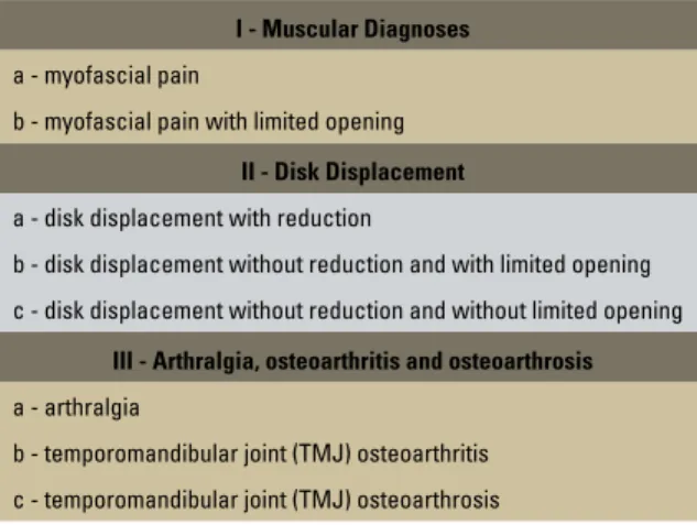
RDC/TMD Axis I addresses the physical conditions of TMD and aims to establish stan-dardized diagnostic criteria for use in scientific research. The suggested system is hierarchical, allowing not only group diagnosis but also the possibility of multiple diagnoses for the same individual. It is thus divided into three major groups representing the vast majority of clinical TMD cases, i.e.: myofascial pain; articular disk displacement; and arthralgia, osteoarthritis and osteoarthrosis (Table 1).
The purpose of this study was to address possible etiologic factors involved in the devel-opment of temporomandibular disorders of ar-ticular origin (groups II and III according to the RDC/TMD) and suggest implications for diag-nosis and treatment.
InternAl tMJ derAngeMents
mandibular movements. Articular disk displace-ment is only a subset of these disorders. When it is called articular disk displacement with reduction, it can be recognized by a ‘pop’ or ‘click’ sound in opening and closing the mouth, which only subsides when the mouth is open and maintained at maximum protrusion (RDC/ TMD Axis I, Group IIa).
Patients presenting with articular disk dis-placement have been characterized in terms of occlusion by the presence of unilateral poste-rior crossbite and long shifts from centric rela-tion (CR) to maximal habitual intercusparela-tion (MHI).26 This correlation, however, was
estab-lished without sufficient and unequivocal evi-dence to support the fact that this malocclusion is a risk factor for disk displacement.
Whereas the anterior articular disk displace-ment asymptomatic and unaccompanied by any other TMD indication (RDC/TMD Axis I, Group IIa) is quite common, with a prevalence of 20-35% of the population, on the other hand, disk displacement without reduction—which need not necessarily to be associated with pain, but may be associated with limitations in mouth opening (RDC/TMD Axis I groups IIb
and IIc)—is relatively rare, with occurrence fre-quency ranging from 1-5% according to studies conducted in TMD clinics around the world.30
In some animal studies, where anterior dis-placements of the articular disk were surgically created in rabbits—keeping the ligament intact in the posterior condyle—their mandibles be-came significantly smaller in the side where the disk had been displaced, resulting in a midline shift in the affected side. Mandibular asymme-try was not observed in the group that had their articular disk displaced.16 These results suggest
that displacement of the articular disk may pre-cede the development of mandibular asymme-try and can therefore be considered as a risk fac-tor for the development of transverse malocclu-sion. Whether or not this sequence of events is relevant to the growth and development of the human mandible has not yet been established.
For appropriate treatment protocols to be implemented, however, it is first necessary to determine under what conditions and for which individuals it might prove wise to control and prevent these diseases. Future investigations are required, preferably focusing on the study of the biomechanical and biochemical events that can trigger disk displacement, such as changes in joint lubricating,22,23 to determine whether
there are specific conditions for the emergence of specific malocclusions.
Biomechanical analyses of TMJ hard and soft tissues have revealed that these tissues are normally capable of withstanding and adapting to the functional loads and pressures that oc-cur during physiological mandibular movement. These tissues, however, cannot withstand com-pression for a long period of time, such as that associated with clenching in some individuals and at certain levels.22
In assessing the levels of intra-articular pres-sure in the TMJ of awake patients undergoing arthrocentesis procedures, Nitzan22 found that
voluntary clenching produced high levels of
I - Muscular Diagnoses
a - myofascial pain
b - myofascial pain with limited opening
II - Disk Displacement
a - disk displacement with reduction
b - disk displacement without reduction and with limited opening
c - disk displacement without reduction and without limited opening
III - Arthralgia, osteoarthritis and osteoarthrosis
a - arthralgia
b - temporomandibular joint (TMJ) osteoarthritis
c - temporomandibular joint (TMJ) osteoarthrosis
intra-articular pressure (as high as 200 mm Hg). Intra-articular pressure above 40 mmHg exceeds peripheral capillary pressure and can cause tem-porary intra-articular hypoxia followed by re-oxygenation as soon as the compression subsides, resulting in the release of free radicals.
A variety of effects caused by free radicals in articular tissue has been described22, including
the degradation of hyaluronic acid, which, once degraded, loses the ability to inhibit enzyme phospholipase A2 and break the active surface of phospholipids, which are primarily respon-sible for the process of TMJ lubrication. Poten-tially, any increase in friction accompanied by a lack of proper lubrication is aimed at preventing the smooth functioning of the articular disk in conjunction with the mandibular condyle during normal functional movements. This condition may hypothetically trigger the anterior displace-ment of the articular disk, as described in detail by Nitzan23. However, these hypotheses have not
hitherto been scientifically confirmed.
Theories and clinical observation have as-cribed to articular disk displacement the oc-currence of joint pain, limited mandibular movement, joint noises and degenerative TMJ changes. These reports are not at present sup-ported by longitudinal data of any kind and suggest the possibility that the articular disk effectively protects the underlying tissues and that its displacement might expose these tis-sues to an additional, excessive pressure, there-by causing degenerative changes. This assumed sequence of events has led to the use of surgi-cal procedures seeking to restore normal man-dibular anatomy and movements, often result-ing in serious complications20 and eventually
forcing professionals to question their belief in a necessary relationship between articular disk displacement and TMD related pain.5
Clinical observation has shown that artic-ular disk displacement may be present in as-ymptomatic as well as sas-ymptomatic patients.14
Likewise, the drainage of the upper TMJ com-partment during arthrocentesis—in the pres-ence of articular disk displacement without reduction—proved, in the short term, to be able to relieve pain and restore function with-out modifying the mandibular relationship be-tween condyle and articular disk.24
Thus, as the symptoms associated with disk displacement are not always the outcome of this internal TMJ derangement, the concept of second stage therapy—whereby irreversible changes such as occlusion adjustment, prosthet-ics, orthodontics or orthognathic surgery are in-dicated—does not appear justified at this time2.
tMJ degenerAtIve chAnges
TMJ degenerative changes are characterized by the presence of clinical signs of continuous crackling noises (crepitus) in the joint. Accord-ing to the RDC/TMD, cracklAccord-ing may be accom-panied by arthralgia. It is named osteoarthritis or, in the absence of pain, osteoarthrosis.6
Tem-poromandibular arthralgia is characterized by spontaneous pre-auricular pain or palpation and/or function induced pain, which is occa-sionally referred to the temporal region.
Patients with osteoarthritis are more consis-tently characterized by long shifts from CR to MHI, increased overjet and a tendency towards anterior open bite. An increased risk for these dis-orders is predominantly associated with extremes of these conditions.26 Practitioners, however, are
confronted with a dilemma to determine whether these malocclusions are etiological factors or con-sequences of dysfunctional joint remodeling.
extremely low. The possible relationship between osteoarthritis and anterior open bite does not seem to be frequent but may be a clinical finding that does not necessarily correlate to TMJ pain.
Morphological changes in the TMJ that are not associated with any significant change in joint dynamics or occlusion are features of func-tional remodeling. This remodeling becomes dysfunctional when it adversely affects mechan-ical joint function or occlusion and is therefore characterized by reduced condyle head volume, ramus size decrease, progressive mandibular re-trusion in adult patients or perhaps a reduction in growth rate between children. This condition can be generated by excessive mechanical stress applied to or sustained by joint structures to the extent that the pressure exceeds the joint’s abil-ity to adapt to such changes.1
Again, although there is radiological evidence of extensive TMJ remodeling, this remodeling may be within a normal biological variation be-cause the occurrence of pain or TMJ pathology requiring treatment is a relatively rare phenom-enon in older people.
In some cases, extensive remodeling of the mandible can lead to occlusal instability reflect-ed in open bite, increasreflect-ed overjet and some-times, in cases where the mandibular muscles manage to secure an MHI position, an increase in the distance between this position and the so-call centric relation position. These relations were demonstrated by Pullinger and Seligman,26
although the hypothesis that the degenerative process is an etiological factor for malocclusion still remains inconclusive.
Multiple variables, including genetic and en-vironmental factors, such as behaviors or harmful breathing habits, have been shown to influence facial growth rate.12 The data mentioned above
suggest that dysfunctional remodeling can also produce defects in mandibular growth, which to-gether with the other variables mentioned, could be contributing factors to the final mandibular
position and may cause specific malocclusions, such as, for example, anterior open bite.
The balance between anabolic and catabolic events appears to be highly individual and sub-ject to a wide range of functional and genetic factors.17 There is a need, however, to enhance
the understanding of normal, biological and bio-mechanical TMJ function, including the identi-fication of variables associated with changes and increases in joint pressure levels. These variables can lead to microtraumatic stimuli to the tissue and, consequently, can trigger a series of events that could lead to degeneration and joint pain.
Proinflammatory cytokines have been iso-lated in samples of synovial fluid drawn from the TMJ of symptomatic patients, since recent evidence shows that free radicals can stimulate the synthesis of cellular proteins by increased expression of specific genes.27
The cytokines predominantly involved in intra-articular degenerative processes are interleukin-1 beta (IL-1beta), interleukin-6 (IL-6) and tumor necrosis factor alpha (TNF-alpha).21 Together,
these cytokines stimulate the breakdown of ara-chidonic acid thus causing a major proinflamma-tory effect and triggering the synthesis and activa-tion of metalloproteinases, which are responsible for the breakdown of extracellular structure, ac-celerating the joint degeneration process.
therApeutIc IMplIcAtIons
As regards therapies, clinical trials are es-pecially useful and, therefore, required by the U.S. NIH as the gold standard to evaluate treat-ment effectiveness. Clinical trials play an even more important part in conditions such as TMD, where pain intensity can vary over time and pla-cebo and nonspecific effects can be just as im-portant as in other chronic pain conditions.13
Dworkin et al8,9 conducted randomized
After monitoring the groups for one year, both showed improvements in all clinical categories as well as those observed by the patients them-selves. Patients undergoing alternative treat-ment programs, however, exhibited a more sat-isfactory response, defined as greater reduction in (a) pain intensity, (b) level of interference in daily activities and (c) number of masticatory muscles painful to palpation.
These results indicate that the use of psy-chosocial assessment criteria such as, for ex-ample, those included in Axis II of the RDC/ TMD, can contribute to the success of clinical decision making regarding the control of TMD, especially muscle generated TMD. Conversely, predominantly articular disorders appear to suf-fer greater influence of localized phenomena.
In light of the wide array of studies that eval-uate the efficacy of stabilizing plates in TMD pain control, Ekberg et al11 argues that the
dif-ferences raised in these studies may be due to the inclusion of different painful TMD sub-groups, such as myofascial pain3 and
temporo-mandibular arthralgia.11 The latter group has
been shown to achieve significant therapeutic results in short11 and long-term10 follow-up.
In the study by Dao et al3, a randomized
group used stabilizing plates only in the dental office during consultations. No significant effect was found on any clinical parameter that could distinguish it from other groups in the random-ized study, i.e.: one group that used a stabilizing plate 24 hours a day and another that used a plate with no flat occlusion surface. In a ran-domized clinical trial scheduled for publication in the near future, a comparison was made be-tween a group using a flat acrylic plate, another using a prefabricated soft device and a control group with no plates. No difference was found between the groups in course of pain, mandibu-lar function or emergence of side effects after a one year follow-up.
Flat surface stabilizing acrylic plates may,
however, continue to be recommended for the treatment of TMJ arthralgia, although they still require further clarification as to the physiologi-cal mechanisms involved in their therapeutic effect, such as the reduction of parafunction-related mechanical stress.
Another study22 that evaluated intra-articular
pressure during functional and parafunctional movements also investigated 22 patients for intra-articular pressure against an interocclusal device, which uniformly increased the occlusion plane, reducing the force applied to the TMJ. A decrease in intra-articular pressure was observed at around 80% within a range of 0-40 mmHg.
The functional integrity of articular carti-lage is determined by the balance between the synthesis of extracellular structure by chondro-cytes and the breakdown of said structure. Glu-cosamine is normally found in human tissues and is directly involved in the synthesis of sub-stances that are essential to maintaining joint function integrity, such as glycosaminoglycans, proteoglycans and hyaluronic acid,19 although
the precise mechanism behind this function has not yet been determined.
In osteoarthritis, this balance is disrupted by the increased presence of enzymes such as metal-loproteinases, which are capable of breaking down the extracellular structure. Preliminary results of laboratory experiments4 indicate that the dietary
supplement glucosamine sulfate can stimulate the protein levels of the extracellular structure while simultaneously inhibiting the enzymatic produc-tion and activity of metalloproteinases in the chondrocytes of osteoarthritic joints.
three years of follow-up,19 although these
find-ings have not yet been carefully evaluated. Thie et al28 compared the therapeutic
po-tential of glucosamine sulfate with ibuprofen in patients with TMJ osteoarthritis. Both groups showed a significant improvement in the vari-ables studied when these data were compared with those at the beginning of treatment. A comparison between these two groups showed that during the time period that patients used glucosamine sulfate they had a significant pain reduction in the affected joint and a decreased influence of pain on the patients’ daily activi-ties, thus reducing their related disability.
The specific effects of pain relief associ-ated with the use of glucosamine sulfate are probably due to their anabolic properties in the cartilage. These effects, which change the degenerative condition of the disease, are not observed with the use of routine analgesics and can yield substantial benefits.
conclusIons
Although historically malocclusions have been identified as risk factors for the develop-ment of TMD—including those predominantly joint-related—in many cases the association
established between these variables seems to wide of the mark. Thus, prospective clinical and laboratory investigations addressing issues related to the etiology of these conditions, es-pecially in the early stages of development, can shed light on the future of therapy.
According to Legrell and Isberg,16 the
find-ings on induced mandibular ramus reduction— secondary to articular disk displacement—indi-cate that the repositioning of the disk in chil-dren and young adolescents may make more sense than previously believed.
In view of the above, the use of orthopedic devices for mandibular advancement, such as the Herbst appliance, which has demonstrated effectiveness in improving the prior positioning of disks displaced in the early stages of this pro-cess,25 should be tested by means of appropriate
randomized clinical trials.
1. Arnett GW, Milam SB, Gottesman L. Progressive mandibular retrusion-idiopathic condylar resorption. Part II. Am J Or-thod Dentofacial Orthop. 1996 Aug;110(2):117-27. 2. Dao TT, Lavigne GJ. Oral splints: the crutches for
temporo-mandibular disorders and bruxism? Crit Rev Oral Biol Med. 1998;9(3):345-61.
3. Dao TT, Lavigne GJ, Charbonneau A, Feine JS, Lund JP. The efficacy of oral splints in the treatment of myofascial pain and jaw muscles: a controlled clinical trial. Pain. 1994 Jun;56(1):85-94.
4. Dodge GR, Jimenez SA. Glucosamine sulfate modulates the levels of aggrecan and matrix metalloproteinase-3 synthesized by cultured human osteoarthritis articular chon-drocytes. Osteoarthritis Cartilage. 2003 Jun;11(6):424-32. 5. Dolwick MF. Intra-articular disc displacement. Part I: its
questionable role in temporomandibular joint pathology. J Oral Maxillofac Surg. 1995 Sep;53(9):1069-72.
6. Dworkin SF, Le Resche L. Research diagnostic criteria for temporomandibular disorders: review, criteria, examina-tions and specificaexamina-tions, critique. J Craniomandib Disord. 1992; 6:301-55.
7. Dworkin SF, Le Resche L, De Rouen T, Von Korff M. As-sessing clinical signs of temporomandibular disorders: reliability of clinical examiners. J Prosthet Dent. 1990 May;63(5):574-9.
8. Dworkin SF, Sherman J, Mancl L, Ohrbach R, Le Resche L, Truelove E. Reliability, validity, and clinical utility of the re-search diagnostic criteria for temporomandibular disorders axis II scales: depression, non-specific physical symptoms, and graded chronic pain. J Orofac Pain. 2002;6:207-20. 9. Dworkin SF, Turner JA, Mancl L, Wilson L, Massoth D, Huggins
KH, et al. A randomized clinical trial of a tailored comprehen-sive care treatment program for temporomandibular disorders. J Orofac Pain. 2002;16:259-76.
10. Ekberg E, Nilner M. A 6- and 12-month follow-up of appliance therapy in TMD patients: a follow-up of a controlled trial. Int J Prosthodont. 2002 Nov-Dec;15(6):564-70.
11. Ekberg EC, Vallon D, Nilner M. Occlusal appliance therapy in patients with temporomandibular disorders. A double-blind controlled study in a short-term perspective. Acta Odontol Scand. 1998 Apr;56(2):122-8.
12. English JD. Early treatment of skeletal open bite malocclusions. Am J Orthod Dentofacial Orthop. 2002 Jun;121(6):563-5.
references
13. Forssell H, Kalso E. Application of principles of evidence-based medicine to occlusal treatment for temporomandibular disorders: are there lessons to be learned? J Orofac Pain. 2004 Winter;18(1):9-22.
14. Kircos LT, Ortendahl DA, Mark AS, Arakawa M. Magnetic reso-nance imaging of the TMJ disc in asymptomatic volunteers. J Oral Maxillofac Surg. 1987;45(10):852-4.
15. Le Resche L. Epidemiology of temporomandibular disorder: implications for the investigation of etiologic factors. Crit Rev Oral Biol Med. 1997;8:291-305.
16. Legrell PE, Isberg A. Mandibular length and midline asym-metry after experimentally induced temporomandibular joint disk displacement in rabbits. Am J Orthod Dentofacial Orthop. 1999 Mar;115(3):247-53.
17. Lobbezoo F, Drangsholt M, Peck C, Sato H, Kopp S, Svensson P. Topical review: new insights into the pathology and diagno-sis of disorders of the temporomandibular joint. J Orofac Pain. 2004 Summer;18(3):181-91.
18. Magnusson T, Egermark I, Carlsson GEA. Longitudinal epide-miologic study of signs and symptoms of temporomandibular disorders from 15 to 35 years of age. J Orofac Pain. 2000 Fall;14(4):310-9.
19. Matheson AJ, Perry CM. Glucosamine: a review of its use in the management of osteoarthritis. Drugs Aging. 2003;14:1041-60. 20. Mercuri LG, Wolford LM, Sanders B, White RD, Hurder A, Hen-derson W. Custom CAD/CAM total temporomandibular joint reconstruction system: preliminary multicenter report. J Oral Maxillofac Surg. 1995 Feb;53(2):106-15.
21. Milam SB, Zardeneta G, Schmitz JP. Oxidative stress and degenerative temporomandibular joint disease: a proposed hypothesis. J Oral Maxillofac Surg. 1998 Feb;56(2):214-23. 22. Nitzan DW. Intraarticular pressure in the functioning
hu-man temporohu-mandibular joint and its alteration by uniform elevation of the occlusal plane. J Oral Maxillofac Surg. 1994 Jul;52(7):671-9.
23. Nitzan DW. The process of lubrication impairment and its involvement in temporomandibular joint disc displace-ment: a theoretical concept. J Oral Maxillofac Surg. 2001 Jan;59(1):36-45.
contact address
Aline Vettore Maydana
Rua Marechal Deodoro 46 sala 207 – Centro CEP: 25.620-150 – Petrópolis / RJ, Brazil E-mail: [email protected] Submitted: September 2006
Revised and accepted: November 2008 25. Popowich K, Nebbe B, Major PW. Effect of Herbst
treat-ment on temporomandibular joint morphology: a systematic literature review. Am J Orthod Dentofacial Orthop. 2003 Apr;123(4):388-94.
26. Pullinger AG, Seligman DA. Quantiication and validation of
predictive values of occlusal variables in temporomandibular disorders using a multifactorial analysis. J Prosthet Dent. 2000 Jan; 83(1):66-75.
27. Remacle J, Raes M, Toussaint O, Renard P, Rao G. Low levels of reactive oxygen species as modulators of cell function. Mutat Res. 1995 Feb;316(3):103-22.
28. Thie NM, Prasad NG, Major PW. Evaluation of glucosamine sulfate compared to ibuprofen for the treatment of tem-poromandibular joint osteoarthritis: a randomized double blind controlled 3 month clinical trial. J Rheumatol. 2001 Jun;28(6):1347-55.
29. Wahlund K, List T, Dworkin SF. Temporomandibular disorders in children and adolescents: reliability of a questionnaire, clinical examination, and diagnosis. J Orofac Pain. 1998 Win-ter;12(1):42-51.