Extraction of upper second molars for treatment
of Angle Class II malocclusion
Maurício Barbieri Mezomo*, Manon Pierret**, Gabriella Rosenbach***, Carlos Alberto E. Tavares****
The purpose of this article is to present an alternative approach to the orthodontic treat-ment of Angle Class II malocclusion. According to a literature review it was observed that the extraction of upper second molars has proven to be a viable alternative for the treat-ment of this type of malocclusion. This therapeutic option enables faster first molar retrac-tion and requires less patient compliance. However, the level of development, intraosseous position and morphology of the third molar should be carefully evaluated to ensure its correct positioning in place of the extracted second molar. Two clinical case reports will demonstrate that the sequence of diagnosis and treatment used with this mechanics yields satisfactory functional and aesthetic results.
Abstract
Keywords: Orthodontic treatment. Second molars. Extractions. Class II.
* Specialist in Orthodontics, Brazilian Orthodontics Association, Rio Grande do Sul State (ABO/RS). MSc in Orthodontics, PUC/RS. Professor, School of Dentistry, UNIFRA-Santa Maria/RS.
** Specialist in Orthodontics ABO-RS.
LITERATURE REVIEW
The extraction of permanent teeth as part of the orthodontic treatment has given rise to con-flicting opinions since it was first performed by Angle and Tweed. Currently, the extraction of premolars, especially the first, is a routine part of orthodontic planning. Such tooth extractions are indicated in cases of crowding, biprotrusion and presence of an unsightly profile (when the retraction of anterior teeth is desirable). These teeth are positioned near the center of each arch quadrant and usually near the site of the crowding. Under certain circumstances, how-ever, extracting other teeth may prove more ap-propriate and convenient.
Molar extractions are not a recent practice. As early as 1939, Chapin6 suggested the
remov-al of these teeth as an remov-alternative to premolar extraction. Several authors have recommended the removal of the second molar for the correc-tion of Class II, division 1 malocclusions with excessive buccal inclination of the incisors, no diastema, minimal overjet and the presence of conveniently positioned and shaped third mo-lars.3,8 Patients with dolichocephalic facial
pat-tern, a tendency towards vertical growth and the need for first molar retraction particularly benefit from second molar extraction thanks to a decreased likelihood of open bites.22
Despite clear indications for this treat-ment approach, some criteria must be satisfied. The presence of third molars is vital and these teeth must feature appropriate size and shape, with crowns partly or wholly formed and cusps clearly identified. Adequate axial inclination is also required to allow for proper tooth eruption. The best age to assess these teeth is between 12 and 14 years when their crowns are almost completely calcified and their position relative to the second molar has been established. The ideal procedure to ensure compliance with these requirements is a radiographic analysis since in most cases third molars have not yet erupted at
the beginning of treatment, thereby rendering impossible any clinical assessment.1,7,16,18,20,22,25
Second molars may also be indicated for ex-traction in the case of existing pathologies—such as buccal eruption, crown or root anomalies, caries or extensive restorations and enamel de-fects—and be replaced by healthy third molars.20
Extraction timing
The findings of most studies agree about the right time to carry out the extractions. The best outcomes are achieved when second molars are removed and third molars are in a stage of devel-opment where the crown is fully developed, with little or no root formation.3,5,7,16,18,20,22,25
Advantages
Second molar extraction is followed by dis-talization of the first molars of the same arch to achieve a Class I relationship. Some authors have reported that this distalization movement is ren-dered easier after second molar extraction.18,28
Besides facilitating first molar distalization, because this is a bodily movement (transla-tion) it requires the delivery of lighter forc-es.2,18 Intraoral mechanics can be used in first
molar distalization and rapid correction of molar relationship.11
One of the concerns of orthodontic treat-ment is with the effects of orthodontic mechan-ics on the patient’s profile. It is a known fact that tooth movement has effect on it, especially after anterior segment retraction or projection. When second molars are extracted, the impact on patient profile is minimal compared with conventional treatments performed with first premolar extraction.11,13,15,17,18,20,21,25,26,28
Third molar eruption is facilitated by second molar extraction. This fact is widely discussed in the literature and can be regarded as a ma-jor advantage of this treatment approach. When the second molar is extracted and the possibility of third molar impaction is decreased, the third molar usually comes into occlusion and in most cases spontaneously assumes a favorable posi-tion relative to the first molar.3,5,14,17,19,20,22
One of the goals of any orthodontic treat-ment is ensuring the stability of the results ob-tained at the end of therapy. The authors agree that second molar extraction provides stability that is unequaled by other forms of treatment. Since there is no need for space closure in this treatment modality, the issue of space reopen-ing (relapse) in the middle of the arch is suc-cessfully addressed.14,16,20,21,25,29 Some authors,
after comparing groups with and without sec-ond molar extraction, ascribed their result sta-bility to the fact that—unlike the non-extrac-tion group—no lower incisor proclinanon-extrac-tion was observed in the extraction group.27
Second molar extraction for the correc-tion of Class II, division 1 malocclusions often streamlines therapy and significantly shortens treatment time by making first molar distaliza-tion easier and faster.4,9,16,18,28
Overbite control is facilitated when second molar extraction is performed. The increment pattern of facial height is in opposition to the mechanics deployed, i.e., even though the poste-rior teeth move distally, facial height is decreased, rather than increased, as would be expected.3,28
Disadvantages
Supraeruption of the second molar can occur while third molar eruption is still on its way. This problem is mainly related to the distal portion of these teeth, which have no contact with the first molar. The use of a fixed orthodontic appliance, a lingual arch or a removable plate can prevent this undesirable lower second molar movement.2,9,23
When orthodontic treatment is completed, the third molar, which will take up the position previously occupied by the extracted second mo-lar, is usually not yet erupted. After the eruption of this tooth, should it be in a position considered less than ideal for a satisfactory occlusion from the functional point of view, resumption of the orthodontic treatment is required in order to en-sure successful treatment results.3,4,11,13,16,20,21,25,28
Basdra, Stellzig and Komposch3, after
ana-lyzing models of cases treated with second mo-lar extractions, found that all reexamined third molars had erupted with a mesial contact point, adequate mesiodistal axial inclination and no periodontal damage.
Some authors argue that second molar ex-traction creates space away from the region where crowding is common, and that this might be a disadvantage.2,10,21
Haas10 remarked that the extraction of these
teeth creates much more space than is necessary to solve crowding problems. However, the space created by extraction is not entirely used by first molar distalization. The first molar is moved dis-tally only to the extent that molar relationship is corrected and the remaining space is occupied by the subsequent third molar eruption.3,9
Patient compliance
Patient compliance is of paramount impor-tance during orthodontic treatment. Treatment requires patient participation in all its different aspects and, in cases where maxillary first molar distalization is needed, headgear use requires pa-tient compliance, especially in the early treatment stages.13 In view of this factor, some authors have
proposed the use of intraoral distalization devices to achieve first molar distalization since these de-vices do not rely on patient compliance.11,22
Risks
One of the major risks of this alternative treat-ment lies in the possible non-eruption of the third molar or its improper root formation.2,5,13,18,21,25
It should be emphasized that predicting third molar eruption with absolute certainty is a daunting task. Moreover, the ideal time to ex-tract the second molar is when the crown of the third molar is fully developed but the root is not formed, which implies the risk of small, too short or malformed roots that can compromise the re-placement of the extracted tooth.12
Haas10 found that the third molar may
erupt with irregular size and shape. Haas also mentioned the limitation of bone growth in this region as yet another problem arising from second molar extraction.
Contraindications
Contraindications for second molar extrac-tion are as follows: Third molars with small or malformed roots; exceedingly large-sized third molars; missing third molars; the possibility of third molars involving the sinus area; hori-zontally positioned third molars; congenital absence of premolars or incisors; severe space
deficiency and the possibility of third molar eruption failure. Additionally, patients with se-vere anterior space deficiency or patients with minimal space problem and patients with pro-nounced incisor protrusion.4,7,20
CLINICAL CASE STUDY 1
Female patient aged 17 years and 01 month, who sought orthodontic treatment complaining of lack of space for her canines.
Diagnosis
A clinical examination showed a slightly asymmetrical face; lip asymmetry (increased muscle contraction on the left side); lip seal at rest; a low smile line and asymmetry when rais-ing the lips; mesocephalic facial pattern; bal-anced facial thirds; and convex profile (Fig 1).
An intraoral examination revealed parabolic shaped arches; Class II relationship of molars and canines; 4 mm overjet; 50% overbite; teeth 25 and 34 in crossbite; light curve of Spee; low-er midline shifted 0.5 mm to the right; sevlow-ere crowding in the upper arch (-11 mm discrep-ancy) and crowding in the lower arch (-5 mm discrepancy) (Fig 2).
FIGURE 2 - Initial intraoral photographs.
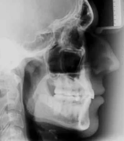
FIGURE 3 - Initial panoramic radiograph.
FIGURE 4 - Initial lateral cephalometric radiograph.
Measurements Pre-treatment values
Post-treatment
values
SNA 84º 81º
SNB 77º 76º
ANB 7º 5º
SND 73º 73º
1.NA 19º 19º
1-NA 4.5 mm 3 mm
1.NB 42º 37º
1-NB 10.5 mm 7 mm
Pog-NB 0 1.5
Pog-1NB 10.5 mm 5.5 mm
1:1 112º 118º
Ocl:SN 22º 22º
GoGn:SN 35º 34º
S – Ls 1 mm -3 mm
S – Li 1 mm -2.5 mm
Y axis 58º 58º
Facial Angle 88º 87º
Convexity Angle 17º 9º
Wits 3 mm 1 mm
FMA 29º 24º
FMIA 41º 50º
IMPA 110º 106º
of the crowding. We used 0.016-in Multiloop “Tweed” style archwires to correct canine me-siobuccal inclination.
After alignment and leveling, the canines were retracted with chain elastics. Brackets were then bonded to the lateral incisors fol-lowed by realignment and releveling.
Any residual space was then closed by re-traction of the upper and lower incisors using rectangular archwires with bull loops.
Twenty-two months after the extraction of the second molars, third molars were erupted and ready for banding or bonding.
After treatment completion, an upper wraparound removable appliance and a fixed lower canine-to-canine lingual arch were in-stalled for retention.
Results
The patient’s extraoral aspect remained as it was initially (Fig 5), except for her profile, which had its convexity reduced.
Intraorally, a Class I relationship was achieved for molars and canines as well as appropriate overbite and overjet. The crossbite was corrected, the curve of Spee leveled and the lower midline corrected, with the upper and lower midlines co-inciding with the facial midline. Both upper and lower crowding were eliminated (Fig 6).
The radiographs disclosed adequate root parallelism. Moreover, upper third molars were found to be appropriately positioned. At this time the removal of the supernumerary upper molar was performed (Fig 7).
From a cephalometric standpoint, the skel-etal pattern was maintained. The most significant changes occurred in the upper and lower inci-sors and lips. The upper and lower inciinci-sors were retracted. Thus, correction of the dental double protrusion was achieved by moving the incisors to their original position. Due to these dental chang-es, the lips were retracted, reducing the patient’s profile convexity (Figs 5 and 8 and Table 1). The radiographs confirmed the presence of
intraosseous third molars with normal anatomy. The upper third molars had fully formed crowns with two-thirds of root formation. The lower third molars were impacted. Supernumerary teeth were also present (Fourth right and left lower molars, and fourth right upper molar), and visible lack of space for correct positioning of the upper canines (Fig 3).
Cephalometric analysis revealed a skeletal Class II (ANB = 7º; Wits = 3 mm); a predominant-ly vertical facial growth pattern (Ocl-SN = 22º; GoGn-SN = 35º); mandibular deficiency (SNB = 77º); proclined lower incisors (1.NB = 42º; IMPA = 110º); and dental double protrusion (1-NA = 4.5 mm, 1-NB = 10.5 mm) (Fig 4 and Table 1).
Treatment
In order to establish a Class I molar rela-tionship as soon as possible and because the patient did not exhibit any growth potential, we opted for upper second molar extraction to facilitate distalization of the upper first molar and Class II correction.
Additionally, we also extracted the lower third molars that were impacted and the low-er suplow-ernumlow-erary teeth. We decided against extracting the upper supernumerary molar given the possibility of damage to the third molar when doing so. The extraction of this tooth was postponed to a future, more conve-nient occasion.
After extraction, the upper first molars were banded and a cervical traction headgear was installed (350 g - 16 h / day) for first molar distalization, which was achieved after a pe-riod of four months.
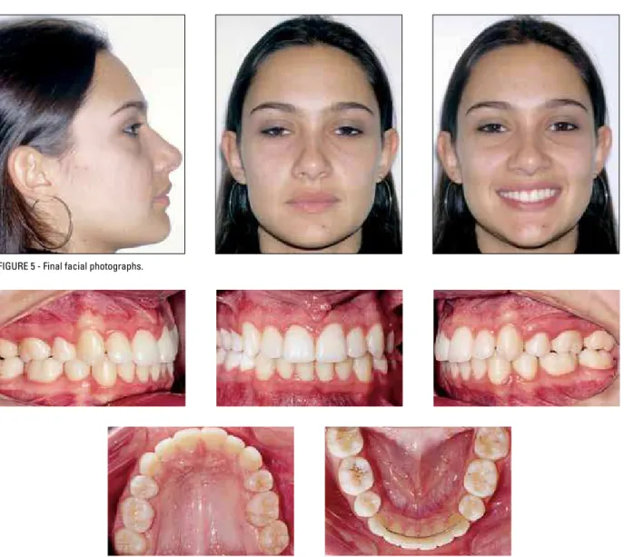
FIGURE 5 - Final facial photographs.
FIGURE 6 - Final intraoral photographs.
CLINICAL CASE STUDY 2
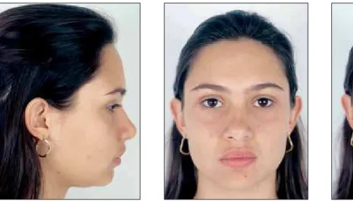
Male patient aged 16 years and 05 months, who sought orthodontic treatment complain-ing of unsightly smile caused by the position of the canines.
Diagnosis
A clinical examination revealed a symmetri-cal face. The patient’s nearly expressionless smile reduced his upper incisor exposure. He had a brachycephalic facial pattern, well balanced fa-cial thirds and convex profile (Fig 9).
The intraoral examination revealed parabolic shaped arches; Class II canine and molar rela-tionship; 5.5 mm overjet; 30% overbite; reverse crossbite between teeth 17 and 47; mild curve of Spee; lower midline shifted 0.5 mm to the left; severe crowding in the upper arch (discrepancy of -11 mm) and moderate crowding in the lower arch (discrepancy of -6 mm) (Fig 10).
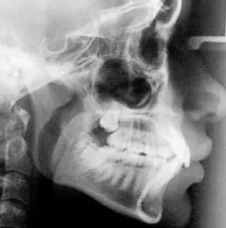
The radiographs confirmed the presence of intraosseous third molars with normal anatomy.
The upper third molars had fully formed crowns with two-thirds of root formation. Space was also lacking for the correct positioning of the upper canines (Fig 11).
The cephalometric analysis revealed a skeletal Class I (ANB = 2º; Wits = 2 mm), horizontal facial growth pattern (GoGN-SN = 24º); mandibular deficiency (SNB = 78º) compensated by maxillary retrusion; incisor proclination (1.NB = 33º; IMPA = 110º); and dental double protrusion (1-NA = 10 mm; 1-NB = 6 mm) (Fig 12 and Table 2).
Treatment
Since the patient had low growth poten-tial, we opted for extracting the upper second molars to facilitate first molar distalization and Class II correction.
After extraction, the upper first molars were banded and a cervical traction headgear was installed (350 g - 16 h / day) for first molar distalization, which was achieved after a pe-riod of five months.
FIGURE 10 - Initial intraoral photographs.
FIGURE 11 - Initial panoramic radiograph.
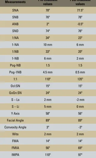
FIGURE 12 - Initial lateral cephalometric radiograph. TABLE 2 - Pre and post-treatment cephalometric data of patient (clinical case study 2).
Measurements Pre-treatment values
Post-treatment
values
SNA 78º 77.5º
SNB 76º 78º
ANB 2º -0.5º
SND 74º 76º
1:NA 34º 23º
1-NA 10 mm 6 mm
1:NB 33º 20º
1-NB 6 mm 2 mm
Pog-NB 1.5 1.5
Pog-1NB 4.5 mm 0.5 mm
1:1 110º 135º
Ocl:SN 15º 15º
GoGn:SN 24º 24º
S – Ls 2 mm -2 mm
S – Li 5 mm 0 mm
Y Axis 58º 56º
Facial Angle 89º 89º
Convexity Angle 3º -3º
Wits 2 mm 2 mm
FMA 14º 14º
FMIA 56º 69º
The first upper and lower premolars were ex-tracted because of the severe upper crowding and the lower protrusion and proceeded to bond the lower fixed appliance. Initially, no brackets were bonded to the upper incisors. Firstly, the canines were retracted to create enough space to accom-modate all teeth in the arch.
After treatment completion, an upper wrap-around removable appliance and a fixed lower canine-to-canine lingual arch were installed for retention.
Results
Extraorally we observed significant changes in the patient’s expression when smiling, with proper exposure of the upper incisors and significant im-provement in the appearance of the profile (Fig 13). Intraorally, a Class I relationship was achieved for molars and canines as well as appropriate over-bite and overjet. The crossover-bite was corrected, the curve of Spee leveled and the lower midline cor-rected, with the upper and lower midlines coincid-ing with the facial midline (Fig 14).
The radiographs presented adequate root par-allelism. Moreover, upper third molars were found to be properly positioned. Tooth 48 was extracted and tooth 38 had already been removed (Fig 15).
From a cephalometric standpoint, we observed a small retraction of point A due to a retraction in the upper incisors while the mandible (point B) advanced by 2º, which decreased facial convexity. The upper and lower incisors were moved back to their original sites, which improved lip positioning (Fig 13 and 16 and Table 2).
FINAL CONSIDERATIONS
When properly indicated, second molar ex-traction can prove a beneficial treatment option for patients. It can shorten treatment time and
FIGURE 15 - Final panoramic radiograph. FIGURE 16 - Final lateral cephalometric ra-diograph.
simplify treatment mechanics. It is essential, however, that all available diagnostic resources be used for an accurate selection of cases best suited for this kind of therapy.
In the clinical cases presented in this article, second molar extraction was performed to enable first molar distalization and, consequently, Class II correction in patients not undergoing facial growth. First molar extraction was performed to improve the facial profile and correction of ante-rior discrepancy caused by either severe crowd-ing or excessive protrusion of the incisors.
These clinical cases serve as examples of how a proper diagnosis coupled with a compliant patient can result in a treatment that enhances both the patient’s aesthetics and function.
1. Aras A. Class II correction with the modiied sagittal appliance and maxillary second molar extraction. Angle Orthod. 2000 Aug;70(4):332-8.
2. Basdra EK, Komposch G. Maxillary second molar extraction treatment. J Clin Orthod. 1994 Aug;28(8):476-81.
3. Basdra EK, Stellzig A, Komposch G. Extraction of maxillary second molars in the treatment of Class II malocclusion. Angle Orthod. 1996;66(4):287-91.
4. Bishara SE, Burkey PS. Second molar extractions: a review. Am J Orthod. 1986 May;89(5):415-24.
REFERENCES
5. Cavanaugh JJ. Third molar changes following second molar extractions. Angle Orthod. 1985 Jan;55(1):70-6.
6. Chapin WC. The extraction of maxillary second molars to reduce growth stimulation. Am J Orthod Oral Surg. 1939;11:1072-8. 7. Chipman MR. Second and third molars: their role in orthodontic
therapy. Am J Orthod. 1961 Jul;47(7):498-520. 8. Graber TM. The role of upper second molar extraction in
orthodontic treatment. Am J Orthod. 1955;41:354-61. 9. Graber TM. Maxillary second molar extraction in Class II
21. Romanides N, Servoss JM, Kleinrock S, Lohner J. Anterior and posterior dental changes in second molar extraction cases. J Clin Orthod. 1990 Sep;24(9):559-63.
22. Rondeau BH. Second molar extraction technique: overrated or under utilized? Funct Orthod. 1999 Oct-Dec;16(4):4-14. 23. Smith R. The effects of extracting upper second permanent
molars on lower second permanent molar position. Br J Orthod. 1996 May;23(2):109-14.
24. Staggers JA. A comparison of results of second molar and irst premolar extraction treatment. Am J Orthod Dentofacial Orthop. 1990 Nov;98(5):430-6.
25. Stellzig A, Basdra EK, Komposch G. Skeletal and dentoalveolar changes after extraction of the second molars in the upper jaw. J Orofac Orthop. 1996 Oct;57(5):288-7.
26. Thomas P. Second molar extraction. Br Dent J. 1994 Nov; 177(9):324.
27. Waters D, Harris EF. Cephalometric comparison of maxillary second molar extraction and nonextraction treatments in patients with Class II malocclusions. Am J Orthod Dentofacial Orthop. 2001 Dec;120(6):608-13.
28. Whitney EF, Sinclair PM. An evaluation of combination second molar extraction and functional appliance therapy. Am J Orthod Dentofacial Orthop. 1987 Mar;91(3):183-92.
29. Zanelato RC, Trevisi HJ, Zanelato ACT. Extração dos segundos molares superiores. Uma nova abordagem para os tratamentos da Classe II, em pacientes adolescentes. Rev Dental Press Ortod Ortop Facial. 2000 mar-abr;5(2):64-75.
Contact address
Maurício Barbieri Mezomo Rua Francisco Manuel 28 / 404 CEP: 97.015-260 – Santa Maria/RS, Brazil E-mail: [email protected]
Submitted: December 2006 Revised and accepted: September 2009
10. Haas AJ. Let’s take a rational look at permanent second molar extraction. Am J Orthod Dentofacial Orthop. 1986 Nov;90(5):361-3. 11. Harnick DJ. Case report: Class II correction using a modiied
Wilson bimetric distalizing arch and maxillary second molar extraction. Angle Orthod. 1998 Jun; 68(3):275-80. 12. Henriques JFC, Janson G, Hayasaki SM. Parâmetros para a
extração de molares no tratamento ortodôntico: considerações gerais e apresentação de um caso clínico. Rev Dental Press Ortod Ortop Facial. 2002 jan-fev;7(1):57-64.
13. Jäger A, El-Kabarity A, Singelmann C. Evaluation of orthodontic treatment with early extraction of four second molars. J Orofac Orthop. 1997 Feb; 58(1):30-43.
14. Jones H. Second molar extraction therapy - two case reports. Funct Orthod. 2000 Winter;17(1):17-20.
15. Liddle DW. Second molar extraction in orthodontic treatment. Am J Orthod. 1977 Dec;72(6):599-616.
16. Light A. Second molar extractions in orthodontic therapy. Penn Dent J. 1986;86(1):14-6.
17. Little RM. Stability and relapse of mandibular anterior alignment: University of Washington Studies. Seminars Orthod. 1999 Sep;5(3):191-204.
18. Magness WB. Extraction of second molars. J Clin Orthod. 1986 Aug; 20(8):519-22.
19. Orton-Gibbs S, Crow V, Orton HS. Eruption of third permanent molars after the extraction of second permanent molars. Part 1: assessment of third molar position and size. Am J Orthod Dentofacial Orthop. 2001 Mar;119(3):226-37.