Evaluation of maxillary atresia associated
with facial type
Marina Gomes Pedreira*, Maria Helena Castro de Almeida**, Katia de Jesus Novello Ferrer***, Renato Castro de Almeida****
Objectives: To associate maxillary atresia with facial types, investigating whether dimorphism occurs between males and females and evaluating the percentage of such dimorphism accord-ing to gender and facial type. Methods: Initially, the sample consisted of 258 lateral cepha-lometric radiographs. After analyzing Ricketts’ VERT index, 108 radiographs were excluded for not meeting the selection criteria. Therefore, the sample consisted of 150 lateral cepha-lometric radiographs and 150 models of 150 Caucasian individuals aged 14 years to 18 years and 11 months, regardless of malocclusion type. The sample was divided into 50 mesofacials, 50 brachyfacials and 50 dolichofacials. The Schwarz’s analysis was applied to all 150 models. Results: The presence of maxillary atresia in the sample consisted of 64% in dolichofacials, 58% in brachyfacials and 52% in mesofacials. Conclusions: There was no evidence showing that atresia is in any way associated with facial type. Gender dimorphism was proportionally greater in dolichofacial males while females did not exhibit different proportions.
Abstract
Keywords: Maxillary atresia. Schwarz’s analysis. Facial types.
* MSc in Orthodontics, CPO São Leopoldo Mandic. Head and Professor of Specialization and Improvement in the area of Orthodontics, Funorte/SOEBRÁS, Alfenas/MG.
** Specialist in Orthodontics, CFO. Professor of Orthodontics, FOP/UNICAMP (retired). Professor of the Masters in Dentistry Program CPO São Leopoldo Mandic.
*** Specialist in Orthodontics, UNICASTELO. MSc in Dentistry in the area of Orthodontics, UNICASTELO. PhD in Orthodontics, FOP / UNI-CAMP. Professor of the Masters in Dentistry Program, CPO São Leopoldo Mandic.
**** Specialist in Orthodontics, CFO. Specialist in Radiology, FOP/UNICAMP. MSc and PhD in Orthodontics, FOP/UNICAMP. Professor and Head of the Masters in Dentistry Program in the Orthodontics area, CPO São Leopoldo Mandic.
INTRODUCTION AND LITERATURE REVIEW
Dental arch shape is essential for the diagnosis of malocclusion given the fact that ideal stability and function require perfect dental intercuspation.
Maxillary atresia is a dentofacial deformity
It may be hidden due to the sagittal position of the maxilla and mandible with no apparent transverse deficiency.³
Witzig and Spahl10 affirm that Pont, in
1909, after assessing Basque individuals of southern France, established a fixed constant for the ideal shape of the dental arches in the premolar (80 mm) and molar (64 mm) re-gions using the formula: SI x 100 divided by 80 or 64, respectively.
Later, however, in disagreement with Pont, Schwarz and Gratzinger12 developed a formula
for each facial type.
For a better diagnosis of maxillary atresia Schwarz’s analysis system is commonly used to determine the magnitude of the discrepancy, in millimeters, by measuring the actual arch width versus the ideal width of the upper and low-er dentitions, thus indicating whethlow-er thlow-ere is more need for anterior or posterior expansion.12
Arch morphology can assume different forms given their relationship with face width. Brachyfacials feature a larger transverse axis than do dolichofacials, whose faces are longer and narrower.4
The combined analysis of models and facial pattern can assist in choosing the mechani-cal procedure to be adopted by professionals, thereby optimizing the chances of a successful treatment.
By analyzing the maxilla transversely using Ricketts analysis and Schwarz’s analysis, we re-alized it is possible to contribute with more evi-dence to orthodontic treatment diagnosis and planning, thereby increasing the likelihood of stability and successful results.
OBJECTIVE
The purpose of this study was to employ Ricketts vertical growth (VERT) analysis and Schwarz’s model analysis to evaluate:
• The percentage of maxillary atresia in the dolichofacial, mesofacial and brachyfacial
facial types.
• Gender dimorphism, considering these fa-cial types.
• Association of atresia with these facial types.
MATERIAL
Initially, our sample consisted of 258 lateral cephalometric radiographs. When performing cephalometry using Ricketts (VERT) analysis we selected 150 lateral cephalometric radiographs, i.e., 50 of brachyfacials, 50 of mesofacials and 50 of dolichofacials. Inclusion criteria required that all subjects should have complete permanent den-tition with no agenesis, supernumerary teeth, ex-tractions or extensive restorations.
The sample also comprised 150 stone casts of maxillary arches of 150 Caucasian individuals of both genders, aged 14 years to 18 years and 11 months, regardless of malocclusion type.
The models were analyzed using Schwarz’s analysis to determine the extent of maxillary atresia.
METHODS
On the lateral cephalometric radiographs we highlighted the landmarks to perform Ricketts’ (VERT) analysis and determine the facial pattern of each individual in the sample.
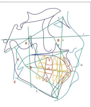
The following measurements were evaluated (Fig 1, Tables 1 and 2): lower facial height (an-gle formed by lines Xi-ENA and Xi-Pm), facial axis (posterior angle formed by the basion-nasi-on line and Pt-Gn), facial depth (angle formed by the intersection of the facial and Frankfurt planes), mandibular plane angle (formed by the intersection of the Frankfurt and mandibular planes), and the mandibular arch [obtained by extending the Xi-Pm and Xi-DC lines (condyle axis)]. With the resulting measurements we cal-culated the VERT index using the age standard, obtained according to the growth prediction method used by Ricketts to determine normal values for 9 year-old children.
10 7
1 2
9
8
3
4
6
5 C
D E
A B
in a Radiology Center with the aid of a com-puter program (CFX 2000, Cuiabá, Mato Grosso, Brazil).
In maxillary arch dental casts a pencil was used to mark landmarks on the occlusal surfaces of the following teeth: distal fossae of the first premolars and distal fossae of the first molars (Fig 2).
A bow divider was positioned over the land-marks on the first right and left premolars and subsequently, on the landmarks of the first right and left molars (Fig 5). The measurements (in mm) were recorded. With this procedure we ob-tained the transverse measurements between the first premolars and first molars in order to deter-mine the presence of maxillary atresia.
Using a bow divider we measured the mesio-distal widths of the central and lateral maxillary incisors (in mm) (Fig 3 and 4).
The sum total of the mesiodistal diameters of the four incisors was represented by SI. The standard formulas of Schwarz used to compare models and cephalometric radiographs were: SI+6 = ideal premolar width and SI+12 = for mo-lars (for leptoprosopics or dolichofacials), SI+7 = ideal premolar width and SI+14 = for molars (for mesoprosopics or mesofacials), SI+8 = ideal premolar width and SI+16 = for molars (for euri-prosopics or brachyfacials).
The value of SI, added to the value for each facial type, resulted in the ideal width of the transverse distances between first maxillary pre-molars and first maxillary pre-molars.
Ub and um acronyms were used: the optimal distance measured in a linear fashion directly on the arch between the distal fossae of the first pre-molars was represented by ub and the ideal arch distance between the central fossae of the first molar was defined as um.
The actual distances between the distal fossae of the first premolars and the distal fossae of the first molars were measured with a bow divider.
The actual values were subtracted from the ideal values. When ub and um were identical in TABLE 1 - Ricketts’ VERT angles.
FIGURE 1 - Ricketts cephalometric analysis with lines, planes and angles in the CFX 2000 software.
TABLE 2 - Lines and planes in Ricketts’ cephalogram.
Angles
A Lower facial height
B Facial axis
C Facial depth
D Mandibular plane angle
E Mandibular arch
Lines and Planes
1 Horizontal Frankfurt plane
2 Cranium-base plane
3 Xi-ENA line
4 Occlusal plane
5 Mandibular plane
6 Axis of the mandibular body
7 Facial axis
8 Long axis of the upper incisors
9 Facial plane
terms of discrepancies, it indicated that they re-quired identical lateral expansion of the maxil-lary arch, when discrepancy ub>um it indicated that it required further anterior lateral expansion, and when discrepancy ub<um it indicated that it required more posterior lateral expansion. All of these results were defined as maxillary atresia.
On the other hand, when the two discrepancies equaled zero, or when the actual distance was greater than the ideal distance, such discrepan-cies were not defined as maxillary atresia.
To investigate the association of atresia and gender with facial type the Pearson’s chi-square test was used. For the comparison between the FIGURE 2 - Landmarks (distal fossa of the first
premolars and distal fossa of the first upper molars).
FIGURE 3 - Measurement of mesiodistal widths of upper central incisors.
FIGURE 4 - Measurement of mesiodistal widths of upper lateral incisors.
Facial Types Atresia Total (%)
Yes (%) No (%)
Dolichofacial 32 (64.00) 18 (36.00) 50 (100.00)
Mesofacial 26 (52.00) 24 (48.00) 50 (100.00)
Brachyfacial 29 (58.00) 21 (42.00) 50 (100.00)
Total 87 (58.00) 63 (42.00) 150 (100.00)
TABLE 3 - Facial types and atresia. TABLE 4 - Atresia in males and facial types.
TABLE 5 - Atresia in females and facial types.
Facial Types Male Atresia Total (%)
Yes (%) No (%)
Dolichofacial 19 (70.37) 8 (29.63) 27 (100.00)
Mesofacial 10 (38.46) 16 (61.54) 26 (100.00)
Brachyfacial 11 (44.00) 14 (56.00) 25 (100.00)
Total 40 (51.28) 38 (48.72) 78 (100.00)
Facial Types Female Atresia Total (%)
Yes (%) No (%)
Dolichofacial 13 (56.52) 10 (43.48) 23 (100.00)
Mesofacial 16 (66.67) 8 (33.33) 24 (100.00)
Brachyfacial 18 (72.00) 7 (28.00) 25 (100.00)
Total 47 (65.28) 25 (34.72) 72 (100.00) mean deviations of the premolars and molars in
relation to gender for each facial type the Stu-dent’s t test was used when the data approached a normal distribution (Shapiro-Wilk test) and the Mann-Whitney U test was used for data without normal distribution. P < 0.05 values were consid-ered significant.
As reference the computer software Statis-tica (version 6, from StatSoft Inc., 2001, www. statsoft.com) was employed.
The presence of maxillary atresia in the sam-ple consisted of 64% in dolichofacials, 58% in brachyfacials and 52% in mesofacials.
No evidence was found (p = 0.4776) of any association between atresia and facial type (Table 3).
Regarding gender dimorphism, however, Ta-ble 4 shows that the presence of atresia in men is proportionally higher in dolichofacials (p = 0.0455), while women, as shown in Table 5, did not show different proportions (p = 0.5229).
DISCUSSION
In this study we found 32 dolichofacial in-dividuals with maxillary atresia, 26 mesofacials with maxillary atresia and 29 brachyfacials with maxillary atresia (Fig 6) in a total of 50 indi-viduals for each facial type. We found that 64% of dolichofacials, 52% of mesofacials and 58% of brachyfacials presented with maxillary atre-sia. However, there was no evidence indicating that maxillary atresia is in any way associated with facial type. These results confirm findings showing no statistically significant differences
between the three facial types in a study7 that
used transverse maxillary measurements. A later study8 eventually found no correlation between
the asymmetry of the maxillary hemiarches and the three facial types, and no statistical differ-ence between the asymmetries.
By comparing Pont’s index with mesofacials and dolichofacials, no differences were found in the interpremolar and intermolar widths associ-ated with the facial types. These findings, how-ever, disagreed with the report5 in which the
transverse measurements were correlated with the mandibular plane angle because it was found that any increase in this angle (in dolichofacials) contributed to a higher incidence of atretic arches. It was also observed that in dolichofacial individuals with nasal obstruction there was a greater prevalence of maxillary atresia.9
When distributing the sample by gender (Figs 7 and 8) we found that 51.28% presented with maxillary atresia with a significant propor-tion of dolichofacials (70.37%). This disagrees with the study1 in which the Class I and Class
when compared with females. The transverse, intercanine, interpremolar and inter-first-mo-lar dimensions of the male patients exhibited higher values than females.2
A total of 65.28% of female patients had maxillary atresia, although different proportions were not found in terms of facial types, which disagrees with a study7 which found a statistically
FIGURE 6 - Association of maxillary atresia with facial type.
FIGURE 7 - Association of maxillary atresia with facial types in males.
FIGURE 8 - Association of maxillary atresia with facial types in females. Dolichofacial Mesofacial Brachyfacial
with without 18 16 14 8 12 6 10 4 2 0
amount of individuals
35 20 30 18 25 16 20 14 8 15 12 6 10 10 4 2 5 0 0 Dolichofacial Dolichofacial Mesofacial Mesofacial Male Female Brachyfacial Brachyfacial with with without without
amount of individuals
amount of individuals
atresia
atresia atresia
signifi cant difference when comparing the maxilla of the mesofacial and dolichofacial groups (males and females). The male group showed larger di-mensions than the female, while in brachyfacials no signifi cant differences were found.
A thorough analysis of the three facial types disclosed that 62.28% of females and 51.28% of males presented with maxillary atresia. No different proportions were found between the genders.
Regarding the presence of maxillary atresia associated with gender,11 the results confirmed
the aforementioned study since we demon-strated that there is a difference in maxillary interpremolar and intermolar widths, which are smaller—indicating maxillary atresia—for both males and females, with no differences between them.6
Therefore the study sample did not show an association between maxillary atresia and facial type, but in dolichofacial males, where a statistically significant value was found, it became clear that measuring the transverse width of the maxilla—in both genders—is of paramount importance since it contributes to diagnosis and planning, thereby avoiding un-necessary expansion and ensuring improved orthodontic treatment results.
CONCLUSIONS
The results and discussion of this study indi-cate that:
1. In our sample, 64% of dolichofacials, 58% of brachyfacials and 52% of mesofacials present-ed with maxillary atresia.
2. There was no gender dimorphism in terms of facial types and presence of atresia, but in males the percentage of dolichofacials presenting with atresia was proportionally higher. Women, on the other hand, did not show different pro-portions between facial types.
Contact address Marina Gomes Pedreira
Rua Amélio da Silva Gomes, 106, Centro CEP: 37.130-000 – Alfenas / MG, Brazil E-mail: [email protected]
1. Albuquerque CM, Vigorito JW. Estudo comparativo do índice de Pont com os tipos faciais, em brasileiros apresentando oclusão normal e maloclusão de Classe I e de Classe II divisão 1ª. [dissertação]. São Paulo: Universidade de São Paulo; 1995. 2. Araújo AM, Ursi WJS. Estudo comparativo das dimensões
transversais em más-oclusões de Classe I e II, de Angle. Rev Dental Press Ortod Ortop Facial. 1997 nov-dez;2(6):69-74. 3. Capelozza Filho L, Silva Filho OG. Expansão rápida da maxila:
considerações e aplicações clínicas. In: Interlandi S. Ortodontia: bases para a iniciação. 4ª ed. São Paulo: Artes Médicas; 1999. p. 285-328.
4. Filho LA. Arcos dentais. In: Madeira MC. Anatomia do dente. São Paulo: Sarvier; 2001. p.17-9.
5. Howes AE. Arch width in the premolar region - still the major problem in orthodontics. Am J Orthod. 1957;43(1):5-31. 6. Kageyama T, Domínguez-Rodríguez GC, Vigorito JW, Deguchi
T. A morphological study of the relationship between arch di-mensions and craniofacial structures in adolescents with Class II division 1 malocclusions and various facial types. Am J Orthod Dentofacial Orthop. 2006 Mar;129(3):368-75.
7. Kanashiro LK, Vigorito JW. Estudo das formas e dimensões das arcadas dentárias superiores e inferiores em leucodermas, brasileiros, com maloclusão de Classe II, divisão 1ª e diferentes tipos faciais. Ortodontia. 2000;33(2):8-18.
REfERENCES
8. Kanashiro LK, Vigorito JW. Estudo comparativo das dimen-sões transversais dos hemi-arcos dentários superiores nas maloclusões de Classe II divisão 1ª, em diferentes tipos faciais. Ortodontia. 2004;37(2):8-13.
9. Mocellin M, Fugmann EA, Gavazzoni FB, Ataíde AL, Ou-riques FL, Herrero F. Estudo cefalométrico-radiográico e otorrinolaringológico correlacionando o grau de obstrução nasal e o padrão de crescimento facial em pacientes não tratados ortodonticamente. Rev Bras Otorrinolaringol. 2000; 66(2):116-20.
10. Witzig JW, Spahl TJ. Ortopedia maxilofacial clínica e aparelhos. 3ª ed. São Paulo: Ed. Santos; 1995. p. 286-93.
11. Rejman R, Martins DR, Scavone H, Ferreira FAC, Ferreira FV. Estudo comparativo das dimensões transversais dos arcos dentários entre jovens com oclusão normal e má oclusão de Classe II, 1ª divisão. Rev Dental Press Ortod Ortop Facial. 2006;11(4):118-25.
12. Schwarz AM, Gratzinger M. Removable orthodontic appliances. Philadelphia: WB Saunders; 1966. p. 61-83.
Submitted: August 2008