Oscillation mechanics of the respiratory system in
never-smoking patients with silicosis:
pathophysio-logical study and evaluation of diagnostic accuracy
Paula Morisco de Sa´,I Agnaldo Jose´ Lopes,IIJose´ Manoel Jansen,IIPedro Lopes de MeloIII
IUniversidade do Estado do Rio de Janeiro, Institute of Biology and Faculty of Engineering Biomedical Instrumentation Laboratory, Rio de Janeiro/RJ,
Brazil.IIUniversidade do Estado do Rio de Janeiro, Faculty of Medical Sciences Pulmonary Function Laboratory, Rio de Janeiro/RJ, Brazil.IIIUniversidade do Estado do Rio de Janeiro, Institute of Biology and Faculty of Engineering, Laboratory of Clinical and Experimental Research in Vascular Biology, BioVasc, Biomedical Center Biomedical Instrumentation Laboratory, Rio de Janeiro/RJ, Brazil.
OBJECTIVES:Silicosis is a chronic and incurable occupational disease that can progress even after the cessation of exposure. Recent studies suggest that the forced oscillation technique may help to clarify the changes in lung mechanics resulting from silicosis as well as the detection of these changes. We investigated the effects of airway obstruction in silicosis on respiratory impedance and evaluated the diagnostic efficacy of the forced oscillation technique in these patients.
METHODS:Spirometry was used to classify the airway obstruction, which resulted in four subject categories: controls (n = 21), patients with a normal exam (n = 12), patients with mild obstruction (n = 22), and patients with moderate-to-severe obstruction (n = 12). Resistive data were interpreted using the zero-intercept resistance (R0), the resistance at 4 Hz (Rrs4), and the mean resistance. We also analyzed the mean reactance (Xm) and the dynamic compliance. The total mechanical load was evaluated using the absolute value of the respiratory impedance (Z4Hz). The diagnostic potential was evaluated by investigating the area under the receiver operating characteristic curve. ClinicalTrials.gov: NCT01725971.
RESULTS:We observed significant (p,0.0002) increases in R0, Rrs4, Rm, and Z4Hz and significant reductions in Crs,dyn (p,0.0002) and Xm (p,0.0001). R0, Rrs4, Rm, and Z4Hz performed adequately in the diagnosis of mild obstruction (area under the curve.0.80) and highly accurately in the detection of moderate-to-severe obstruction (area under the curve.0.90).
CONCLUSIONS:The forced oscillation technique may contribute to the study of the pathophysiology of silicosis and may improve the treatment offered to these patients, thus representing an alternative and/or complementary tool for evaluating respiratory mechanics.
KEYWORDS: Silicosis; Respiratory Physiopathology; Forced Oscillations; Respiratory Impedance; Diagnostic; Respiratory Mechanics.
Sa´ PM, Lopes AJ, Jansen JM, Melo PL. Oscillation mechanics of the respiratory system in never-smoking patients with silicosis: pathophysiological study and evaluation of diagnostic accuracy. Clinics. 2013;68(5):644-651.
Received for publication onNovember 26, 2012;First review completed onDecember 12, 2012;Accepted for publication onJanuary 18, 2013 E-mail: [email protected]
Tel.: 55 21 2334-0705
& INTRODUCTION
Silicosis refers to pulmonary fibrosis caused by inhalation of dust that contains crystalline silica (1,2). Silicosis is an occupational disease that is chronic and incurable; it starts in the peripheral airways and can progress even after the cessation of exposure (2). Although the incidence of silicosis
has decreased since the Second World War, the disease continues to be a major cause of occupational lung disease in exposed workers (3).
Currently, the diagnosis of silicosis is based on a patient’s occupational history and radiological findings. However, many cases are detected several years after exposure. The advent of high-resolution computed tomography has enabled the earlier identification of radiological signs of silicosis (2). However, this method is expensive and less commonly available, and trained professionals are neces-sary to perform the examination (4). Several authors have observed that changes in airway resistance, as measured by plethysmography, significantly influence the prognosis of individuals who are exposed to dust (5), but these measurements are difficult to obtain for claustrophobic patients and demand high levels of subject cooperation (6,7).
Copyrightß2013CLINICS– This is an Open Access article distributed under the terms of the Creative Commons Attribution Non-Commercial License (http:// creativecommons.org/licenses/by-nc/3.0/) which permits unrestricted non-commercial use, distribution, and reproduction in any medium, provided the original work is properly cited.
No potential conflict of interest was reported.
The carbon monoxide diffusion technique may also be used; however, this technique requires a high level of expertise and patient cooperation (8). Currently, spirome-try, which is a test that also requires a high degree of patient cooperation, is commonly used to assess pulmon-ary function in individuals with silicosis. This test, which requires patients to perform forced expirations and inspirations, can generate changes in bronchial tone that modify airway patency and rendering the indices obtained hardly physiologic (9).
There is general agreement in the literature regarding the necessity of developing new, accurate, and non-invasive tests of lung function (10-12). One of the possible methods, namely, the forced oscillation technique (FOT), may over-come the aforementioned inconveniences and may be conducted during spontaneous breathing. It was recently noted that the FOT represents the current state-of-the-art technique in the assessment of lung function (13). Several authors have argued that the FOT has the potential to improve diagnosis and the tracking of treatments and that further studies are necessary in this area (7,13,14). Previous studies from our group suggest that this technique may contribute to the detection of respiratory changes in smokers (15,16) and in patients suffering from sarcoidosis (17) and rheumatoid arthritis (18). Therefore, the FOT may facilitate the diagnosis of patients with occupational diseases (8) and enable simple, routine evaluations in these patients. However, few studies that utilize the FOT have been conducted to analyze the changes in respiratory mechanics that are associated with silicosis (6,8,19,20,21).
Based on the above-mentioned considerations, the aims of the present study were 1) to use the FOT to analyze how respiratory mechanics are affected in patients with silicosis, 2) to evaluate the potential of the FOT for detecting mild and moderate-to-severe alterations in patients with silicosis, and 3) to identify the best FOT parameters for the diagnosis of respiratory changes in silicosis patients.
& MATERIALS AND METHODS
The present work is a transverse study that was developed at the Biomedical Instrumentation Laboratory and Pulmonary Function Laboratory at the Universidade do Estado do Rio de Janeiro (UERJ). The study was approved by the Ethics Committee of the Pedro Ernesto University Hospital (HUPE). Informed consent was obtained from all of the volunteers before inclusion in the study, which was registered at ClinicalTrials.gov (identifier: NCT01725971).
Subjects
A total of 67 subjects were analyzed. The control group (CG) included 21 subjects with normal spirometric data and without a history of smoking or pulmonary disease.
Smoking has been established as a potentially important confounding factor in the study of the pathophysiology of respiratory diseases. To eliminate this factor, 46 silicosis patients who had never smoked were analyzed. These patients were selected after an initial interview of 470 patients, 424 of whom were excluded due to their smoking history. The diagnosis of silicosis was established based on a history of substantial exposure to silica dusts and compa-tible radiological features, together with the exclusion of other competing diagnoses.
All of the subjects had a body mass index (BMI) of less than 29.9 kg/m2. Spirometry was used to classify the airway obstruction into one of three categories (19,23): normal upon spirometric examination (NE, n = 12), mild obstruction (MO, n = 22), or moderate-to-severe obstruction (MSO = 12).
Spirometry
Measurements of forced expiratory volume in the first second (FEV1), forced vital capacity (FVC), FEV1/FVC, and forced expiratory flow (FEF) between 25 and 75% of FVC (FEF/FVC) were obtained for all of the patients according to the Brazilian Consensus on Spirometry (22). These para-meters are presented as raw data and as a percentile of the predicted values, which were obtained from Knudson et al. (23) and Pereira et al. (24). The forced expiratory maneuvers were repeated until three sequential measurements were obtained. The indices were obtained from the best curve, which was associated with the highest value of FEV1plus FVC. Quality control measures for the spirometry were provided by the ATS criteria, with software used to detect non-acceptable maneuvers.
Forced Oscillation Technique
The FOT has previously been described in detail (25) and meets the international standards (26). The equipment was a standard multifrequency FOT test system that operated within the most commonly used frequency range (26,27). Briefly, a pseudorandom sinusoidal signal, which had a 2-cmH2O peak-to-peak amplitude and contained all the harmonics of 2 Hz between 4 and 32 Hz, was applied by a loudspeaker. The pressure input was measured using a Honeywell 176 PC pressure transducer (Microswitch, Boston, MA, USA). The airflow was measured with a screen pneumotachometer coupled to a similar transducer with a matched frequency response. The signals were digitized at a rate of 1024 Hz for periods of 16 s by a personal computer, and a fast Fourier transform was computed using blocks of 4096 points with 50% overlap. To perform the FOT analysis, the subject remained in a sitting position, maintaining the head in a normal position and breathing at functional reserve capacity through a mouthpiece. During the mea-surements, the subject wore a nose clip and firmly supported his/her cheeks and mouth floor using both hands. A minimal coherence function of 0.9 was considered to be adequate (15-18,28). Three measurements were obtained, and the result was expressed as the mean of these three measurements.
The reactive properties were described by the following three parameters: the resonance frequency (fr), which is defined as the frequency at which Xrs equals zero; the mean reactance (Xm), which is a property usually related to respiratory system non-homogeneity (32) and that is calculated based on the entire studied frequency range (4 to 32 Hz); and the dynamic compliance (Crs,dyn). Crs,dyn includes the compliances of the lung and bronchial walls, the chest wall/abdominal compartment, the upper airways (associated with the soft tissues of the mouth, cheeks, and pharynx), and thoracic gas compression (30). Crs,dyn was estimated using Xrs at 4 Hz (Crs,dyn = -1/(2pfXrs)) (33). The
total mechanical load of the respiratory system, including the resistive and elastic effects (27,30), was estimated using the absolute value of respiratory impedance at 4 Hz (Z4Hz). This parameter is associated with the necessary work to promote the movement of air within the respiratory system.
Data processing, data presentation, and statistical analysis
The results are presented as the mean ¡ SD. A
commercial software package (STATISTICA for Windows, release 5.0) was used to compare the differences between the groups. A one-way ANOVA with Tukey’s test was performed to analyze the normally distributed data; con-versely, a non-parametric analysis (Kruskal-Wallis) with a Mann-Whitney test was performed for the non-normally distributed data. The results withp,0.05 were considered to
be statistically significant. Correlations between the spiro-metry and FOT indices were determined using Pearson’s correlation coefficient.
The clinical potential of the FOT indices for the early detection of respiratory alterations due to silicosis was evaluated using receiver operation characteristic (ROC) analyses, which were performed using Med Calc 8.2 (MedCalc Software, Mariakerke, Belgium).
& RESULTS
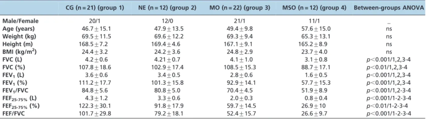
Table 1 lists the subjects’ biometric and spirometric characteristics. No significant differences were found between the groups with respect to the biometric character-istics. The spirometric parameters exhibited significant reductions with increases in airway obstruction.
The mean courses of Rrs and Xrs as a function of frequency in the normal patients and the patients with
silicosis are presented in Figure 1. Parameters derived from Rrs are described in Figure 2, and significant increases in Rrs4 (ANOVA, p,0.0002), R0 (p,0.0001), and Rm
(p,0.0001), as well as decreases in S (p,0.0004), were
associated with the worsening of airway obstruction. Regarding the parameters derived from Xrs, we observed that Xm (Figure 3A) became more negative and that Crs,dyn (Figure 3B) decreased with airway obstruction (p,0.0001
andp,0.0002, respectively).
Table 1 -Biometric and spirometric characteristics of the studied individuals.
CG (n = 21) (group 1) NE (n = 12) (group 2) MO (n = 22) (group 3) MSO (n = 12) (group 4) Between-groups ANOVA Male/Female 20/1 12/0 21/1 11/1 _
Age (years) 46.7¡15.1 47.9¡13.5 49.4¡9.8 57.6¡15.0 ns
Weight (kg) 69.5¡11.5 69.6¡12.2 69.3¡9.4 65.3¡13.1 ns
Height (m) 168.5¡7.2 169.4¡4.6 167.1¡9.1 165.2¡8.9 ns
BMI (kg/m2) 24.4
¡3.2 24.2¡3.6 24.8¡2.9 23.7¡4.0 ns
FVC (L) 4.2¡0.6 4.21¡0.7 4.1¡1.0 3.1¡0.8 p,0.001/1,2,3-4
FVC (%) 107.8¡18.6 102.9¡17.4 108.5¡15.3 88.7¡17.1 p,0.01/1,2,3-4
FEV1(L) 3.6¡0.6 3.4¡0.5 2.8¡0.6 1.6¡0.5 p,0.001/1,2,3-4
FEV1(%) 111.2¡17.7 101.3¡15.8 92.9¡14.1 57.7¡15.3 p,0.001/1,2,3-4
FEV1/FVC 84.8¡5.6 80.8¡5.0 70.4¡4.5 51.9¡8.9 p,0.001/1,2-3-4
FEF25-75%(L) 4.3¡1.2 3.3¡0.6 2.0¡0.3 0.8¡0.4 p,0.001/1-2-3-4
FEF25-75%(%) 122.3¡30.1 91.8¡17.9 59.7¡14.5 26.9¡10 p,0.01/1-2-3-4
FEF/FVC 101.7¡29.8 79.2¡18.1 52.4¡15.7 26.6¡9.7 p,0.001/1-2-3-4 n: number of subjects; ns: non-significant (p.0.05); %pred: percentage of the predicted values; comparisons of the three groups/comparisons between adjacent groups: the dashes indicate a significant difference.
Figure 2 -Resistive parameters at 4 Hz (Rrs4; A), total resistance (R0; B), mean resistance (Rm; C), and resistance slope (S; D) in the normal subjects and in the patients with silicosis. The bottom and top of the box plot represent the 25thand 75thpercentile values, respectively, while the circle represents the mean value, and the bar across the box represents the 50thpercentile value. The whiskers
outside the box represent the 10thto 90thpercentile values.
Higher resonance frequencies were found to be associated with increased airway obstruction (Figure 3C;p,0.0004). A
similar behavior was found for Z4Hz (Figure 3D;p,0.0002).
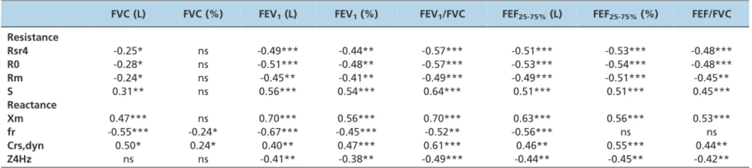
The correlations between the FOT and spirometric parameters are presented in Table 2. Rrs4, R0, and Rm had significant inverse correlations with the spirometric parameters (except for FVC), whereas S was directly correlated with almost all of the spirometric parameters, excluding FVC. Xm, Crs,dyn, and Z4Hz were directly correlated with all of the spirometric parameters, excluding FVC. The resonance frequency was inversely associated with the spirometric indices and did not present a significant correlation with FEF/FVC.
The parameters obtained from the ROC analysis, includ-ing the sensitivity (Se), specificity (Sp), area under the curve (AUC), and cut-off points, are described in Table 3. Rrs4, R0, Rm, and Z4Hz presented the highest AUC values (0.933, 0.905, 0.905, and 0.933, respectively).
& DISCUSSION
The main findings of the present study are the following: 1) in the initial phases of airway obstruction in silicosis (NE and mild) there is a predominance of the obstructive component; 2) in contrast to earlier stages, in the late stages of airway obstruction increase (moderate-to-severe) a clear mixed pattern is developed; and 3) four of the studied FOT parameters showed great potential to enable easier detec-tion of the abnormal respiratory effects of silicosis.
The forced oscillation parameters did not demonstrate systematic changes that were strictly related to spirometry (Table 2). The linear correlation coefficients between all of the spirometric and FOT parameters were evaluated, and, similar to previous studies (34-36), these coefficients were low, ranging between20.57 and 0.70. This result indicates that the FOT and spirometry data are complementary, each providing unique data, thereby confirming the ability of FOT to provide additional information on the mechanical characteristics of the respiratory system.
Pathophysiology
The FOT exams allowed us to obtain additional informa-tion from parameters that are easily associated with mechanical and structural properties of the respiratory system, including respiratory (R0) and airway (R4Hz, Rm) resistance, elastance (Crs,dyn), homogeneity (S, Xm, fr), and work of breathing (Z4Hz) (26-33). Based on these para-meters, FOT may provide insight into both the obstructive and restrictive components of silicosis, thereby improving our understanding of the lung mechanics and how they change during this disease. However, this understanding may be impaired by the inclusion of data from smoking patients, which is an important confounding factor. We expended considerable effort to perform this pathophysio-logical study in a sample of never-smoking patients, thus eliminating this important confounding factor and precisely describing the effects of silicosis. The results of the present study demonstrated that the initial phases of airway obstruction in silicosis (NE and mild obstruction) cause a decrease in respiratory system homogeneity, which is primarily associated with changes in the resistive properties of the respiratory system (Figure 2C). This disease stage was also characterized by a decrease in Crs,dyn (Figure 3B) and an increase in the respiratory work (Figure 3D). These results provide evidence that, although the obstructive component predominates during this stage of the disease, changes in the elastic properties also exist, which suggests that a mixed pattern of disease develops.
The subsequent increase in airway obstruction (mild to moderate) is characterized by a significant rise in respira-tory resistances (Figures 2A, B, C) and a decrease in the homogeneity associated with imbalances in the resistive (Figure 1D) and elastic (Figure 3A) airway properties. A further decrease in Crs,dyn (Figure 3B) and increased values of respiratory work (Figure 3D) are also characteristics of this more advanced disease state. These findings provide clear evidence that a mixed disease pattern develops during the latter stages of silicosis. Below, we discuss in detail the
Table 2 -Correlations between the forced oscillation technique parameters and spirometric indices.
FVC (L) FVC (%) FEV1(L) FEV1(%) FEV1/FVC FEF25-75%(L) FEF25-75%(%) FEF/FVC
Resistance
Rsr4 -0.25* ns -0.49*** -0.44** -0.57*** -0.51*** -0.53*** -0.48***
R0 -0.28* ns -0.51*** -0.48** -0.57*** -0.53*** -0.54*** -0.48***
Rm -0.24* ns -0.45** -0.41** -0.49*** -0.49*** -0.51*** -0.45**
S 0.31** ns 0.56*** 0.54*** 0.64*** 0.51*** 0.51*** 0.45***
Reactance
Xm 0.47*** ns 0.70*** 0.56*** 0.70*** 0.63*** 0.56*** 0.53***
fr -0.55*** -0.24* -0.67*** -0.45*** -0.52** -0.56*** ns ns
Crs,dyn 0.50* 0.24* 0.40** 0.47*** 0.61*** 0.46** 0.55*** 0.44**
Z4Hz ns ns -0.41** -0.38** -0.49*** -0.44** -0.45** -0.42**
*p,0.05; **p,0.01; ***p,0.0001; ns: non-significant (p.0.05); %: percentage of the predicted values. The moderate correlations are displayed in blue.
Table 3 -Values of the AUC, sensitivity, and specificity for the optimal cut-off points for the forced oscillation technique indices describing their performance in the detection of patients with mild and moderate-to-severe obstruction.
AUC Se (%) Sp (%) Cut-off Rrs4 (cmH2O/L/s) 0.885/0.933 72.7/83.3 90.5/100.0 2.19/2.72
R0 (cmH2O/L/s) 0.847/0.905 81.8/83.3 81.8/90.5 2.22/2.86
Rm (cmH2O/L/s) 0.829/0.905 90.9/83.3 71.4/90.5 2.19/2.73
S (cmH2O/L/s2) 0.719/0.865 68.2/75.0 66.7/90.5 3.85/25.91
Xm (cmH2O/L/s) 0.716/0.897 63.6/75.0 76.2/95.2 0.07/0.28
fr (Hz) 0.662/0.841 68.2/83.3 61.9/76.2 13.54/15.25
Crs,dyn (L/cmH2O) 0.747/0.889 72.7/75.0 71.4/95.2 0.018/0.013
pathophysiological changes that can be described by the FOT.
Silica is a mineral oxide dust that exists as a crystal structure of silicon dioxide (SiO2). Once silica particles reach the alveoli, they are ingested by alveolar macrophages. Silica causes toxicity by directly stimulating the inflamma-tory process and the proliferation of fibroblasts (3). This process may partly explain the increase in Rrs4 together with the worsening of airway obstruction, as depicted in Figure 2A. In agreement with the present study, Yang et al. (21) observed higher levels of Rrs, measured at 3 Hz, in coal miners compared with healthy subjects.
Similar to Rrs4, R0 (Figure 2B) increased with increasing obstructive disturbances, which is a finding that confirms the results of previous studies (8). Pham et al. (8) analyzed coal miners and obtained results similar to ours, with higher levels of R0 in symptomatic workers compared with asymptomatic workers.
In agreement with the results obtained regarding obstructive diseases (28,36) and subjects with silicosis (6,8,19), Rm increased with airway obstruction (Figure 1C). This increase describes the typical pathophysiology of silicosis, which includes inflammation, tissue damage, fibrosis, and lung remodeling. Rm presented reasonable but significant correlation with the spirometric parameters related to the more peripheral airways (Table 2). As respiratory alterations begin at these points of the bronchial tree, we speculate that Rm may be useful in the early identification of silicosis.
The slope of the resistive component reflects the homo-geneity of the respiratory system. In the present study, S was significantly more negative in the silicosis subjects with worse airway obstruction (Figure 1D). Similar results are described in the literature (6,8,21). The extensive replace-ment of the pulmonary parenchyma by fibrous tissue and the thickened alveolar septum in patients with silicosis may contribute to local changes in elasticity. This shift, in turn, may cause an imbalance in the time constants of the respiratory system and contribute to the increase in system heterogeneity (3).
The significant modification of the Xm values with airway obstruction (Figure 3A) was consistent with a previous pilot study (20) and previous studies of obstructive diseases (26,28,36). Xm is generally associated with respiratory heterogeneity. In silicosis, heterogeneity may result from a combination of reduced respiratory system compliance and higher peripheral resistance. Inflammation and airway wall remodeling may also be involved in this process (3,21). In contrast with the present study, Faria et al. (17) found significant changes in Xm when comparing a control group with subjects with sarcoidosis and normal spirome-try values. These discrepant findings may be explained by differences in the pathophysiology of silicosis and sarcoidosis.
Crs,dyn reflects the combined effects of the pulmonary and chest wall tissue, as well as the compliance of the airways (27-29). The values of Crs,dyn diminished signifi-cantly with airway obstruction (Figure 3B). In agreement with our results, Pham et al. (8) obtained lower Crs,dyn values in symptomatic individuals with pneumoconiosis. Using the esophageal balloon technique, Van Noord et al. (37) observed a reduction in the pulmonary static compli-ance in patients with interstitial lung disease and a predicted TLC of ,50%. In the same study, the authors
found decreased lung volumes in conjunction with increased resistances (Rrs) and decreased reactances (Xrs), and this trend was more obvious at low frequencies. Because tissue resistance is inversely related to lung compliance (37), these results are consistent with the findings of our study, namely the increase in R0 and a decrease in Crs,dyn.
The description of the pathophysiology of silicosis using Crs,dyn suggests that the FOT may be an alternative method to the use of esophageal balloons for evaluating these individuals. Thus, because the measurement is noninvasive, Crs,dyn may be useful as a functional parameter in monitoring silicosis.
In the present study, we observed higher values of fr in individuals with silicosis compared with the control group. These results were more pronounced with increased airway obstruction (Figure 3C). Similar results have been described in obstructive diseases (28,36). In contrast with our findings, Faria et al. (17) found no significant difference for fr in sarcoidosis. It is possible that fr, which expresses the inertial and elastic properties of the respiratory system and acts as an indicator of the homogeneity of this system (37), is not the most suitable parameter for identifying obstructive changes associated with this disease. Pleuropulmonary involvement in silicosis (3) produces changes in elasticity and dynamic compliance that contribute to a loss of ventilation homogeneity (19). These factors may explain our findings.
We observed a progressive increase in Z4Hz with airway obstruction (Figure 3D). The changes in the resistive and elastic properties of the respiratory system resulting from the pathophysiology of silicosis may explain the significant increase in this parameter. Similar results have been described in the literature regarding sarcoidosis (17). This parameter is related to the total mechanical load of the respiratory system (27,30) and may therefore be associated with fatigue and shortness of breath, which are common symptoms in patients with silicosis (1-3).
Diagnostic accuracy in patients with mild and moderate-to-severe obstruction
An AUC$0.80 is generally considered to be adequate for clinical use (38,39). In the comparison between the control group and patients with mild obstruction (Table 3), Rsr4, R0, Rm, and Z4Hz all had an AUC$0.80, making them suitable for clinical use. As expected, we observed higher AUCs in the analysis that compared the control group with patients exhibiting moderate-to-severe obstruction (3). S, Xm, fr, and Cdyn,rs presented sufficient accuracy for clinical use (AUC.0.80), while Rsr4, R0, Rm, and Z4Hz presented high diagnostic accuracy (AUC.0.90). These results confirm the preliminary findings from our group (20), namely that R0 and Rm presented high accuracy (AUC = 0.90) in the analysis comparing the control and MO groups. In close agreement with these findings, Pham et al. (8) determined that R0 and Rm are the more accurate parameters for identifying individuals with obstructive coal miner’s pneumoconiosis.
encountered during daily activities (40); thus, extrapolation to patient symptoms may be difficult. Another important characteristic of the FOT is that it provides information about the mechanical properties of the respiratory system that is complementary to that provided by traditional lung function tests (7,13,26). However, the routine clinical use of the FOT has yet to gain full acceptance. Although interest in the approach is growing and the technique shows consider-able promise (6-9,13-21,25-37,41), realizing this promise remains a work in progress.
Limitations of the study
Similar to other functional assessment techniques, the limitations of the FOT and its consequences must be recognized. First, an important source of errors is related to the process of spontaneous breathing in patients, which introduces random and systematic errors. These errors are reduced by using excitation frequencies that are at least 10 times higher than those present during the spontaneous ventilation process and by using appropriate acceptance criteria (13,25,26,30). In the present study, these errors were minimized by using a minimal coherence function (c2) value
of 0.9, which is usually considered to be adequate (30). Second, the measurement of respiratory impedance may be influenced by the shunt effect of the upper airway walls (26,27). This effect becomes progressively stronger as the resistance of the pulmonary airways increases because fewer of the imposed flow oscillations are able to move beyond the upper airway shunt and make their way into the lungs. In practice, this drawback is minimized by asking the patient to firmly support his/her cheeks and mouth floor (26,27).
Although this study focused on the effect of airway obstruction on the resistive and reactive properties of the respiratory system, inclusion of the diffusion capacity and lung volumes in the analysis would have been beneficial. These factors may contribute to a more detailed study of the pathophysiology of silicosis and should be addressed in future research.
One of the primary objectives of the present work was to increase our understanding of the pathophysiological changes that are observed when patients with silicosis are examined using the most common, current pulmonary function method. This aim explains why spirometry was used as the gold standard. Another interesting possibility for future work is the comparison of the accuracies of the FOT and spirometry using computer tomography (CT) as a reference technique. In a previous study (19), we evaluated the associations between the CT and pulmonary function findings that were obtained by the FOT and spirometry. However, the present study did not employ CT as a gold standard. We are planning to perform this analysis in the next step of this research.
FOT analysis contributed further insights into how the respiratory system is affected by silicosis. The initial phases of airway obstruction in silicosis were observed to be mainly characterized by the increase in the respiratory system’s resistive parameters. More advanced phases of airway obstruction are characterized by a significant reduction in the respiratory system’s dynamic compliance, which is consistent with a mixed disease pattern. Rrs4, R0, Rm, and Z4Hz were shown to be useful for accurately and non-invasively identifying mild and mild-to-moderate modifica-tions of the respiratory mechanics of patients with silicosis.
These results indicate that the FOT may contribute to the study of the pathophysiology of silicosis and to an improvement of the treatment that is offered to these patients. Therefore, this technique may represent an alter-native and/or complementary tool for evaluating respira-tory mechanics via forced expiration.
& ACKNOWLEDGMENTS
The authors thank A. C. Faria and I. A. Miranda for their technical assistance. This study was financed by the Brazilian Council for Scientific and Technological Development (CNPq) and the Rio de Janeiro State Research Supporting Foundation (FAPERJ).
& AUTHOR CONTRIBUTIONS
Sa´ PM conducted the measurements for this study, analyzed the data, and drafted the manuscript. Lopes AJ provided data and subject identification. Jansen JM mentored Sa´ PM and participated in data analysis process. Melo PL mentored Sa´ PM, organized the study and helped to draft the manuscript. All the authors have read and approved the manuscript.
& REFERENCES
1. Greenberg MI, Waksman J, Curtis J. Silicosis - A Review. Dis Mon. 2007;53(8):394-416.
2. Leung CC, Yu ITS, Chen W. Silicosis. Lancet. 2012;379:2008-18. 3. Sirajuddin A, Kanne JP. Occupational Lung Disease. J Thorac Imaging.
2009;24(4):310-20.
4. Meireles GSP, Kavakma JI, Rodrigues RT. Imaging in occupational lung diseases. J Bras Pneumol. 2006;32:suppl 2.
5. Mandi A, Galgoczy G, Galambos E, Nemeth L, Dombos K. Changes in clinical status and lung functions of patients with chronic respiratory diseases over 10 years. Respiration. 1984;46(2):151-9.
6. Mesquita Jr JA, Lopes AJ, Jansen JM, Melo PL. Using the forced oscillation technique to evaluate respiratory resistance in individuals with silicosis. J Bras Pneumol. 2006;32(3):213-20.
7. Kaczka DW, Dellaca´ RL. Oscillation Mechanics of the Respiratory System: Applications to Lung Disease. Critical ReviewsTMin Biomedical
Engineering. 2011;39(4):337-59.
8. Pham QT, Bourgkard E, Chau N, Willim G, Megherbi SE, Teculescu D, et al. Forced oscillation technique (FOT): a new tool for epidemiology of occupacional lung diseases? Eur Respir J. 1995;8(8):1307-13.
9. Kaminsky AD, Irvin CG. New insights from lung function. Curr Opin Allergy Clin Immunol. 2001;1(3):205-9.
10. Enright PL, Crapo RM. Controversies in the use of spirometry for early recognition and diagnosis of chronic obstructive pulmonary disease in cigarette smokers. Clin Chest Med. 2000;21(4):645-52.
11. Croxton TL, Weinmann GG, Senior RM, Hoidal JR. Future research directions in chronic obstructive pulmonary disease. Am J Respir Crit Care Med. 2002;165(6):838-44.
12. Polkey MI, Farre´ R, Dinh-Xuan AT. Respiratory monitoring: revisiting classical physiological principles with new tools. Eur Resp J. 2004;24(5):718-9.
13. Bates JHT, Irvin CG, Farre´ R, Hantos Z. Oscillation mechanics of the respiratory system. Compr Physiol. 2011;1:1233-72.
14. King GG. Cutting edge technologies in respiratory research: lung function testing. Respirology. 2011;16(6):883-90.
15. Faria ACD, Lopes AJ, Jansen JM, Melo PL. Evaluating the forced oscillation technique in the detection of early smoking-induced respiratory changes. Biomed Eng Online. 2009;25:8-22.
16. Faria ACD, Costa AA, Lopes AJ, Jansen JM, Melo PL. Forced oscillation technique in the detection of the smoking-induced respiratory altera-tions: diagnostic accuracy and comparison with spirometry. Clinics. 2010;65(12):1295-304.
17. Faria ACD, Lopes AJ, Jansen JM, Melo PL. Assessment of respiratory mechanics in patients with sarcoidosis using forced oscillation: correla-tions with spirometric and volumetric measurements and diagnostic accuracy. Respiration. 2009;78(1):93-104.
18. Faria ACD, Lopes AJ, Pinheiro GRC, Melo PL. Contrasting diagnostic performance of Forced oscillation and spirometry in patients with rheumatoid arthritis and respiratory symptoms. Clinics. 2012;67(9):987-94.
20. Sa´ PM, Faria ACD, Ferreira AS, Lopes AJ, Jansen JM, Melo PL. Validation of the forced oscillation technique in the diagnosis of respiratory changes in patients with silicosis. Conf Proc IEEE Eng Med Biol Soc. 2011;398-401. 21. Yang SC, Lin YF. Airway Function and Respiratory Resistance in Taiwanese Coal Workers with Simple Pneumoconiosis. Chang Gung Med J. 2009;32(4):432- 46.
22. Brazilian Society of Pneumology and Tisiology. Standards for conduct-ing pulmonary function tests. J Bras Pneumol. 2001;28:suppl 3. 23. Knudson RJ, Lebowitz MD, Holberg CJ, Burrows B. Changes in the
normal maximal expiratory flow-volume curve with growth and aging. Am Rev Respir Dis. 1983;127(6):725-34.
24. Pereira CAC, Barreto SP, Simo˜es JG, Pereira FWL, Gerstler JG, Nakatani J. Valores de refereˆncia para espirometria em uma amostra da populac¸a˜o brasileira adulta. J Bras Pneumol. 1992;18:10-22.
25. Melo PL, Werneck MM, Giannella-Neto. A New impedance spectro-meter for scientific and clinical studies of respiratory system. Rev Sci Inst. 2000;71(7):2867-72.
26. Oostveen E, MacLeod D, Lorino H, Farre´ R, Hantos Z, Desager K, et al. The forced oscillation technique in clinical practice: methodology, recommendations and future developments. Eur Respir. 2003; 22(6):1026-41.
27. Navajas D, Farre´ R. Forced oscillation technique: from theory to clinical applications. Monaldi Arch Ches Dis. 2001;56(6):555-62.
28. Cavalcanti JV, Lopes AJ, Jansen JM and Melo PL. Detection of changes in respiratory mechanics due to increase degrees airway obstruction in asthma by the forced oscillation technique. Resp Med. 2006;100(12):2207-19. 29. Lorino AM, Zerah F, Mariette A, Harf A, Lorino H. Respiratory resistive
impedance in obstructive patients: linear regression analysis vs viscoelastic modelling. Eur Respir J. 1997;10(1):150-5.
30. Macleod D, Birch M. Respiratory input impedance measurements: forced oscillation methods. Med Biol Eng Comput. 2001;39(5):505-16. 31. Peslin R, Hannhart B, Pino J. Mechanical impedance of the chest in
smokers and non-smokers. Bull Europ Physiopath Resp. 1981;17(1):93-115.
32. Ying Y, Peslin R, Duvivier C, Gallina C, Felicio da Silva J. Respiratory input and transfer mechanical impedances in patients with chronic obstructive pulmonary disease. Eur Respir J. 1990;3(10):1186-92. 33. Nagels J, La`ndse´r FJ, Van Der Linden L, Clement J, Van de Woestijne KP.
Mechanical properties of lungs and chest wall during spontaneous breathing. J Appl Physiol. 1980;49(3):408-16.
34. Chalker RB, Celli BR, Habib CH, Jackson AC. Respiratory input impedance from 4 to 256 Hz in normals and Chronic Airflow obstruction. comparisons and correlations with spirometry. Am Rev Resp Dis. 1992;146(3):570-6.
35. Wouters EF, Mostert R, Polko AH, Visser BF. Forced expiratory flow and oscillometric impedance measurement in evaluating airway obstruction. Respir Med. 1990;84(3):205-9.
36. Di Mango AMGT, Lopes AJ, Jansen JM, Melo PL. Changes in respiratory mechanics with degrees of airway obstruction in COPD: detection by forced oscillation technique. Resp Med. 2006;100(3):399-410.
37. Van Noord JA, Cle´ment J, Cauberghs M, Mertens I, Van de Woestijne KP, Demedts. M. Total respiratory resistance and reactance in patients with diffuse interstitial lung disease. Eur Respir J. 1989;2(9):846-52. 38. Swets JA. Measuring the accuracy of diagnostic systems. Science. 1988;
240:1285-93.
39. Golpe R, Jime´nez A, Carpizo R, Cifrian JM. Utility of home oximetry as a screening test for patients with moderate and severe symptoms of obstructive sleep apnea. Sleep. 1999;22(7):932-7.
40. Bauer TT, Heyer CM, Duchna HW, Andre´as K, Weber A, Schmidt EW, et al. Radiological findings, pulmonary function and dyspnea in underground coal miners. Respiration. 2007;74(1):80-7.