ABSTRACT
Objective: To evaluate the diagnostic accuracy of CT-guided percutaneous core needle biopsy (CT-CNB) of pulmonary nodules ≤ 2 cm, as well as to identify factors infl uencing the accuracy of the procedure and its morbidity. Methods: This was a retrospective, single-center study of 170 consecutive patients undergoing CT-CNB of small pulmonary nodules (of ≤ 2 cm) between January of 2010 and August of 2015. Results: A total of 156 CT-CNBs yielded a defi nitive diagnosis, the overall diagnostic accuracy being 92.3%. Larger lesions were associated with a higher overall accuracy (OR = 1.30; p = 0.007). Parenchymal hemorrhage occurring during the procedure led to lower accuracy rates (OR = 0.13; p = 0.022). Pneumothorax was the most common complication. A pleura-to-lesion distance > 3 cm was identifi ed as a risk factor for pneumothorax (OR = 16.94), whereas performing a blood patch after biopsy was a protective factor for pneumothorax (OR = 0.18). Conclusions: Small nodules (of < 2 cm) represent a technical challenge for diagnosis. CT-CNB is an excellent diagnostic tool, its accuracy being high.
Keywords: Image-guided biopsy; Neoplasms; Lung.
CT-guided percutaneous core needle biopsy
of pulmonary nodules smaller than 2 cm:
technical aspects and factors infl uencing
accuracy
Juliano Ribeiro de Andrade1,a, Rafael Dahmer Rocha1,b, Priscila Mina Falsarella1,c, Antonio Rahal Junior1,d, Ricardo Sales dos Santos2,e, Juliana Pereira Franceschini3,f, Hiran Chrishantha Fernando4,g, Rodrigo Gobbo Garcia1,h
Correspondence to:
Priscila Mina Falsarella. Departamento de Radiologia Intervencionista, Hospital Israelita Albert Einstein, Avenida Albert Einstein, 627, Morumbi, CEP 05652-900, São Paulo, SP, Brasil.
Tel.: 55 11 2151-1233. E-mail: [email protected] Financial support: None.
INTRODUCTION
Recent widespread availability of CT and advances in low-dose CT screening techniques have enabled the
identifi cation of an increasing number of small pulmonary nodules (of ≤ 2 cm).(1-3) These small nodules represent a diagnostic challenge. In addition, stage IA lesions represent an excellent opportunity to perform lung-sparing resections for non-small cell lung cancer, with excellent 5-year survival and low local recurrence rates.(4)
Lung lesions can be considered benign when imaging fi ndings suggest stability or when consistent clinical and laboratory fi ndings are available. In contrast, lesions that have CT features suggestive of malignancy require further investigation. The options for managing such lesions include surveillance CT imaging, CT-guided biopsy, (navigational or non-navigational) bronchoscopic biopsy, and surgical resection.(5-7) Follow-up CT requires ionizing radiation and provides results after considerable delay. This can cause anxiety in some patients, a more rapid diagnostic method therefore being preferable. CT-guided biopsy can be performed on an outpatient basis and constitutes a viable option in such cases.(8)
CT-guided percutaneous transthoracic core needle biopsy
(CNB) is a safe and accurate technique that has been widely used in order to evaluate pulmonary nodules. (9-11) Although some studies have evaluated the accuracy of CT-guided CNB of pulmonary nodules,(12-15) only a few have tested the accuracy of 20-gauge coaxial CNB performed exclusively for lesions of ≤ 2 cm in size.(16,17)
The primary objective of the present study was to
evaluate the overall diagnostic accuracy of CT-guided
percutaneous 20-gauge CNB of small pulmonary nodules, as well as to identify factors infl uencing the accuracy of the procedure. A secondary objective was to evaluate morbidity and the factors infl uencing it.
METHODS
Patients
This was a retrospective, single-center study. The study was approved by the local research ethics committee. Between January of 2010 and August of 2015, 174 CT-guided percutaneous CNBs of small pulmonary
1. Departamento de Radiologia Intervencionista, Hospital Israelita Albert Einstein, São Paulo (SP) Brasil.
2. Instituto do Tórax, Hospital Israelita
Albert Einstein, São Paulo (SP) Brasil.
3. Centro Universitário São Camilo, São Paulo (SP) Brasil.
4. Department of Surgery, Inova Fairfax
Hospital, Falls Church (VA) USA.
a. http://orcid.org/0000-0002-8274-1034 b. http://orcid.org/0000-0001-7599-583X c. http://orcid.org/0000-0003-3063-9174 d. http://orcid.org/0000-0002-9701-020X
e. http://orcid.org/0000-0002-7972-0355 f. http://orcid.org/0000-0002-6166-0235
g. http://orcid.org/0000-0002-5330-7036 h. http://orcid.org/0000-0002-1968-9595
Submitted: 26 July 2017. Accepted: 28 January 2018.
CT-guided percutaneous core needle biopsy of pulmonary nodules smaller than 2 cm: technical aspects and factors infl uencing accuracy
nodules (of ≤ 2 cm) were performed in 170 patients, all of whom gave written informed consent before the procedure.
All 170 patients had undergone CT before recommendation of CT-guided CNB. Platelet count and prothrombin time were determined before the procedure, which was not performed if the platelet count was < 50,000 or if the international normalized ratio was > 1.5. Patients were admitted to the interventional radiology department on the same day of the procedure.
Biopsy procedure
Patients underwent local anesthesia, sedation,
or general anesthesia depending on the size and
location of the lesion. Lower lobe lesions were most commonly biopsied under general anesthesia, whereas large lesions were most commonly biopsied under local anesthesia. Therefore, the type of anesthesia
used varied according to the nodule features and the
professional team. The choice of patient position was made in order to facilitate access to the target lesion and avoid target lesion motion, given that lying on the
side of the lung to be biopsied reduces the respiratory
motion of the lung.
All biopsies were guided by a multislice CT scanner (Somatom Defi nition AS 40-slice; Siemens Healthcare GmbH, Erlangen, Germany). Each biopsy was performed by one of seven interventional radiologists with more than 5 years of experience.
An initial ultra-low-dose noncontrast chest CT was performed for biopsy planning. The imaging parameters were as follows: tube voltage, 80 kVp; tube current, 8 mA; collimation, 1.2 mm; and slice thickness, 2.4-3.0 mm. The CT scanner gantry laser lights and radiopaque landmarks indicated the site of needle entry on the patient’s skin. After needle insertion through the thoracic wall, thin-section CT images were obtained in order to guide the needle. All biopsies were performed by
using a coaxial technique with a 19-gauge introducer
needle (Argon Medical Devices Inc., Frisco, TX, USA or Cook Medical LCC, Bloomington, IN, USA). With the introducer needle in the correct position, samples were collected through a 20-gauge semiautomatic core needle (SuperCore™; Argon Medical Devices Inc., or Quick-Core®; Cook Medical LCC), which can obtain specimens of 10 or 20 mm in length. Initially, one to three specimens were collected. One of fi ve on-site pathologists with more than 5 years of experience was present for all biopsies. The specimens were gently rolled onto a glass slide and immediately sent for cytopathology (imprint cytology). If the specimen was insuffi cient, another specimen was taken until a diagnosis was made. When pathology showed that the specimens contained cells consistent with the lesion (suffi cient for later analysis), at least three more nodule samples were obtained through the coaxial needle. At the end of the biopsy, all specimens were placed in a container with 10% formalin.
Prior to removal of the coaxial needle, a CT scan of the chest was performed to assess immediate complications. When no pneumothorax was present, a blood patch was performed in the biopsy path, at the discretion of the interventional radiologist, by injecting 1 ml of patient peripheral blood for every 1 cm of needle withdrawn. Immediately after biopsy, a follow-up CT scan of the chest was performed to detect complications. After recovery from anesthesia, all patients were monitored closely, and an expiratory posteroanterior chest X-ray was obtained 1 h after the procedure. After 4-6 h, patients with no complaints or complications were discharged.
Cases of pneumothorax during or after biopsy were classifi ed as mild, moderate, or severe on the basis of the extent of retraction of the lung parenchyma from the chest wall: < 2 cm, mild; 2-4 cm, moderate; and > 4 cm, severe. Symptomatic or increasing pneumothorax despite monitoring or aspiration was treated with placement of a 14-French chest tube (Cook Medical LCC) connected to a Heimlich valve. Hemodynamically stable patients were discharged and returned 2-3 days later for chest tube removal. Parenchymal hemorrhage was classifi ed as mild when it compromised the same lung segment as the nodule; moderate when it compromised distant segments or when associated with small-volume hemoptysis; and severe when associated with large-volume hemoptysis or hemodynamic instability.
Histopathological fi ndings
Final pathology results were used in order to evaluate
the accuracy of the CNBs. Because the surgical management of premalignant lesions (such as atypical adenomatous hyperplasia) and malignant lesions is similar, they were grouped together.
CNB fi ndings of malignancy were considered true positives when 1) there was surgical confi rmation; 2) the histological fi ndings were consistent with the known primary malignancy; and 3) the subsequent clinical course was consistent with malignancy. Malignant CNB fi ndings were considered false positives when 1) there was no surgical confi rmation; and 2) the subsequent clinical course was inconsistent with malignancy. Benign CNB fi ndings were considered true positives when 1) there was surgical confi rmation; 2) the lesion
disappeared or decreased in size with or without
antibiotics; and 3) the lesion remained stable for at least 1 year after biopsy. Benign CNB fi ndings were considered false positives when 1) surgical fi ndings showed malignancy; and 2) the subsequent clinical course was inconsistent with a benign diagnosis. Finally, inadequate or paucicellular CNB specimens were considered false negatives (nondiagnostic). No lesions were classifi ed as true negatives, because the CT fi ndings were conclusive. Patients without a fi nal
diagnosis (because of cancer-unrelated death during
follow-up, loss to follow-up, or a follow-up period of < 1 year) were excluded. Diagnostic accuracy was
calculated as the sum of all true positives divided by the sum of all included patients.
Statistical analysis
Categorical variables were described as absolute
and relative frequencies. Nodule size was described as median and interquartile range. In order to analyze the association between dichotomous outcomes and other study variables, we used generalized estimating equations to take into account the dependence between measurements on each individual. Binomial
distribution and exchangeable correlation structure
were used. Initially, crude adjustments were made, variables being compared two by two. Associations showing p < 0.200 were considered for inclusion in the multiple regression model, and, after a stepwise process of exclusion and inclusion of variables, only variables that had a signifi cant association with the outcome remained in the model. All statistical analyses were performed with the R software, version 3.1.3 (The R Foundation for Statistical Computing,
Vienna, Austria), and the level of signifi cance was set at 5%.
A safety analysis was conducted in all of the patients who underwent CNB. An accuracy analysis was conducted in all of the patients in whom the CNB diagnosis was confi rmed by surgery or clinical follow-up (≥ 1 year).
RESULTS
A total of 174 CNBs were performed in 170 patients, for whom there were data available for safety analysis. Of the 170 patients analyzed, 89 were male and 81 were female, their mean age being 61.5 years (range, 4-87). The mean lesion size was 1.25 cm (range, 0.4-2.0). Most (80.4%) of the lesions were predominantly solid. Eighteen patients with nonmalignant results were excluded for the following reasons: no follow-up (in 9), less than 1 year of follow-up after biopsy (in 8), and death from other causes during the fi rst year of follow-up (in 1).
Lesion size was associated with increased accuracy
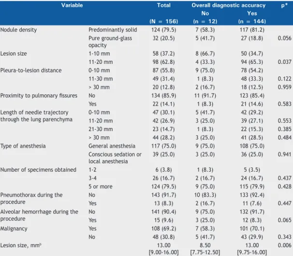
Table 1. Variables associated with the overall diagnostic accuracy of CT-guided percutaneous core needle biopsy of
small pulmonary nodules.a
Variable Total Overall diagnostic accuracy p*
No Yes
(N = 156) (n = 12) (n = 144) Nodule density Predominantly solid 124 (79.5) 7 (58.3) 117 (81.2)
Pure ground-glass opacity
32 (20.5) 5 (41.7) 27 (18.8) 0.056
Lesion size 1-10 mm 58 (37.2) 8 (66.7) 50 (34.7)
11-20 mm 98 (62.8) 4 (33.3) 94 (65.3) 0.037 Pleura-to-lesion distance 0-10 mm 87 (55.8) 9 (75.0) 78 (54.2)
11-30 mm 49 (31.4) 1 (8.3) 48 (33.3) 0.122
> 30 mm 20 (12.8) 2 (16.7) 18 (12.5) 0.959
Proximity to pulmonary fi ssures No 134 (85.9) 11 (91.7) 123 (85.4)
Yes 22 (14.1) 1 (8.3) 21 (14.6) 0.583
Length of needle trajectory through the lung parenchyma
0-10 mm 47 (30.1) 5 (41.7) 42 (29.2)
11-20 mm 42 (26.9) 3 (25.0) 39 (27.1) 0.553
21-30 mm 23 (14.7) 1 (8.3) 22 (15.3) 0.385
> 30 mm 44 (28.2) 3 (25.0) 41 (28.5) 0.484 Type of anesthesia General anesthesia 117 (75.0) 9 (75.0) 108 (75.0)
Conscious sedation or local anesthesia
39 (25.0) 3 (25.0) 36 (25.0) 0.941
Number of specimens obtained 1-2 6 (3.8) 1 (8.3) 5 (3.5)
3-4 26 (16.7) 2 (16.7) 24 (16.7) 0.437
5 or more 124 (79.5) 9 (75.0) 115 (79.9) 0.428 Pneumothorax during the
procedure
No 143 (91.7) 10 (83.3) 133 (92.4)
Yes 13 (8.3) 2 (16.7) 11 (7.6) 0.447
Alveolar hemorrhage during the procedure
No 141 (90.4) 9 (75.0) 132 (91.7)
Yes 15 (9.6) 3 (25.0) 12 (8.3) 0.065
Malignancy Yes 108 (69.2) 7 (58.3) 101 (70.1)
No 48 (30.8) 5 (41.7) 43 (29.9) 0.343
Lesion size, mmb 13.00
[9.00-16.00]
8.50 [7.75-12.50]
13.00 [9.75-16.00]
0.006
CT-guided percutaneous core needle biopsy of pulmonary nodules smaller than 2 cm: technical aspects and factors infl uencing accuracy
(p = 0.037), whereas pleura-to-lesion distance, proximity to pulmonary fi ssures, length of needle trajectory through the lung parenchyma, and number of specimens obtained were not. Table 1 shows all of
the variables analyzed for their effect on diagnostic
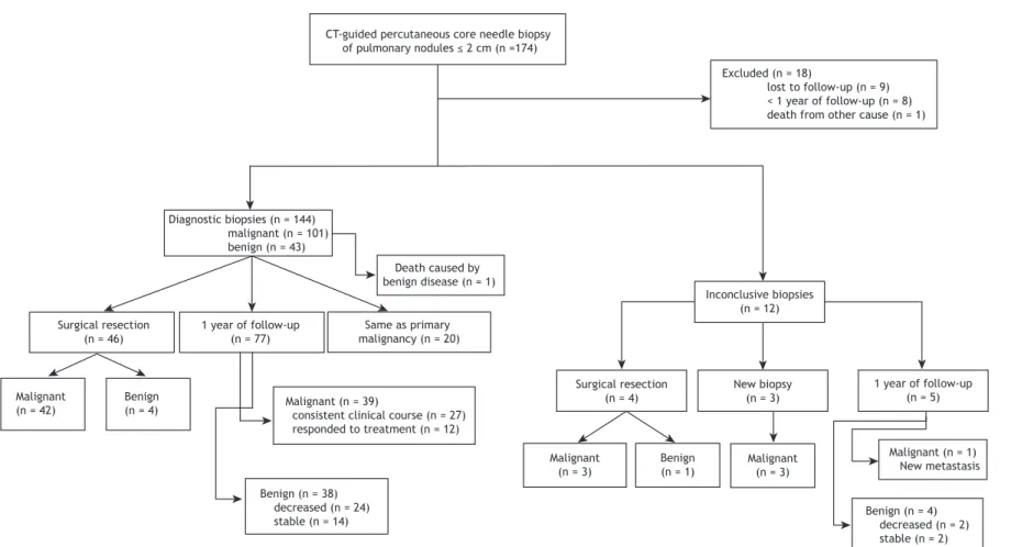
accuracy. Accuracy analysis was possible in 156 CNBs (Figure 1). The histopathological fi ndings of the CNBs are shown in Table 2. The overall diagnostic accuracy of those 156 CNBs was 92.3%. All 144 conclusive CNB results were confi rmed as benign or malignant by follow-up imaging or surgery. In the multivariate analysis, larger lesions were associated with higher overall accuracy (OR = 1.30; 95% CI: 1.08-1.57; p = 0.007), whereas parenchymal hemorrhage during the procedure had lower accuracy rates (OR = 0.13; 95% CI: 0.02-0.75; p = 0.022). Some of the features of the 12 biopsied nodules that were misdiagnosed are shown in Table 3.
Pneumothorax was the most common complication, having occurred in 25 (16.0%) of 156 CNBs. Pneumothorax was mild in 5 cases, moderate in 13, and severe in 7. Of the 25 pneumothoraces, 10 decreased in size or remained stable, whereas 15 moderate-to-severe pneumothoraces required chest tube placement. Of those, 1 occurred before biopsy, i.e., during intubation, being associated with massive mediastinal emphysema secondary to tracheal injury, and 3 were delayed pneumothoraces, which were identifi ed 24 h after the procedure.
In the multivariate analysis, a pleura-to-lesion distance > 3 cm was identifi ed as a risk factor for pneumothorax (OR = 16.94; 95% CI: 2.39-120.26), whereas performing a blood patch after biopsy (n = 88/156; 56.4%) was a protective factor for pneumothorax (OR = 0.18; 95% CI: 0.04-0.86).
With regard to bleeding complications of the procedure, alveolar hemorrhage occurred in 15 CNBs (9.6%), being mild in 10 and moderate in 5. In addition, there was 1 case of mild hemothorax. No additional treatment was required in any of the aforementioned cases. In the multivariate analysis, there were no risk factors associated with bleeding complications.
Uncommon complications included myocardial infarction, in 1 patient, and cerebral air embolism, in 1 patient (Figure 2). The former was due to the anesthetics, and the patient made a full recovery; the
latter was possibly due to positive pressure ventilation
during biopsy, and the patient developed cognitive
dysfunction despite having received specialized
treatment.
DISCUSSION
Recent advances in imaging techniques and screening protocols have allowed the identifi cation of an increasingly large number of small nodules in many parts of the body, such as the prostate, breasts, and lungs.(16) CT-CNB plays a key role in the evaluation of small pulmonary nodules. Several studies using
different guidance techniques and needle sizes have
shown that CNB has a high diagnostic accuracy.(9,15-21) In the present study, 170 patients underwent CT-guided 20-gauge CNB. Eighteen patients were
lost to follow-up or were followed for less than 1
year, which is one of the limitations of the study. The
diagnostic accuracy of CT-CNB in the present study
was 92.3%. This fi nding is similar to those of other
studies reporting the diagnostic accuracy of CNBs
of nodules smaller than 2 cm (i.e., 87-95%).(12,16,17) Unlike most other studies, in which accuracy was measured for malignancy only, our study measured “overall accuracy”, which includes accuracy for benign CNB results. This is especially important in Brazil, where tuberculosis remains a health problem. CNB can provide a defi nitive diagnosis in cases of infection or benign disease, avoiding an unnecessary pulmonary resection and its associated morbidity.
There are several reasons why diagnostic accuracy
was high in the present study. First, we used a coaxial technique, which allows multiple biopsies once the lesion has been targeted. Second, we performed CNB rather than fi ne-needle biopsy. Third, we had an experienced pathologist in our team. Finally, we
used a biopsy protocol that consisted of an additional three biopsies after adequate diagnostic tissue had
been obtained (as determined by the pathologist). The coaxial technique is the most widely recommended
technique for percutaneous lung biopsy(16) because it has been shown to reduce procedure time and complications. In addition, larger specimens can be
collected with core needles that are inserted into the
coaxial introducer, thus facilitating histopathology.(22) Finally, the presence of an experienced pathologist is extremely helpful because it ensures that the biopsy is performed in the target lesion and that the specimen collected is suffi cient for diagnosis, especially when the lesion has a necrotic component.
With regard to the coaxial introducer and core needles, we always use 19- and 20-gauge needles, respectively. For percutaneous biopsies, the smallest gauge needle that can obtain suffi cient tissue for diagnosis should be used. It has been reported that a larger gauge needle translates to a higher risk of complications.(8) We found that, for small pulmonary nodules, 20-gauge core needles can be used not only for histopathology and immunohistochemistry but also for simultaneous molecular and genetic analyses. This fi nding is important because of the ever-growing trend toward personalized cancer therapies.
Pneumothorax and bleeding were the most common complications in our sample, with an incidence of 14.3% and 7.4%, respectively. A chest tube was placed in 8.7% of the patients in our sample. These results are similar to those of previous studies.(12,16) According to the guidelines proposed by Gupta et al.,(8) all except one of the complications observed in the present study were minor. The only major complication was
Andrade JR, Rocha RD, Falsarella PM, Rahal A Jr, Santos RS, Franceschini JP, Fernando HC, Garcia RG
Figure 1. Flow chart of 174 patients undergoing CT-guided percutaneous core needle biopsy of pulmonary nodules of ≤ 2 cm in size, together with the fi nal diagnosis.
lost to follow-up (n = 9) < 1 year of follow-up (n = 8) death from other cause (n = 1)
Diagnostic biopsies (n = 144) malignant (n = 101) benign (n = 43)
Death caused by
benign disease (n = 1)
Surgical resection
(n = 46)
Malignant
(n = 42) Benign(n = 4)
1 year of follow-up (n = 77)
Same as primary
malignancy (n = 20)
Malignant (n = 39)
consistent clinical course (n = 27) responded to treatment (n = 12)
Benign (n = 38) decreased (n = 24) stable (n = 14)
Malignant
(n = 3) Benign(n = 1) Malignant(n = 3)
Benign (n = 4) decreased (n = 2) stable (n = 2)
Malignant (n = 1)
New metastasis New biopsy
(n = 3)
Surgical resection
(n = 4)
Inconclusive biopsies
(n = 12)
1 year of follow-up (n = 5)
311
CT-guided percutaneous core needle biopsy of pulmonary nodules smaller than 2 cm: technical aspects and factors infl uencing accuracy
Table 2. Histopathological fi ndings of 156 biopsied lesions.
Biopsy diagnosis Patients, n (%)
Malignant/premalignant 100 (64.1)
Primary adenocarcinoma 48
Metastatic adenocarcinoma 21
Other metastasis 13
Carcinoid tumor 7
Primary squamous cell carcinoma 6
Atypical adenomatous hyperplasia 5
Benign 44 (28.2)
Fungal infection 12
Organizing pneumonia 10
Caseating granuloma 5
Noncaseating granuloma 5
Other non-specifi c chronic infl ammation 5
Hamartoma 4
Abscess 1
Fibrosis 1
Pulmonary infarction 1
Inconclusive/insuffi cient material 12 (7.7)
Total 156 (100)
Table 3. Features of the nodules that were initially misdiagnosed, together with the fi nal diagnosis.
Case Size, mm
Density Distance from the pleura, mm
Specimen, n Complications during biopsy
Final diagnosis
1 4 subsolid 3 > 7 none Benign - unknown
2 7 solid 2 > 7 mild pneumothorax Benign - unknown
3 7 subsolid 6 6 none Primary
adenocarcinoma
4 8 subsolid 8 7 none Primary
adenocarcinoma
5 8 solid 7 > 7 none Metastasis
6 8 subsolid 2 > 7 none Benign - fi brosis
7 9 solid 4 1 mild bleeding Metastasis
8 10 solid 2 > 7 moderate
pneumothorax
Metastasis
9 12 solid 0 3 hemodynamic
instability*
Metastasis
10 14 solid 31 3 moderate bleeding Benign - unknown
11 14 subsolid 15 > 7 moderate bleeding Benign - unknown
12 14 solid 35 7 none Carcinoid tumor
*The patient presented with myocardial infarction during biopsy, which was immediately interrupted.
Figure 2. In A, pulmonary nodule with slightly jagged edges, measuring 0.6 cm and located in the lateral basal segment
of the right lower lobe. In B, coaxial needle placement guided by CT. In C, alveolar hemorrhage after collection of the fi rst specimen.
A B C
cerebral air embolism (in 1 patient), a relatively rare complication.(23)
Several factors have been found to affect the accuracy
of CT-guided CNB of pulmonary lesions, including lesion size ≤ 1 cm,(9) lesion size ≤ 1.5 cm,(24) lower lobe lesions,(9) ≤ 2 specimens,(9) and malignant lesions. (9) In studies analyzing the aforementioned factors in patients with nodules of ≤ 2.0 cm in size, the only factor that was found to have a positive infl uence on the diagnostic accuracy of CT-CNB was a lesion size ≥ 0.8 cm.(17) In the present study, two factors were found to have infl uenced the overall diagnostic accuracy of CT-CNB: lesion size and parenchymal hemorrhage. To the best of our knowledge, ours is the fi rst study in which parenchymal hemorrhage was associated with a lower diagnostic accuracy.
Some studies have shown that smaller lesions lead to lower biopsy accuracy,(9-12) whereas others have not.(1,25) In the present study, a smaller lesion translated to a lower diagnostic accuracy. Given that biopsies of pulmonary nodules smaller than 1 cm are challenging, our choice of patient position was made so as to minimize target lesion motion and provide greater stability to the coaxial and biopsy needles.
This is partic ularly advantageous for nodules of < 1
cm in size.
Parenchymal hemorrhage was another factor that negatively infl uenced the overall diagnostic accuracy. We found that parenchymal hemorrhage usually occurs after the fi rst sample collection and results in poor visualization on subsequent imaging and biopsy. On CT, alveolar hemorrhage has an attenuation coeffi cient that is very similar to that of a pulmonary nodule, which is therefore obscured by it. An attempt should therefore be made to obtain the best possible specimen at the fi rst collection, with CT confi rmation of the cutting needle crossing the largest diameter of the target lesion.
In conclusion, the overall diagnostic accuracy of CT-guided percutaneous 20-gauge CNB of pulmonary nodules smaller than 2 cm is high. Lesion size and parenchymal hemorrhage are associated with reduced accuracy. Pneumothorax is the most common complication of CT-CNB, being associated with a pleura-to-lesion distance > 3 cm, whereas performing
a blood patch after biopsy appears to be a protective
factor in selected cases.
REFERENCES
1. Henschke CI, McCauley DI, Yankelevitz DF, Naidich DP, McGuinness G, Miettinen OS, et al. Early lung cancer action project: overall design and fi ndings from baseline screening. Lancet. 1999;354(9173):99-105. https://doi.org/10.1016/S0140-6736(99)06093-6
2. Diederich S, Wormanns D, Semik M, Thomas M, Lenzen H, Roos N, et al. Screening for early lung cancer with low-dose spiral CT: prevalence in 817 asymptomatic smokers. Radiology. 2012;222(3):773-81. https://doi.org/10.1148/radiol.2223010490 3. Henschke CI, Naidich DP, Yankelevitz DF, McGuinness G,
McCauley DI, Smith JP, et al. Early lung cancer action project: initial fi nding on repeat screenings. Cancer. 2001;92(1):153-9. https://doi.org/10.1002/1097-0142(20010701)92:1<153::AID-CNCR1303>3.0.CO;2-S
4. El-Sherif A, Gooding WE, Santos R, Pettiford B, Ferson PF, Fernando HC, Urda SJ, et al. Outcomes of sublobar resection versus lobectomy for stage I non-small cell lung cancer: a 13-year analysis. Ann Thorac Surg. 2006;82(2):408-15; discussion 415-6. https://doi.org/10.1016/j.athoracsur.2006.02.029
5. Libby DM, Smith JP, Attorki NK, Pasmantier MW, Yankelevitz D, Henschke CI. Managing the small pulmonary nodule discovered by CT. Chest 2014;125(4):1522-9. https://doi.org/10.1378/ chest.125.4.1522
6. MacMahon H, Austin JH, Gamsu G, Herold CJ, Jett JR, Naidich DP, et al. Guidelines for management of small pulmonary nodules detected on CT scans: a statement from the Fleischner society. Radiology. 2005;237(2):395-400. https://doi.org/10.1148/ radiol.2372041887
7. Naidich DP, Bankier AA, MacMahon H, Schaefer-Prokop CM, Pistolesi M, Goo JM, et al. Recommendations for the management of subsolid pulmonary nodules detected at CT: a statement from the Fleischner society. Radiology. 2013;266(1):304-17. https:// doi.org/10.1148/radiol.12120628
8. Gupta S, Wallace MJ, Cardella JF, Kundu S, Miller DL, Rose SC; et al. Quality improvement guidelines for percutaneous needle biopsy. J Vasc Interv Radiol. 2010;21(7):969-75. https://doi. org/10.1016/j.jvir.2010.01.011
9. Hiraki T, Miruma H, Gobara H, Iguchi T, Fujiwara H, Sakurai J, et al. CT fl uoroscopy-guided biopsy of 1,000 pulmonary lesions performed with 20-gauge coaxial cutting needles: diagnostic yield
and risk factors for diagnostic failure. Chest. 2009;136(6):1612-1617. https://doi.org/10.1378/chest.09-0370
10. Tsukada H, Satou T, Iwashima A, Souma T. Diagnostic accuracy of CT-guided automated needle biopsy of lung nodules. AJR Am J Roentgenol. 2000;175(1):239-43. https://doi.org/10.2214/ ajr.175.1.1750239
11. Takeshita J, Masago K, Kato R, Hata A, Kaji R, Fujita S, et al. CT-guided fi ne-needle aspiration and core needle biopsies of pulmonary lesions: a single-center experience with 750 biopsies in Japan. AJR Am J Roentgenol. 2015;204(1):29-34. https://doi. org/10.2214/AJR.14.13151
12. Laurent F, Latrabe V, Vergier B, Montaudon M, Vernejoux JM, Dubrez J. CT-guided transthoracic needle biopsy of pulmonary nodules smaller than 20 mm: results with an automated 20-gauge coaxial cutting needle. Clin Radiol. 2000;55(4):281-7. https://doi. org/10.1053/crad.1999.0368
13. Choi SH, Chae EJ, Kim JE, Kim EY, Oh SY, Hwang HJ, et al. Percutaneous CT-guided aspiration and core biopsy of pulmonary nodules smaller than 1 cm: analysis of outcomes of 305 procedures from a tertiary referral center. AJR Am J Roentgenol. 2013;201(5):964-970. https://doi.org/10.2214/AJR.12.10156
14. Yamauchi Y, Izumi Y, Nakatsuka S, Inoue M, Hayashi Y, Kohno M, et al. Diagnostic performance of percutaneous core-needle lung biopsy under CT scan fl uoroscopic guidance for pulmonary lesions measuring ≤10 mm. Chest. 2011;140(6):1699-1670. https://doi.org/10.1378/chest.11-1821
15. Jae LI, June IH, Miyeon Y, Kwanseop L, Yul L, Hoon BS. Percutaneous core needle biopsy for small (≤ 10 mm) lung nodules: accurate diagnosis and complication rates. Diagn Interv Radiol. 2012;18(6):527-30.
16. Li Y, Du Y, Yang HF, Yu JH, Xu XX. CT-guided percutaneous core needle biopsy for small (≤20 mm) pulmonary lesions. Clin Radiol. 2013;68(1):e43-8. https://doi.org/10.1016/j.crad.2012.09.008 17. Yoshimura N, Takeda K, Tada H, Kudoh S, Hirata K, Takifuji N,
et al. The factors determining diagnostic accuracy in CT-guided percutaneous needle biopsy of small pulmonary nodules [Article in Japanese]. Nihon Kokyuki Gakkai Zasshi. 2002;40(2):101-5. 18. Hayashi N, Sakai T, Kitagawa M, Kimoto T, Inagaki R, Ishii
CT-guided percutaneous core needle biopsy of pulmonary nodules smaller than 2 cm: technical aspects and factors infl uencing accuracy
frozen-section pathologic diagnosis. AJR Am J Roentgnol. 1998;170(2):329-31. https://doi.org/10.2214/ajr.170.2.9456939
19. Choo JY, Park CM, Lee NK, Lee SM, Lee HJ, Goo JM. Percutaneous transthoracic needle biopsy of small (≤ 1 cm) lung nodules under C-arm cone-beam CT virtual navigation guidance. Eur Radiol. 2013;23(3):712-9. https://doi.org/10.1007/s00330-012-2644-6
20. Yeow KM, Tsay PK, Cheung YC, Lui KW, Pan KT, Chou AS. Factors affecting diagnostic accuracy of CT-guided coaxial cutting needle lung biopsy: retrospective analysis of 631 procedures. J Vasc Interv Radiol. 2013;14(5):581-8. https://doi.org/10.1097/01. RVI.0000071087.76348.C7
21. Lee SM, Park CM, Lee KH, Bahn YE, Kim JI, Goo JM. C-arm cone-beam CT-guided percutaneous transthoracic needle biopsy of lung nodules: clinical experience in 1108 patients. Radiology. 2014;271(1):291-300. https://doi.org/10.1148/radiol.13131265 22. Laurent F, Latrabe V, Vergier B, Michel P. Percutaneous
CT-guided biopsy of the lung: comparison between aspiration and automated cutting needles using a coaxial technique. Cardiovasc Intervent Radiol. 2000;23(4):266-72. https://doi.org/10.1007/ s002700010067
23. Rocha RD, Azevedo AA, Falsarella PM, Rahal A Jr, Garcia RG. Cerebral air embolism during CT-guided lung biopsy. Thorax. 2015;70(11):1099-100. https://doi.org/10.1136/ thoraxjnl-2015-207205
24. Kothary N, Lock L, Sze DY, Hofmann LV. Computed tomography-guided percutaneous needle biopsy of pulmonary nodules: impact of nodule size on diagnostic accuracy. Clin Lung Cancer. 2009;10(5):360-3. https://doi.org/10.3816/CLC.2009.n.049
25. Hwang HS, Chung MJ, Lee JW, Shin SW, Lee KS. C-arm cone-beam CT-guided percutaneous transthoracic lung biopsy: usefulness in evaluation of small pulmonar y nodules. AJR Am J Roentgenol. 2010;195(6):W400-7. https://doi.org/10.2214/ AJR.09.3963