Treatment of dental and skeletal bimaxillary
protrusion in patient with Angle Class I
malocclusion
» The author reports no commercial, proprietary or financial interest in the products or companies described in this article.
» The patient displayed in this article previously approved the use of her facial and intraoral photographs.
Contact address: Claudio José Ramos
SGAS 607, Centro Clínico Metrópolis, Sala 217 – Brasília /DF – CEP: 70.200-670 E-mail: [email protected]
INTRODUCTION
Caucasian female patient, 33 years and 5 months old, with the chief complaint: “I want to correct my teeth, because they are sticking out, and also improve my esthetics”. The clinical exam showed the need of closing preexisting spaces, due to dental extractions, and reducing bimaxillary and dental protrusion. An-amnesis showed good general health.
DIAGNOSIS
The patient’s facial aspect, in frontal view, did not present visible asymmetry, but absence of passive lip sealing. From the lateral view, the patient presented a convex profile, normal nasolabial angle, lack of lip sealing at rest, and increased lower anterior facial height (Fig 1). The patient presented a Class I mo-lar relationship and 5 mm overjet; slight upper and Claudio José Ramos1
1Former Professor of the Specialization Course in Orthodontics, APCD-DF. Former
President of ABOR-DF. Specialist in Orthodontics, APCD-Bauru/São Paulo. Political Scientist, Graduated in the Federal District University Center (UDF). BBO Diplomate.
* Clinical case approved by the Brazilian Board of Orthodontics.
How to cite this article: Ramos CJ. Treatment of dental and skeletal bimaxil-lary protrusion in patient with Angle Class I malocclusion. Dental Press J Or-thod. 2013 Nov-Dec;18(6):130-7.
In the orthodontic clinic, skeletal and dental bimaxillary protrusion is presented frequently as one of the factors leading patients to seek orthodontic treatment, mainly due to the esthetic involvement it has. The patient of this article illustrates this situation, being deeply uncomfortable with her esthetic appearance, due to the excessive upper incisors exposure and problems with lip sealing. This case was presented to the Brazilian Board of Orthodontics and Facial Orthopedics (BBO), as part of the requisites to become a BBO Diplomate.
Keywords:Class I malocclusion. Corrective orthodontics. Esthetics.
Na clínica ortodôntica, a biprotrusão esquelética e dentária, rotineiramente, aparece como um dos fatores que levam os pacientes a buscarem o tratamento ortodôntico, principalmente pelo envolvimento estético que provoca. A paciente apresentada neste artigo ilustra essa situação, pois estava bastante insatisfeita com sua estética, devido à exposição exces-siva dos incisivos superiores e diiculdade em selar os lábios. Esse caso foi apresentado à Diretoria do Board Brasileiro de Ortodontia e Ortopedia Facial (BBO) como parte dos requisitos para a obtenção do título de Diplomado pelo BBO.
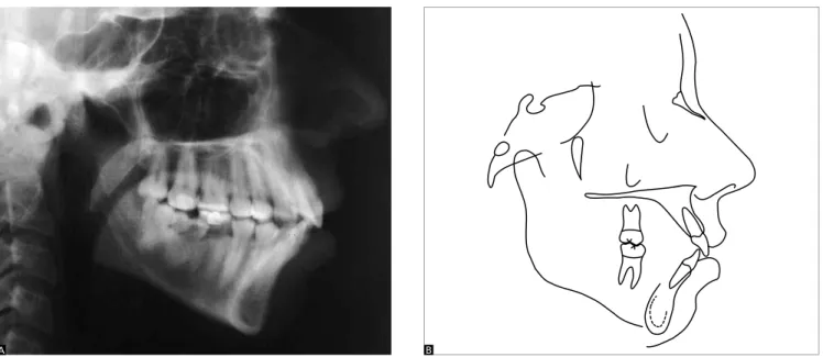
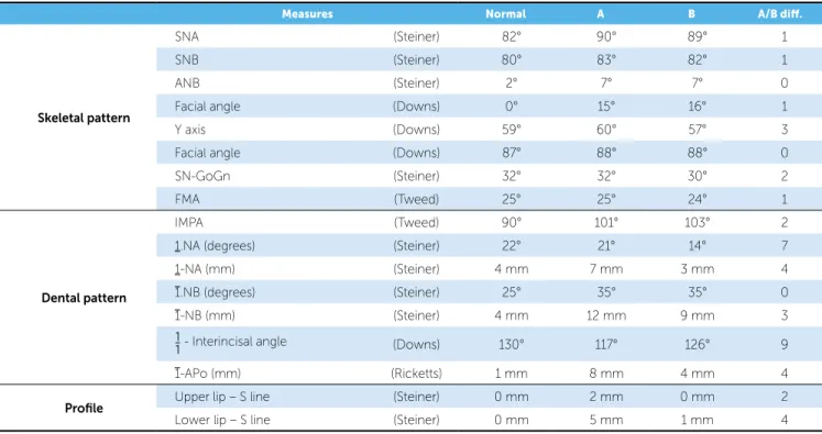
In skeletal terms, according to the lateral cephalomet-ric radiograph and respective tracing (Fig 5), it was ob-served that the patient presented unbalanced skeletal bases, characterized by angles SNA = 90°, SNB = 83° and ANB = 7°, with a dolichofacial pattern, and pre-senting protruded upper incisors, with slight lingual tipping, and protruded lower incisors with buccal tip-ping.1 The functional analysis of mandibular move-ments revealed absence of anterior guides.
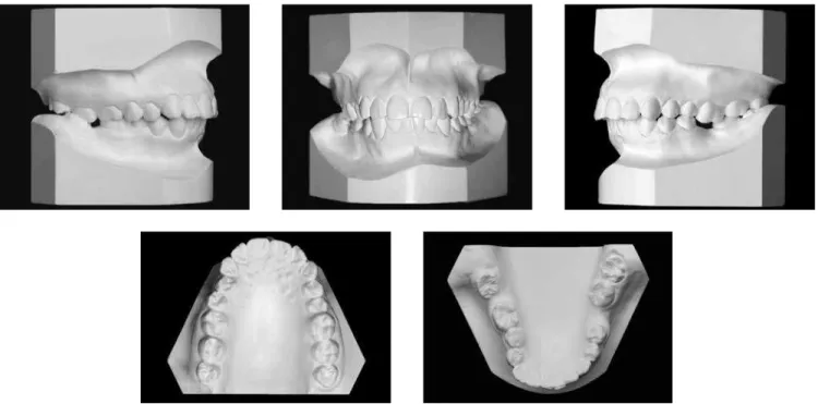
lower crowding; moderate Curve of Spee; absence of teeth #36 and #37; posterior cross bite on the right side, with buccal cross bite between teeth #15 and #45; lower midline deviated 1 mm to the left; and up-per and lower incisors with incisal wear (Fig 2). The periapical and panoramic radiographs demonstrated root shortening of the upper right central incisor, ab-sence of teeth #36 and #47, lower molar mesialization and lack of parallelism between roots (Figs 3 and 4).
Figure 2 - Initial casts.
Figure 3 - Initial periapical radiographs. Figure 4 - Initial panoramic radiographs.
TREATMENT PLAN
Due to compromised facial esthetics and dental and skeletal bimaxillary protrusion, the treatment plan had the following objectives: Maintain canine occlu-sion; align and level the teeth; eliminate posterior cross bites; reduce overbite and overjet; eliminate crowd-ing on both arches; level the Curve of Spee, close the spaces due to extraction of teeth #36 and #47, and also achieve lower molar root parallelism.
For this purpose, extractions of the irst upper and right lower premolars were necessary, besides the re-traction of upper and lower anterior teeth, to reduce bimaxillary protrusion and correct the lower midline. All this very carefully, in order to avoid root shorten-ing of tooth #11.
Figure 5 - Initial lateral cephalometric radiograph (A) and cephalometric tracing (B).
passive lip sealing would be expected and decrease of the lower anterior facial height, in order to reach smile harmony.
TREATMENT PROGRESS
The treatment was performed with the Straight wire technique, using metallic orthodontic brack-ets in both arches, according to Roth’s prescription (0.022 x 0.028-in), with a removable transpalatal bar (stainless steel archwire, 0.032-in) as upper arch an-chorage, and a lip bumper as lower arch anchorage. Leveling and alignment were carried with a sequence of NiTi round archwires, NiTi rectangular archwires 0.017 x 0.025-in and stainless steel rectangular arch-wires 0.019 x 0.025, with loops for retraction of ante-rior teeth and 5/16-in elastics to assist in the inishing. Bands were placed on the irst and second upper lars, as well as on the irst, second and third lower mo-lars, with a transpalatal bar on the irst upper molars and lip bumper on lower molars.
The placement of the ixed orthodontic appliance was completed with the direct bonding of brackets on the remaining upper and lower teeth, except on teeth #14, #24 and #44 — which would be extracted, with
the objective of eliminating crowding and allow the cor-rection of the dental bimaxillary protrusion, by means of retracting the anterior teeth.1,3,4 Alignment and level-ing were carried with 0.012, 0.014, 0.016 and 0.018-in NiTi archwires; and 0.017 x 0.025-in NiTi rectangular, and stainless steel 0.019 x 0.025-in archwires. Then, the retraction of upper and lower anterior teeth, and the correction of the lower midline were performed. The extraction of tooth #18 was suggested because it would be without its antagonist due to the mesial move-ment of tooth #48. For intercuspation, 5/16-in inter-maxillary elastics were used.
A stainless steel 0.028-in fixed lower retainer was bonded to teeth #33 to #45, and to teeth #35 to #37. On the upper arch, a wraparound removable plate was used.
OBTAINED RESULTS
The evaluation of the inal records shows that there was 1° reduction for SNA and SNB angles, a slight decrease in the vertical direction (Table 1). The canine guidance was kept, there was overjet reduction and overbite and Curve of Spee correction. All teeth were aligned and lev-eled, and the posterior cross bites eliminated; the crowding
Figure 6 - Final facial and intraoral photographs.
on both arches was corrected; Curve of Spee was leveled. The spaces for teeth #36 and #47, and also the extraction spaces for teeth #14, #24 and #44 were properly closed; up-per and lower anterior retraction was satisfactory.
The lower midline correction and anterior guides were obtained. However, lip sealing was not completely passive, due to the patient’s vertical growth pattern. The nasolabial angle was preserved, despite the decrease of incisor protrusion, which promoted smile harmony (Figs 6 to 10). The treatment lasted 35 months.
FINAL CONSIDERATIONS
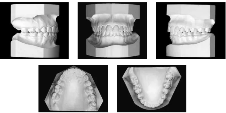
Figure 7 - Final casts.
Figure 10 - Final lateral cephalometric radiograph (A) and cephalometric tracing (B).
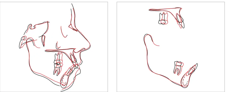
Figure 11 - Total superimposition of initial (black) and inal (red) cephalomet-ric tracings, registered on SN line.
Figure 12 - Partial superimposition of the maxilla (A) and the mandible (B), of ini-tial (black) and inal (red) cephalometric tracings, evidencing tooth movements.
In facial terms, passive lip sealing, initially ex-pected, was not completely obtained, mainly due to increased anterior facial height. Nevertheless, greater smile harmony was obtained, which fully met the pa-tient’s expectations, who always collaborated regard-ing oral hygiene and clinical recommendations.
The level of root resorptions was acceptable, except for the upper right central incisor, in which the root shortening increased signiicantly,7,9 but without any mobility observed in regular evaluation of each treat-ment phase (Fig 8). The patient was pleased with the treatment result, since it improved her facial esthetics
1. Almeida FM, Siqueira VCV. O efeito da exodontia dos primeiros pré-molares sobre a AFAI. Rev Dental Press Ortod Ortop Facial. 2004;9(6):48-62. 2. Bills DA, Handelman CS, BeGole EA. Bimaxillary dentoalveolar protrusion:
traits and orthodontic correction. Angle Orthod. 2005;75(3):333-9. 3. Bravo LA. Soft tissue facial proile changes after orthodontic treatment with
four premolars extracted. Angle Orthod. 1994;64(1):31-42.
4. Erdinc AE, Nanda RS, Dandajena TC. Proile changes of patients treated with and without premolar extractions. Am J Orthod Dentofacial Orthop. 2007;132(3):324-31.
5. Kim TK, Kim JT, Mah J, Yang WS, Baek SH. First or second premolar extraction efects on facial vertical dimension. Angle Orthod. 2005;75(2):177-82.
6. Maltagliati LA, Montes LAP. Análise dos fatores que motivam os pacientes adultos a buscarem o tratamento ortodôntico. Rev Dental Press Ortod Ortop Facial. 2007;12(6):54-60.
7. Mohandesan H, Ravanmehr H, Valaei N. A radiographic analysis of external apical root resorption of maxillary incisors during active orthodontic treatment. Eur J Orthod. 2007;29(2):134-9.
8. Oliveira GF, Almeida MR, Almeida RR, Ramos AL. Alterações dentoesqueléticas e do peril facial em pacientes tratados ortodonticamente com extração de quatro primeiros pré-molares. Rev Dental Press Ortod Ortop Facial. 2008;13(2):105-14. 9. Simplício H. Avaliação da reabsorção radicular apical em incisivos
submetidos à retração [tese]. Araraquara (SP): Universidade Estadual Paulista; 2002.
REFERENCES
substantially,8 with the reduction of dental and skeletal bimaxillary protrusion.2 Ater removing the appliances and placing new retainers, the patient was referred to dental whitening and composite restoration of the in-cisal borders of the upper anterior teeth. Thus, ater the recommended procedures, an even more pleasant smile was obtained, which contributed signiicantly for the patient’s complete satisfaction.6
Measures Normal A B A/B dif.
Skeletal pattern
SNA (Steiner) 82° 90° 89° 1
SNB (Steiner) 80° 83° 82° 1
ANB (Steiner) 2° 7° 7° 0
Facial angle (Downs) 0° 15° 16° 1
Y axis (Downs) 59° 60° 57° 3
Facial angle (Downs) 87° 88° 88° 0
SN-GoGn (Steiner) 32° 32° 30° 2
FMA (Tweed) 25° 25° 24° 1
Dental pattern
IMPA (Tweed) 90° 101° 103° 2
1.NA (degrees) (Steiner) 22° 21° 14° 7
1-NA (mm) (Steiner) 4 mm 7 mm 3 mm 4
1.NB (degrees) (Steiner) 25° 35° 35° 0
1-NB (mm) (Steiner) 4 mm 12 mm 9 mm 3
1
1- Interincisal angle (Downs) 130° 117° 126° 9
1-APo (mm) (Ricketts) 1 mm 8 mm 4 mm 4
Profile Upper lip – S line (Steiner) 0 mm 2 mm 0 mm 2
Lower lip – S line (Steiner) 0 mm 5 mm 1 mm 4