324
https://doi.org/10.1590/0004-282X20170037
IMAGES IN NEUROLOGY
The many faces of demyelinating
diseases: acute disseminated
encephalomyelitis and Guillain-Barré
syndrome in the same patient
As várias faces das doenças desmielinizantes: encefalomielite aguda disseminada e
síndrome de Guillain-Barré no mesmo paciente
Régis Augusto Reis Trindade
1, Amália Izaura Nair Medeiros Klaes
1, Juliana Ávila Duarte
2Acute disseminated encephalomyelitis and Guillain-Barré
syndrome represent distinct demyelinating diseases that
share an autoimmune pathogenesis. A history of viral
infec-tion or vaccinainfec-tion are essential for the diagnosis
1,2,3.
A 26-month-old boy presented with fever 10 days after
vaccination (inactivated polio vaccine and tetravalent),
progressive drowsiness, lower limb strength/sensory loss
and urinary retention. The cerebrospinal fluid showed mild
pleocytosis and an elevated total protein concentration;
it was negative for infections. The initial MRI study was
compatible with acute disseminated encephalomyelitis,
with no spine abnormalities (Figure 1).
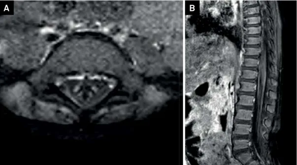
Ten days later, after therapy with corticosteroids,
he experienced acute paraplegia. Follow-up brain and
spine MRI scans demonstrated partial regression of brain
lesions and nerve root thickening with intense
enhance-ment extending along the cauda equina, compatible with
Guillain-Barré syndrome. (Figure 2).
1Hospital de Clínicas de Porto Alegre (HCPA), Radiologia e Diagnóstico por Imagem, Porto Alegre RS, Brasil; 2Hospital de Clínicas de Porto Alegre (HCPA), Ressonância Magnética, Porto Alegre RS, Brasil.
Correspondence:Régis Augusto Reis Trindade; R. Ramiro Barcelos, 2350; 90035-903 Porto Alegre RS, Brasil; E-mail: [email protected] Conflict of interest:There is no conflict of interest to declare.
Received 31 August 2016; Received in final form 29 December 2016; Accepted 03 February 2017.
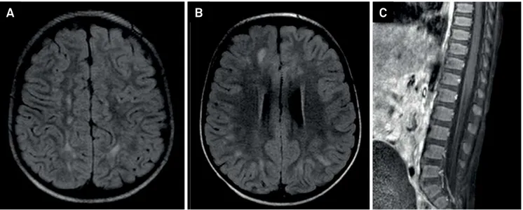
Figure 1.
Brain MRI revealed white matter hyperintensities lesions in T2/fluid- attenuated inversion recovery (FLAIR) (A, B) in the
cerebral hemispheres and cerebellum, without restriction to water molecules diffusivity or gadolinium enhancement, suggestive
of acute disseminated encephalomyelitis . Sagittal T1 SPIR post-gadolinium (C), without cauda equina root enhancement.
325
Trindade RAR et al. The many faces of demyelinating diseases
References
1. Deshmukh IS, Bang AB, Jain MA, Vilhekar KY. Concurrent acute disseminated encephalomyelitis and Guillain-Barré syndrome in a child. J Pediatr Neurosci 2015;10(1):61-3. https://doi.org/10.4103/1817-1745.154357
2. Okumura A, Ushida H, Maruyama K, Itomi K, Ishiguro Y, Takahashi M et al. Guillain-Barré syndrome associated with
central nervous system lesions. Arch Dis Child. 2002;86(4):304-6. https://doi.org/10.1136/adc.86.4.304
3. Mohamed RR, Jan MM. Co-morbid Guillain-Barré syndrome and acute disseminated encephalomyelitis. Neurosciences (Riyadh). 2013;18(2):166-8.