Arq Neuropsiquiatr 2004;62(2-B):519-522
Programa de Cirurgia de Epilepsia, Serviço de Neurologia, Hospital de Clínicas da Universidade Federal do Paraná, Curitíba PR, Brasil (UFPR):1Médico Neurologista; 2Neurofisiologista Clínico;3M édico Residente em Neurologia;4Professor Adjunto de Neurologia;5Professor Adjunto de Neurocirurgia;6Psicóloga;7Professor Titular de Neurologia.
Received 6 October 2003, received in final form 9 January 2004. Accepted 9 February 2004.
Dr. Luciano De Paola - Programa de Cirurgia de Epilepsia/Serviço de Neurologia - Hospital de Clínicas UFPR - Rua General Carneiro 181/3º andar - 80060-900 Curitiba PR - Brasil.
CEREBELLAR HEM ORRHAGE AS A COM PLICATION OF TEM
PO-RAL LOBECTOM Y FOR REFRACTORY M EDIAL TEM POPO-RAL
EPILEPSY
Report of three cases
Luciano de Paola
1,2, André R.Troiano
3, Francisco M .B. Germiniani
1, Patrícia Coral
1,2, M arcus V. Della
Coletta
1, Carlos E.S. Silvado
1,2,4, M arlus M oro
5, João Cândido de Araújo
5, M aria Joana M äder
6, Lineu C.
Werneck
1,7ABSTRACT - Cerebellar hemorrhage is listed among the potential complications following neurosurgical procedures. In this scenario it is usually reported as a rare condition. However, it seems that epilepsy surgery patients are somewhat more prone to this kind of complication, compared to other surgical groups. Head positioning, excessive cerebral spinal fluid draining and the excision of non-expanding encephalic tissue (or combinations among the three) are likely to be cause underlying remote cerebellar hemor-rhage. Out of the 118 ATL/AH performed at our institution, between 1996 and 2002, we identified 3 (2.5%) patients presenting with cerebellar hemorrhage. We report on such cases and review the literature on the topic.
KEY WORDS: cerebellar hemorrhage, epilepsy surgery, neurosurgical complications.
Hemorragia cerebelar como complicação de lobect omia t emporal para epilepsia do lobo t emporal medial: rela-t o de rela-t rês casos
RESUMO - A hemorragia cerebelar faz parte das potenciais complicações dos procedimentos neurocirúrgicos. De forma geral, é considerada uma condição rara. Entretanto, há aparente propensão dos pacientes submetidos ao tratamento cirúrgico de epilep-sia em apresentar este tipo de complicação, quando comparados com outros grupos cirúrgicos. O posicionamento da cabeça, exces-siva drenagem de líquido cefalorraquidiano e a excisão de tecido cerebral não expansível (ou talvez combinações entre os três) con-stituem as potenciais causas da hemorragia cerebelar remota. Entre os 118 pacientes em nossa série de LTA ⁄ AH, identificamos 3(2.5%) casos de hemorragia cerebelar. Relatamos os três casos desta natureza, com revisão da literatura pertinente a esta com-plicação.
PALAVRAS-CHAVE: hemorragia cerebelar, cirurgia de epilepsia, complicações neurocirúrgicas.
Anterior temporal lobectomy (ATL) and amygdalohip-pocampectomy (AH) are effective treatment alternatives in patients w ith temporal lobe epilepsy refractory to medical treatment. Neuropsychological disturbances (language and memory) are the most common post-operatory derangements. Nevertheless, surgical complications are rarely expected, both locally and at remote sites. Recent papers have shed some light on the relative high frequency of cerebellar hemorrhage in patients submitted to anterior temporal lobectomy, w hen compared to other surgical groups. It seems that ATL/AH pa-tients are particularly prone to this kind of complication. At our institution ATL ⁄ AH are always performed using the same surgical technique (that is, a trans-temporal approach). Out
of the 118 ATL ⁄ AH performed at our institution w e w ere able to identify 3(2.5%) patients presenting w ith cerebellar hem-orrhage. Their cases are reported.
CASES
showed left temporal interictal spikes and a brain magnetic resonance image (M RI) disclosed left hippocampal atrophy, consistent w ith the diagnosis of mesiotemporal sclerosis (M TS). During videoeletroen-cephalographic (VEEG) monitoring, 3 complex partial and 1 secondari-ly generalized seizures w ere recorded from the left mesiotemporal lobe.
He was then admitted to undergo left ATL. There w ere neither metabolic nor coagulation disorders, as verified by normal platelets count and PT / aPTT values. While supine w ith his head turned to the right, the patient underw ent a craniotomy for a left 3 cm ATL and 3.5 cm AH. At the end of the surgery, a subgaleal suctor drain was placed. Pathology was confirmatory of hippocampal sclerosis.
In the immediate post-operatory (PO) period, he was observed in the Intensive Care Unit (ICU) w ith an initial Glasgow Coma Scale (GCS) of 10. On the second day at the ICU he was still judged as “ unusually drow sy and dysarthric” . On the 3rdPO day a brain
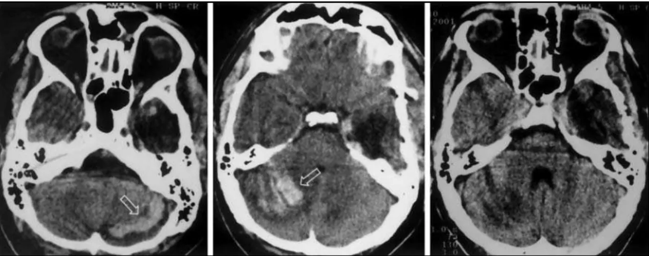
com-puted tomography (CT) scan disclosed multiple foci of cerebellar hemorrhage, predominantly on the right cerebellar hemisphere, but no surgical drainage was necessary (Fig 1). A cerebral angiographic study was normal.
In the 9thPO day the patient developed a liquoric fistula and fever.
A cerebrospinal fluid (CSF) study showed 21 red cells/mm3, 7680
leu-cocytes/mm3, 80% neutrophils, glucose 1mg/dL and proteins 404mg/dL.
He was put on cefepime for 14 days, after w hich CSF show ed 58 red cells/mm3, 19 leucocytes/mm3, 87% lymphocytes, glucose 39 mg/dL
and proteins 124 mg/dL. He was discharged on the 24thPO day,
with-out significant coordination impairment and later returned in the with- out-patient clinic for reevaluation of his seizures. He remained seizure-free on antiepileptic drugs (AEDs) for approximately tw elve months, after w hich he experienced seizure recurrence. Drug adjustments were unsuccessful.A follow-up CT scan performed on the 28thPO day
show ed resolution of the hemorrhage on both cerebellar hemi-spheres.
Patient 2. A 37 year-old female patient with a seizure disorder starting at the age of 13 y/o, consisting of complex partial seizures w ith frequent secondary generalization. Her best AED combination was sodium valproate and carbamazepine, when she presented with complex partial seizures 2 to 3 times per w eek and infrequent sec-ondarily generalized seizures. Other combinations or a regime includ-ing higher doses invariably lead to intolerable side effects. Past and familial history were unremarkable. EEG disclosed right temporal
inter-520 Arq Neuropsiquiatr 2004;62(2-B)
ictal epileptiform discharges. Brain M RI show ed right hippocampal atrophy consistent with MTS. She was submitted to VEEG and two of her typical complex partial seizures were recorded from the right tem-poral lobe.
She then underwent right ATL. Pre-operative metabolic and coag-ulation studies w ere normal. While supine w ith her head turned to the left, a right craniotomy was performed for right ATL and AH, using the vein of Labbé as the posterior margin for the resection, that is, approximately a 6cm resection from the temporal tip . At the end of the surgery, a subgaleal suctor drain was placed.
After surgery, consciousness recovery was normal and she pre-sented w ith no deficits. In the 2ndPO day, the patient presented w ith
headache, nausea and vomiting. Fundoscopy was normal, as well as, motor and coordination tests. Brain CT scan show ed laminar right hemisphere cerebellar hemorrhage (Fig 2). A neurosurgery consult was requested and the option was for clinical treatment. The patient received analgesic and antiemetic treatment and was discharged four days later. At discharge she was asymptomatic with a normal physi-cal and neurologiphysi-cal exam. A control CT scan performed on the 4thPO
day showed partial resolution of the hemorrhage.
Patient 3. This is a 38 year-old male patient, presenting with a seizure disorder starting at the age of 7 y/o, characterized by an aura (“ light-headedness” sensation), shortly follow ed by a complex par-tial seizure w ith secondarily generalization. He was tried on pheny-toin, valproic acid, lamotrigine, clobazam and clonazepam, w ith unsatisfactory seizure control. A combination of carbamazepine and phenobarbital led to a better control of the generalized seizures, where-as complex partial seizures still occurred monthly, usually in clusters of up to four seizures a day. Physical and neurological examinations w ere normal. An EEG show ed left temporal interictal spikes and a brain MRI disclosed left hippocampal atrophy, consistent with the diag-nosis of MTS. During VEEG monitoring, 6 complex partial seizures were recorded from the left mesiotemporal lobe.
He was then admitted to undergo left ATL. There w ere neither metabolic nor coagulation disorders. While supine w ith his head turned to the right, the patient underw ent a craniotomy for a left 3 cm ATL and 3.5 cm AH. A subgaleal suctor drain was placed.
At the ICU, in the immediate PO, he presented w ith a GCS of 8, aphasic and had a generalized tonic-clinic seizure. Diazepam 5 mg was administered intravenously, as well as, a bolus of manitol, with improvement of consciousness. A skull CT scan disclosed laminar
cerebellar hemorrhage, more prominent on the left cerebellar hemi-sphere (Fig 3). Once again, no surgical drainage was necessary. On the 7thPO day he was discharged, w ith no further cerebellar signs.
He later returned in the out-patient clinic for reevaluation of his seizures and he has remained seizure-free on AEDs for seven months. A follow-up CT scan was obtained on the 30th PO day, showing good resolution of the cerebellar hemorrhage.
DISCUSSION
Arq Neuropsiquiatr 2004;62(2-B) 521
Although infrequent, cerebellar hemorrhage may be a potential complication follow ing neurosurgical interventions. Nevertheless, its development at a distant site from the oper-ative incision is definitively a rare situation to which attention has been draw n only in the past few years. In a retrospective survey of 4992 intracranial procedures, Kalfas and Little1found
40 patients (0.8%) to present hemorrhages, 33 of them at the operative site (intracerebral, epidural, subdural or intrasellar) and 7 at a distant location. The most common etiology that led to surgical treatment was brain tumor in 56%, with menin-gioma as the leading pathological type.
A group analysis of 37 cases of remote intracerebral hem-orrhage conducted by Brisman et al.2(5 patients from their
ow n series and 32 review ed from the literature) included supratentorial hemorrhages in infratentorial craniotomies and the reverse. Seventy-eight percent of patients had symptoms suggestive of acute intracranial hypertension in the first few hours after surgery. In the setting of infratentorial hemor-rhage, 81% of the patients underw ent access through the deep sylvian fissure and paraclinoid regions.The cerebellar ver-mis was the focus in 67%. Such derangements were not relat-ed to hypertension, coagulopathies or the volume of cere-brospinal fluid drainage. Fourteen percent of patients from this heterogenic sample w ere disabled, and 32% died.
The specific concern of cerebellar hemorrhage after epilep-sy surgery is illustrated by the reports of Toczek et al. and Yacubian et al.The former presented four patients treated with ATL and/or AH w ho presented uni or bilateral cerebellar hemorrhage 1 to 4 days after surgery3. Blood pressure,
plate-lets count and coagulation w ere normal in all patients except for one, promptly treated with anti-hypertensive drugs.Varying amounts of cerebrospinal fluid drained in the PO period (215 to 525mL) was reported as the only potentially implicated fac-tor. One patient had a ventricle drain placed to compensate hydrocephalus, the others needed nothing but conservative measures. All of them had normal neurological examinations at one month follow -up. A series of three patients submitted to ATL/AH found similar results both on patient profile and clin-ical outcome. One patient did not have his lateral ventricles opened or a suctor drain in the post-operative period4.
Doubt about the timing of cerebellar hemorrhage, whether intra or post-operatory, was fed by discordant data on patients who did not recover consciousness after surgery and those who, after awaking from anesthesia, re-entered coma state or devel-oped cerebellar signs. This issue was addressed by Honegger et al. w ith data from a neurosurgical institution in w hich all patients are routinely CT scanned in the first hour after sur-gery5. Of 1650 patients w ho undergone supratentorial
cran-iotomy over a three-year period, 10 had cerebellar hemorrhage. Seven of these had been submitted to ATL/AH. In addition, out of the original ten, seven patients had an initial normal CT scan, an indicator that posterior fossa bleeding appeared over the next few hours follow ing surgery (mean time for diagnosis: 7
Fig 3. Patient 3: bilateral cerebellar hemorrhage, more prominent on the left hemisphere.
hours and 35 minutes).
Systemic hypertension, the top-ranking etiologic factor of spontaneous cerebellar hemorrhage, is not related to this entity. Apparently, the same applies to coagulation disorders and anticoagulant treatment. Sodium valproate was once bla-med as a cause to peri-operatory excessive bleeding, but even this remote cause has been recently questioned in the litera-ture6.
Positioning of the patient during surgery might play a sec-ondary role in increasing venous pressure over the posterior fossa. Seoane and Rhoton7, in an elegant microsurgical
anatom-ical study in adult cadaveric specimens, show ed unilateral jugular compression, to the point of occlusion, by transverse process of C1 when the head is turned contralateraly.That per-haps w ould be sufficient explanation if remote cerebellar hemorrhage was secondary to virtually any surgical access based on head tilting, w hich certainly does not hold true on neurosurgical routine. Nevertheless, w e still lack a better hypothesis, since evidence show s that most supratentorial approaches other than epilepsy surgery do not present w ith cerebellar bleeding. Plus, according to Kalfas and Little, the recumbent position does not seem safer than the sitting posi-tion in preventing posterior fossa bleeding1.
The most accepted theory to date underlying remote cere-bellar hemorrhage is an association between liquor overdrain-ing and the excision of non-expandoverdrain-ing encephalic tissue. ATL/AH is commonly carried out w ith opening of lateral ven-tricles and subsequent CSF flow. Intracranial drains and suc-tors are part of the post-operative protocol in various institu-tions. M oreover, removal of a brain tumor w ould cause supra-tentorial pressure to return to near normal values, but lobec-tomy is an adjunct factor to create a gradient betw een supra and infratentorial compartments. Such pressure gradient may act as a suction mechanism over the cappilary veins of cere-bellum, w hich are then traumatized, leading to intraparen-quimatous bleeding4,5.The association of fluid overdraining and
removal of non-tumorous tissue is specially prone to happen in epilepsy surgery, and this might be the reason why such com-plication is infrequently seen involving other neurosurgical approaches.
In conclusion, cerebellar hemorrhage in post-operative supratentorial craniotomy for epilepsy surgery constitutes a rare complication and its physiopathology is yet to be entire-ly understood. Some patients may not even present with clear symptomatology, and hyperdense signals on posterior fossa may be an occasional finding in follow-up CT studies.Treatment of such complication is similar to that of nontraumatic cerebel-lar hematomas and must be individualized. Most cases are asso-ciated with an excellent outcome.
REFERENCES
1. Kalfas IH, Little JR. Postoperative hemorrhage: a survey of 4992 intracra-nial procedures. Neurosurgery 1988;23:343-347.
522 Arq Neuropsiquiatr 2004;62(2-B)
2. Brisman MH, Bederson JB, Sen CN, Germano IM, Moore F, Post KD. Intracerebral hemorrhage occurring remote from the craniotomy site. Neurosurgery 1996;39:1114-1121.
3. Toczek MT, Morrell MJ, Silverberg GA, Lowe GM. Cerebellar hemor-rhage complicating temporal lobectomy: report of four cases. J Neurosurg 1996;85:718-722.
4. Yacubian EM, Andrade MM, Jorge CL, Valério RM. Cerebellar hemor-rhage after supratentorial surgery for treatment of epilepsy: report of three cases. Neurosurgery 1999;45:159-161.
5. Honegger J, Zentner J, Spreer J, Carmona H, Schulze-Bonhage A. Cerebellar hemorrhage arising postoperatively as a complication of supratentorial surgery: a retrospective study. J Neurosurg 2002;96:248-254.
6. Ward MM, Barbaro NM, Laxer KD, Rampil IJ. Preoperative valproate administration does not increase blood loss during temporal lobecto-my. Epilepsia 1996;37:98-101.