163
Radiol Bras. 2013 Mai/Jun;46(3):163–167
Cochlear implant: what the radiologist should know
*
Implante coclear: o que o radiologista precisa saber
Natália Delage Gomes1, Caroline Laurita Batista Couto1, Juliana Oggioni Gaiotti1, Ana Maria Doffémond Costa1, Marcelo Almeida Ribeiro2, Renata Lopes Furletti Caldeira Diniz2
Cochlear implant is the method of choice in the treatment of deep sensorineural hypoacusis, particularly in patients where conventional amplification devices do not imply noticeable clinical improvement. Imaging findings are crucial in the indication or contraindication for such surgical procedure. In the assessment of the temporal bone, radiologists should be familiar with relative or absolute contraindication factors, as well as with factors that might significantly complicate the implantation. Some criteria such as cochlear nerve aplasia, labyrinthine and/or cochlear aplasia are still considered as absolute contraindications, in spite of studies bringing such criteria into question. Cochlear dysplasias constitute relative contraindications, among them labyrinthitis ossificans is highlighted. Other alterations may be mentioned as complicating agents in the temporal bone assessment, namely, hypoplasia of the mastoid process, aberrant facial nerve, otomastoiditis, otosclerosis, dehiscent jugular bulb, enlarged endolymphatic duct and sac. The experienced radiologist assumes an important role in the evaluation of this condition.
Keywords: Cochlear aplasia; Labyrinthine aplasia; Sensorineural hypoacusis; Cochlear implant; Computed tomography.
O implante coclear é o método de escolha no tratamento da hipoacusia neurossensorial profunda, notadamente nos pacientes em que os aparelhos de amplificação convencionais não implicam melhora clínica notável. Achados de ima-gem são fatores decisórios na indicação ou contraindicação desse procedimento cirúrgico. Os fatores que contraindi-cam absoluta ou relativamente, assim como os que podem complicar de forma significativa o implante, devem ser fa-miliares aos radiologistas na avaliação do osso temporal. Alguns critérios ainda são considerados contraindicações absolutas, como a aplasia do nervo coclear, a aplasia da cóclea e/ou labiríntica, apesar de já existirem relatos que ques-tionam ou contradizem esses dois últimos. As contraindicações relativas são as displasias cocleares, destacando a la-birintite ossificante. Outros achados podem ser citados como agentes complicadores na avaliação temporal, tais como hipoplasia do processo mastoideo, nervo facial aberrante, otomastoidite, otosclerose, deiscência do bulbo da jugular, alargamento dos ductos e saco endolinfático. O radiologista experiente na avaliação do osso temporal assume papel de destaque no curso dessa doença.
Unitermos: Aplasia coclear; Aplasia labiríntica; Hipoacusia neurossensorial; Implante coclear; Tomografia computado-rizada.
Abstract
Resumo
* Study developed in the Unit of Radiology and Imaging Diagnosis, Hospital Mater Dei, Belo Horizonte, MG, Brazil.
1. MDs., Trainees in Radiology and Imaging Diagnosis at Hospital Mater Dei – Mater Imagem, Belo Horizonte, MG, Brazil. 2. MDs, Radiologists, Preceptors, Unit of Radiology and Imaging Diagnosis, Hospital Mater Dei – Mater Imagem, Belo Horizonte, MG, Brazil.
Mailing Address: Dra. Natália Delage Gomes. Rua Padre Marinho, 480, ap. 302, Santa Efigênia. Belo Horizonte, MG, Brazil, 30140-140. E-mail: [email protected].
Received June 16, 2012. Accepted after revision October 19, 2012.
Gomes ND, Couto CLB, Gaiotti JO, Costa AMD, Ribeiro MA, Diniz RLFC. Cochlear implant: what the radiologist should know. Radiol Bras. 2013 Mai/Jun;46(3):163–167.
tors which might contraindicate surgery or hinder a successful outcome of the proce-dure, by means of a comprehensive clini-cal and imaging evaluation of such factors.
DISCUSSION
Cochlear implant consists in the subcu-taneous implantation of a receptor behind the ear and a cochlear electrode passing through the mastoid cavity, which directly stimulates the acoustic nerve. The receptor sends a signal to the electrode implanted at the basal coil of the cochlea. Such sig-nal is transformed into an electrical stimu-lation which propagates throughout the re-maining auditory pathways until reaching the auditory cortex of the temporal lobe ing factors in the indication or
contraindi-cation for such surgical procedure and the radiologist must be familiar with such al-terations. Thus, multislice computed to-mography (CT) (64 detectors) and high-field thin section magnetic resonance im-aging (MRI) with three-dimensional recon-structions are essential to provide data that previously could not be revealed by other imaging methods. Furthermore, the crease in number of cochlear implants in-creased the demand for investigation with such imaging methods.
Based on a recent literature review, the present study was aimed at highlighting the importance of the radiologist in the evalu-ation of patients eligible for cochlear im-plant, particularly in the definition of
fac-INTRODUCTION
mak-(Figure 1). Thanks to such a process, a great part of sensorineural hypoacusis cases can be reversed by means of the cochlear im-plant, as the sounds are received by a mi-crophone, in the form of codes which are decoded, and directly stimulate the cochlea (implant), being converted into electrical signals. In those cases of cochlear nerve aplasia, where the implant is contraindi-cated, there is the possibility of stimulating the ganglion at the brain stem(1–4).
The procedure consists in a small inci-sion behind the auricle, followed by mas-toidectomy and opening of the facial recess in order to reach the basal coil of the co-chlea, next to the round window, and inser-tion of the cochlear electrode(1–3).
Postoperative imaging studies also con-tribute to define the window created by the surgeon, confirm the location of the elec-trodes, detect the presence of labyrinthitis ossificans in the basal coil of the cochlea, next to the round window, demonstrate fluid collections suggestive of cerebrospi-nal fluid fistula or abscess, and even visu-alize the facial nerve to rule out postopera-tive complications in that structure (many times it is necessary to compare postopera-tive with preoperapostopera-tive images)(1,5).
Absolute contraindications
A detailed radiological study is indi-cated to evaluate criteria which are still considered as absolute contraindications for the implant, such as cochlear nerve aplasia (evidenced by MRI), cochlear and/ or labyrinthine aplasia, in spite of reports questioning the two latter contraindica-tions(2,6–10).
Labyrinthine aplasia is determined by the complete absence of the internal ear, in-cluding the cochlea, the vestibulum and semicircular canals. The symptoms consist of congenital sensorineural deafness, some-times associated with Klippel-Feil syn-drome and exposure to thalidomide. The CT findings in the milder cases demon-strate the petrous bone involving labyrinth structures, petrous apex hypoplasia, narrow internal auditory canal and normal middle ear. On the other hand, in severe cases, absence of the internal auditory canal and of the petrous apex bone, absence or fusion of the ossicles may be observed. The facial nerve canal is prominent and the geniculate ganglion is more posterior than normal. On MRI T2-weighted sequences, the high sig-nal intensity from the fluid contained in the membranous labyrinth is not observed, with absence of the vestibulocochlear com-plex. In cases of unilateral labyrinthine aplasia, the implantation is contralaterally
performed, and, if bilateral, (deep deaf-ness), the cochlear implant is contraindi-cated, and the remaining option is the uti-lization of a hearing aid device(2,9).
In cases of cochlear aplasia, only the co-chlea is absent while the other components of the internal ear are present in various forms (dysmorphic). It is also congenital sensorineural deafness, usually bilateral. CT demonstrates absence of the cochlea, and the vestibulum, semicircular canals and the internal auditory tube may be normal, hypoplastic or dilated (Figure 2). The co-chlear promontory is flat, and the labyrinth, geniculate ganglion and tympanic portion of the facial nerve occupy the space of the cochlea(3). Cochlear aplasia may be associ-ated with cochlear nerve aplasia, which is only visualized at MRI (Figures 3 and 4)(2).
Relative contraindications
Relative contraindications for cochlear implantation include cochlear dysplasias, Figure 1. Illustrative drawing depicting a cochlear
implant comprising an external receptor and the cochlear electrode.
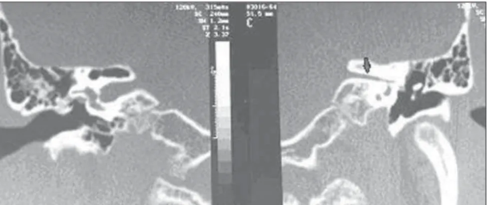
Figure 3. Comparative coronal CT sections. Narrowing of the left internal auditory canal, an indirect sign of cochlear nerve aplasia (arrow).
particularly labyrinthitis ossificans, which may be secondary to infection, inflamma-tion, trauma or previous surgery of the in-ternal ear. Labyrinthitis ossificans affects the fluid-filled spaces of the membranous labyrinth, sometimes with ossification in the form of focal or diffuse plates, with consequential sensorineural deafness and vertigo. CT demonstrates high density bone deposition in the membranous labyrinth. On the other hand, MRI is superior to dem-onstrate the focus that is not yet calcified. Such cochlear calcification does not con-traindicate the implantation, but its imag-ing documentation is required, since such condition could make cochleostomy more difficult to be performed (Figure 5)(6,9).
Complicating factors
Hypoplastic mastoid process is included in the range of complicating factors: when unilateral, such a finding favors the con-tralateral placement of the implant, in the spared side (Figure 6).
Enlargement of the ducts and endolym-phatic sacs is the most common amongst congenital internal ear abnormalities de-tectable at imaging studies. Such condition is generally bilateral, associated with co-chlear dysplasia and abnormalities of the vestibular system semicircular canals. It is more commonly found in children under the age of ten. Sensorineural deafness deep-ens after one year, or fluctuations in the level of such deafness may be observed
after a potentializing trauma event. CT demonstrates the enlargement of the vesti-bular aqueduct and MRI demonstrates the enlarged endolymphatic sac on the poste-rior wall of the temporal bone (Figure 7)(6). Mastoiditis and/or otitis media present as opacification of the middle ear and mas-toid cells, sometimes complicating with mastoid septa erosion (coalescent otomas-toiditis), abscess, meningitis, thrombophle-bitis, sigmoid sinus thrombosis, among other factors (Figure 8)(6,9).
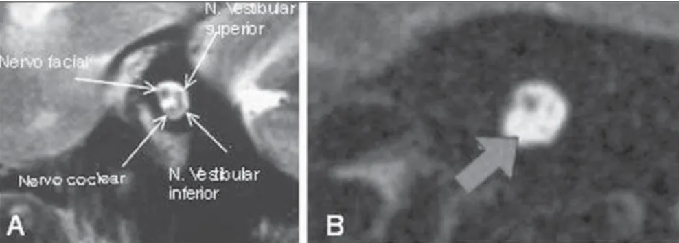
Otosclerosis, a primary disorder of the endochondral layer of the labyrinthine bone, evolves with focal lytic plates pro-gressing to calcification in some cases. Fenestral otosclerosis is most frequently associated with conductive hearing loss, while cochlear otosclerosis may induce sen-sorineural deafness due to involvement of the basilar membrane. The condition is bi-lateral and symmetrical in 95% of the cases, and its causes are still to be established. Co-chlear otosclerosis is generally followed by fenestral otosclerosis. CT is the method of choice, and gadolinium-enhanced MRI demonstrates focal contrast uptake in the Figure 4. Sagittal MRI T2-weighted images. A: Anatomical distribution of the nerves inside the internal
auditory canal. B: Absence of the cochlear nerve (arrow).
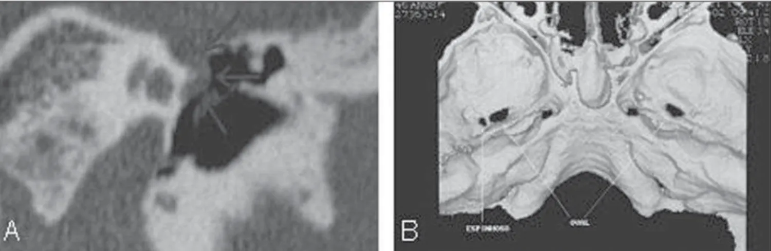
Figure 5. Computed tomography – axial (A) and coronal (B) sections. Increased density within the basal coils of the cochleae, compatible with calcifications (arrows).
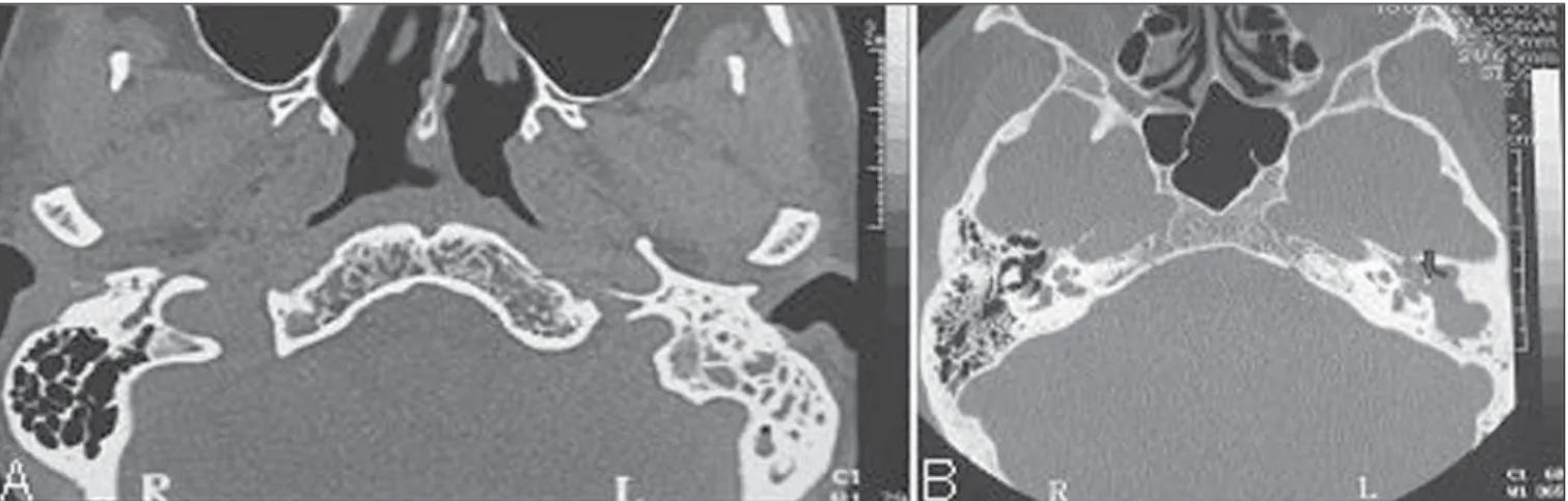
Figure 6. Axial CT section. Bilateral decreased mastoid cells aeration.
Figure 7. Axial CT sections (A,B) and oblique multiplanar reconstruction (C). Normal appearing vestibular aqueducts (A) and bilateral dilation of vestibular aq-ueducts (B,C – arrows and asterisk).
Figure 10.A: Curve reconstruction along the entire normal osseous neural canal of the facial nerve, demonstrating that it is caudally located in relation to the lateral semicircular canal (CAI, internal auditory canal) B: Oblique reconstruction demonstrating part of the aberrant facial nerve (minor arrows) superiorly located in relation to the lateral semicircular canal (major arrow). C: Coronal CT reconstruction showing tympanic segment of the enlarged right facial nerve, positioned anteriorly to the oval window (arrow).
Figure 11. Axial CT image. Aberrant carotid artery at left inside the middle ear.
Figure 8. Axial CT sections. A: Total opacification of mastoid cells at left in association with bony septa sclerosis. B: Chronic cholesteatomatous otomastoiditis at left, with important opacification of the tympanic antrum and membrane in association with ossicular chain erosion (arrow).
Figure 9. Axial CT section. Bilateral pericochlear bone demyelination (retrofenes-tral otosclerosis). active phase. The most common focus is
anteriorly located in relation to the oval window, but it may involve any bone of the medial of the middle ear wall (Figure 9)(6). The preoperative detection of aberrant or dehiscent facial nerve may prevent pos-sible facial palsy secondary to the procedure, as the nerve would be out of its pathway and the surgeon would be aware of this abnor-mality prior to the procedure (Figure 10)(2,9).
Aberrant internal carotid artery results from a congenital vascular abnormality where a small segment of the internal ca-rotid artery is inside the middle ear. CT demonstrates a tubular vascular structure surrounding the cochlear promontory, as-sociated with enlargement of the inferior tympanic canaliculus and absence of the ca-rotid foramen and of the vertical segment of the carotid artery (Figure 11)(6,9).
Persistence of stapedial artery repre-sents another congenital abnormality, usu-ally asymptomatic, whose diagnosis is made intraoperatively or at imaging stud-ies demonstrating enlargement of the ante-rior tympanic segment of the facial nerve canal and absence of the spinous foramen. Association with aberrant internal carotid artery may be observed (Figure 12)(3).
as-ymptomatic anatomical variant, with supe-rior and lateral extension of the jugular bulb into to the middle ear, through the dehis-cent sigmoid plate (Figure 13)(6).
CONCLUSIONS
The correct classification of cochlear conditions and a clear description of such ab-normalities by means of multislice CT and
high-field MRI are determining factors in the surgical planning developed by the co-chlear implantation team, with direct impact on the success of the surgical intervention. Thus the radiologist experienced in the evaluation of the temporal bone plays a major role in the course of this disorder.
REFERENCES
1. Powitzky ES, Hayman LA, Chau J, et al. High-resolution computed tomography of temporal
bone: Part IV: Coronal postoperative anatomy. J Comput Assist Tomogr. 2006;30:548–54. 2. Witte RJ, Lane JI, Driscoll CL, et al. Pediatric and
adult cochlear implantation. Radiographics. 2003; 23:1185–200.
3. Lammers MJ, van der Heijden GJ, Grolman W. Cochlear implants in children and adolescents. Arch Pediatr Adolesc Med. 2012;166:677. 4. Mackeith S, Joy R, Robinson P, et al.
Pre-opera-tive imaging for cochlear implantation: magnetic resonance imaging, computed tomography, or both? Cochlear Implants Int. 2012;13:133–6. 5. Bouccara D, Mosnier I, Bernardeschi D, et al.
Co-chlear implant in adults. Rev Med Interne. 2012; 33:143–9.
6. Harnsberger HR, Wiggins RH, Hudgins PA, et al. Diagnostic imaging: head and neck. 1st ed. Salt Lake City, UT: Amirsys; 2004.
7. Lima Júnior LR, Rocha MD, Walsh PV, et al. Evaluation by imaging methods of cochlear im-plant candidates: radiological and surgical corre-lation. Braz J Otorhinolaryngol. 2008;74:395– 400.
8. Chaturvedi A, Mohan C, Mahajan SB, et al. aging of cochlear implants. Indian J Radiol Im-aging. 2006;16:385–92.
9. Swartz JD, Harnsberger HR. Imaging of the tem-poral bone. 3rd ed. New York, NY: Thieme Med Publ; 1998.
10. Fishman AJ. Imaging and anatomy for cochlear implants. Otolaryngol Clin North Am. 2012;45: 1–24.
Figure 12.A: Coronal CT reconstruction demonstrating persistent stapedial artery at left: enlargement of the facial nerve canal, with projection of the artery towards the middle year (arrows). B: 3D CT reconstruction showing normal appearing spinous foramen at right and absence at left.