143
Lara Filho LA et al. Cranial CT findings in the emergency unit of Hospital Cajuru
Radiol Bras. 2013 Mai/Jun;46(3):143–148
Cranial computed tomography findings in patients admitted
to the emergency unit of Hospital Universitário Cajuru
*
Achados tomográficos de pacientes submetidos a tomografia de crânio no pronto-socorro do Hospital Universitário Cajuru
Lauro Aparecido Lara Filho1, Samir Sari Omar2, Rodrigo Foletto Biguelini2, Rony Augusto de Oliveira
Santos2
Objective: To identify and analyze the prevalence of cranial computed tomography findings in patients admitted to the emergency unit of Hospital Universitário Cajuru. Materials and Methods: Cross-sectional study analyzing 200 consecutive non contrast-enhanced cranial computed tomography reports of patients admitted to the emergency unit of Hospital Universitário Cajuru. Results: Alterations were observed in 76.5% of the patients. Among them, the following findings were most frequently observed: extracranial soft tissue swelling (22%), bone fracture (16.5%), subarachnoid hemorrhage (15%), nonspecific hypodensity (14.5%), paranasal sinuses opacification (11.5%), diffuse cerebral edema (10.5%), subdural hematoma (9.5%), cerebral contusion (8.5%), hydrocephalus (8%), retractable hypodensity /gliosis/ encephalomalacia (8%). Conclusion: The authors recognize that the most common findings in emergency departments reported in the literature are similar to the ones described in the present study. This information is important for professionals to recognize the main changes to be identified at cranial computed tomography, and for future planning and hospital screening aiming at achieving efficiency and improvement in services.
Keywords: Computed tomography; Craniocerebral trauma; Imaging diagnosis.
Objetivo: Reconhecer e analisar a prevalência dos achados tomográficos de pacientes submetidos a tomografia de crânio atendidos no pronto-socorro do Hospital Universitário Cajuru. Materiais e Métodos: Estudo transversal pela análise sequencial de 200 laudos de tomografia de crânio incluindo todos os pacientes admitidos no pronto-socorro do Hospi-tal Universitário Cajuru submetidos a este exame sem contraste intravenoso. Resultados: Observou-se a presença de alterações em 76,5% dos exames. Destas, as mais frequentes foram: aumento de volume de partes moles extracra-nianas (22%), fratura óssea (16,5%), hemorragia subaracnoidea (15%), hipodensidade inespecífica (14,5%), velamento dos seios paranasais (11,5%), edema cerebral difuso (10,5%), hematoma subdural (9,5%), contusão cerebral (8,5%), hidrocefalia (8%), hipodensidade retrátil/gliose/encefalomalácia (8%). Conclusão: Reconhecemos que os achados mais comuns em um serviço de urgência e emergência em outros estudos se assemelham aos nossos. Essas informações são importantes para que os profissionais reconheçam quais são as principais alterações a serem identificadas em uma tomografia de crânio e para um futuro planejamento e triagem hospitalar, a fim de obter eficiência e melhora nos ser-viços prestados.
Unitermos: Tomografia computadorizada; Traumatismos cranioencefálicos; Diagnóstico por imagem. Abstract
Resumo
* Study developed at Hospital Universitário Cajuru – Ponti-fícia Universidade Católica do Paraná (PUCPR), Curitiba, PR, Brazil.
1. Specialist in Radiology and Imaging Diagnosis, Profes-sor, Course of Medicine, Pontifícia Universidade Católica do Pa-raná (PUCPR), Curitiba, PR, Brazil.
2. Students, Course of Medicine, Pontifícia Universidade Católica do Paraná (PUCPR), Curitiba, PR, Brazil.
Mailing Address: Samir Sari Omar. Rua Alferes Poli, 464, ap. 1407, Rebouças. Curitiba, PR, Brazil, 80220-050. E-mail: [email protected].
Received July 10, 2012. Accepted after revision February 19, 2013.
Lara Filho LA, Omar SS, Biguelini RF, Santos RAO. Cranial computed tomography findings in patients admitted to the emergency unit of Hospital Universitário Cajuru. Radiol Bras. 2013 Mai/Jun;46(3):143–148.
ORIGINAL ARTICLE
absorbing the X-rays. The detector trans-forms the emitted photons into analogical signals (by means of voltage) and then a computer transforms such signals into digi-tal ones(12,15,16).
There is a convention utilized to trans-late the detected voltage values into digi-tal units, the absorption coefficient (attenu-ation). It is calculated in relation to the lin-ear coefficient of water, for which the nu-meric value 0 is attributed, in a scale that may range from +1,000 to –1,000 Houns-field units (HU) and is represented by a gray scale comprising a large spectrum of neuroradiology for the diagnosis and
treat-ment of several diseases(1–11).
Computed tomography (CT) was devel-oped by the British physicist Godfrey Hounsfield and was first utilized at the Atkinson Morley Hospital, London, in 1972, and quickly became one of the main methods for the evaluation of structural brain disorders(12–14).
Currently, the method relies on an X-ray tube that spins 360° and is equipped with photon detectors opposite to the X-ray source. The image will depend upon the thickness of the object and its capability of INTRODUCTION
tones representation between white, gray and black (Table 1)(15,16).
With the advent of magnetic resonance imaging, cranial studies by means of CT became better indicated in acute situations such as those encountered at specialized emergency services. CT is the imaging method of choice in the evaluation of head trauma (HT) as it can demonstrate bone and parenchymal changes, besides hemor-rhages. It is a very important method as it is accessible because of its wide availabil-ity, lower cost and low acquisition time. Magnetic resonance imaging, on its turn, presents some limitation in patients with implanted materials, such as clips or pace-makers, claustrophobia or obesity, which do not prevail in CT(17–19).
At the CT unit of Hospital Universitário Cajuru (HUC), the main indications for cranial CT include: decreased conscious-ness level without known causes; alcohol or drug intoxication; focal neurological deficit; suspected penetrating injury or skull fracture; consciousness loss during or after trauma; ages < 2 years and > 65 years with a history of HT; unreliable history; reports of trauma vomiting; post-trauma amnesia; signs of skull base frac-ture; polytrauma; severe facial trauma; sig-nificant extracranial soft tissue swelling; and suspicion of child abuse.
Because of the shortage of local scien-tific studies (Curitiba metropolitan area) evaluating the prevalence of cranial CT findings and, considering that HUC is a specialized emergency service where such imaging method is required, the present in-vestigation became necessary to evaluate the service, as well as to analyze the preva-lence of findings. Thus, the present study was aimed at recognizing and analyzing the prevalence of cranial CT findings in pa-tients admitted to the emergency unit of HUC in Curitiba, PR, Brazil.
MATERIALS AND METHODS
A cross-sectional study was undertaken with sequential analysis of 200 cranial CT reports of patients admitted to the HUC’s emergency unit in the period between Janu-ary 1st and FebruJanu-ary 9, 2008.
All the patients admitted to the HUC’s emergency unit who were submitted to cra-nial non contrast enhanced CT were in-cluded in the present study, regardless of gender and age. Only reports of patients submitted to contrast-enhanced scan were excluded. The scans were performed with the patients in dorsal decubitus, in a Si-emens Somaton Spirit apparatus, with the following acquisition parameters: 130 kVp, 100 mAs, axial plane, 3 mm-thick slices for the posterior fossa, and 10 mm-thick slices for the remaining areas of the skull, with-out intravenous contrast injection.
Two report patterns were considered and recorded as being normal: without vol-ume reduction and with volvol-ume reduction (normal pattern for elderly patients).
Normal pattern without volume reduc-tion – Absence of expansile lesions, extra-axial collections or pathological calcifica-tions in the encephalon; ventricular system with normal morphology, topography and dimensions; anatomical features of the basal cisterns, as well as of the sulci be-tween the cortical gyri in the brain convexi-ties. Diagnostic impression: study within the normality limits.
Normal pattern with volume reduction – Absence of expansile lesions, extra-axial collections or pathological calcifications in the encephalon; dilation of the ventricular system (ex-vacuo); prominence of the cer-ebellar folia, basal cisterns, Sylvian fissure as well as of the sulci between the cortical gyri in the brain convexities. Diagnostic impression: brain and cerebellar volume reduction.
All the other CT findings, such as hypodense lesions or collections, micro-angiopathy, hyperdense lesions or collec-tions, hemorrhages, expansile lesions (tu-mor, hematoma, hemorrhages), soft-tissue volume increase, calcifications, atheromas, fractures, alterations secondary to surgical procedures (craniotomy, aneurysmal clips, shunt catheter), pneumocephalus, signs of intracranial hypertension (hydrocephalus, edema, hernias, ventricular asymmetries, midline structures displacement and focal obliteration of sulci), sinusal alterations, foreign body, metal fragments and ana-tomic variations, were considered abnor-mal and exposed to analysis, establishing a relation between number of findings and total number of scans.
RESULTS
In the sample of this study, 47 patients (23.5%) presented results within the nor-mality standards. Thus, 153 patients (76.5%) presented alterations detected by CT.
The most common finding was intrac-ranial soft-tissue swelling, observed on 44 (22%) of the 200 CT scans.
The bone fractures found in 33 patients (16.5%) are shown on Table 2.
The findings of ectopic air collections included pneumocephalus, observed in 11 patients (5.5%), and intra-orbital emphy-sema, in one patient (0.5%).
Hyperdensities and hypodensities are listed on Tables 3 and 4, respectively.
Ventricular alterations and signs of in-tracranial hypertension are represented on Table 5.
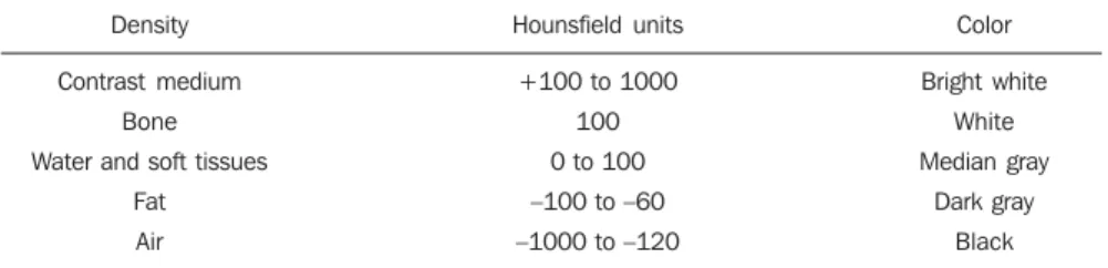
Abnormal findings observed in the nose, nasal cavity and paranasal sinuses Table 1 Relationship between density, attenuation and color.
Density
Contrast medium Bone Water and soft tissues
Fat Air
Hounsfield units
+100 to 1000 100 0 to 100 –100 to –60 –1000 to –120
Color Bright white White Median gray Dark gray Black
Table 2 Bone fractures.
Location Skull – Temporal – Frontal – Occipital – Parietal – Sphenoid – Ethmoid Orbit
– Orbital bones Face
Table 6 Most prevalent findings.
Finding
Extracranial soft-tissue swelling Bone fracture
Subarachnoid hemorrhage Nonspecific hypodensity Veiling of paranasal sinuses Diffuse cerebral edema Subdural hematoma Cerebral contusion Hydrocephalus
Retractile hypodensity/gliosis/encephalomalacia
Quantity
44 33 30 29 23 21 19 17 16 16
Percentage
22% 16.5%
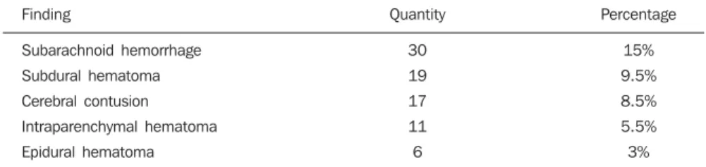
15% 14.5% 11.5% 10.5% 9.5% 8.5% 8% 8% Table 3 Hyperdensities.
Finding
Subarachnoid hemorrhage
Subdural hematoma Cerebral contusion Intraparenchymal hematoma
Epidural hematoma
Quantity
30
19 17 11
6
Percentage
15%
9.5% 8.5% 5.5%
3%
Table 4 Hypodensities.
Finding
Nonspecific
Retractile hypodensity/gliosis/encephalomalacia Microangiopathy
Previous ischemia Recent ischemia
Quantity
29 16 12 7 6
Percentage
14.5% 8% 6% 3.5%
3%
Table 5 Ventricular alterations and signs of intracranial hypertension.
Finding
Diffuse cerebral edema Hydrocephalus
Ventricular hemorrhage Midline structures displacement Focal sulci obliteration
Subfalcine herniation Ventricular asymmetry
Quantity
21 16
11 11 5
2 1
Percentage
10.5% 8%
5.5% 5.5% 2.5%
1% 0.5%
(25.3%), and is inferior to the results re-ported by Palheta et al.(21) (46.4%). It is im-portant to highlight that in those two stud-ies, only CT scans with indication for HT investigation were considered, while in the present study all CT scans performed in the emergency unit were considered, regardless of their indication. Palheta et al.(21) have justified the high index of normal results by the fact that, at those authors’ institution, cranial CT is routinely performed for mild HT, an indication which is not supported by Conselho Federal de Medicina (Federal Medicine Council)(22) for all patients. In the opinion of the authors of the present study, their findings are within the expected lev-els for an emergency unit, considering the current indications for cranial CT.
The most prevalent finding was extrac-ranial soft-tissue swelling (22%). Such finding was also prevalent in the study de-veloped by Rocha(20), with 57%, by Bor-dignon et al.(23), with 47%, and by Palheta et al.(21), with 44.3%. Such an alteration is typical in cases of HT, and for such reason its prevalence was higher in those studies. Bone fracture was found in 33 patients (16.5%), and was the second most preva-lent finding in the present study. The most common one was temporal bone fracture, with 5.5%, a value that is close to the 4.7% reported by Amin et al.(24).
Pneumocephalus is an air collection in-side the cranial cavity that may be second-ary to surgery or procedures, skull base fracture, congenital defects, tumors caus-ing bone erosion and infection by gas pro-ducing bacteria(15,18). Such finding had a prevalence of 5.5% in the present study, while Rocha(20 found 9.3% and Palheta et al.(21) found 7.1%.
were the following: veiling of paranasal sinuses, in 23 patients (11.5%); sinuso-pathy, in 13 patients (6.5%); and septal deviation in 2 patients (1%). Veiling and sclerosis of the mastoid process was ob-served in 4 patients (2%).
The following types of pathological cal-cifications were observed: intraparenchymal calcifications, with 15 findings (7.5%); calcified atheromas in the carotid or verte-bral system, with 14 (7%) findings; and eyeball calcifications with 2 (1%) findings. As regards expansile lesions, there were 5 (2.5%) findings – 3 (1.5%) hypodense and 2 (1%) heterogeneous –; 2 (1%) paren-chymal heterogeneities and one (0.5%) cor-pus callosum lipoma.
Craniotomy was found in 15 (7.5%) pa-tients, one (0.5%) of them with herniation. Ventricular shunt catheters were found in 10 (5%) patients.
In addition, motion artifacts were found on 12 scans, impairing their results, 7 (3.5%) metal fragments, 4 (2%) aneurysmal clips and 2 (1%) orotracheal tubes.
Cavum vergae, an anatomic variation, was found in 2 (1%) patients.
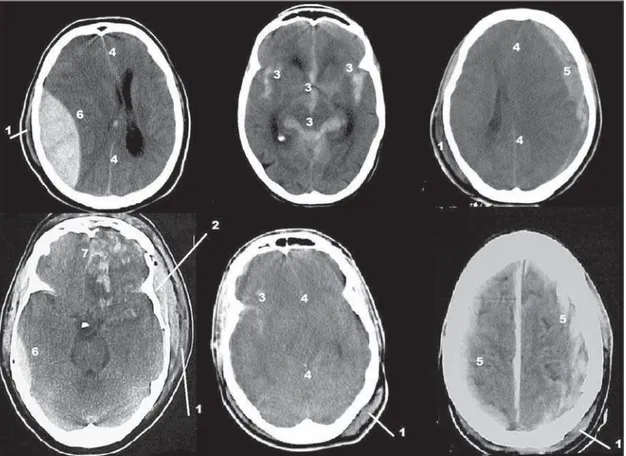
The 10 most prevalent findings are listed on Table 6 and the most relevant images are shown on Figures 1, 2 and 3.
DISCUSSION
Figure 1. 1, extracranial soft-tissue swelling; 2, depressed bone fracture; 3, subarachnoid hemorrhage; 4, midline structures displacement/diffuse cerebral edema; 5, subdural hematoma; 6, epidural hematoma; 7, cerebral contusion in frontal pole (straight and orbital gyri).
Hyperdensity may indicate the presence of hemorrhages, hematomas, collections, among other findings(15,16,18). The most com-mon one was subarachnoid hemorrhage, with 15%, and was the third most prevalent finding in the study. Such a finding was observed by Rocha(20) in 22.4% of his pa-tients, a higher prevalence than that re-ported by Stein et al.(25) and by Palheta et al.(21), with respectively 5.7% and 6.4%. Ce-rebral contusion was found in 8.5% of the CT scans in the present study. Such a find-ing was observed by Rocha(20) in 24.4% of his patients, in 12.9% by Bordignon et al.(23) and in 8.6% by Palheta et al.(21). Sub-dural and epiSub-dural hematomas had a preva-lence of 9.5% and 3%, respectively, the latter with a lower prevalence than that re-ported by Palheta et al.(21), of 4.3%, and by Rocha(20), of 8%. Subdural hematoma was found by Rocha(20) in 7.3% of his patients, by Palheta et al.(21) in 10%, and by Servadei et al.(26), in 11% of the patients. Intraparen-chymal hematoma was found in 5.5% of patients, while Rocha(20) and Palheta et
al.(21) found 6.7% and 11.4%, respectively. Hypodensity may indicate the presence of tumor, abscess, hematoma in resolution, ongoing infarction, chronic hematoma, chronic infarction, encephalomalacia, microangiopathy (microvascular ischemic damage), among others(15,16,18). In the present study, hypodensity of nonspecific type was the most prevalent (14.5%), rep-resenting the fourth most prevalent finding in the study. The second most prevalent type of hypodensity was retractile hypo-dense area/gliosis/encephalomalacia, in 8% of the patients. Microangiopathy was the third most prevalent, with 6%. It is impor-tant to note that, in the present study, the authors considered as nonspecific findings those that were not described as typical is-chemic lesions, microangiopathy or en-cephalomalacia.
Intracranial hypertension may be caused by an expansile process (tumor, hematoma, cysts, abscess, empyema and granuloma), hydrocephalus, cerebral edema, metabolic disorder, and intoxication by prescription
drugs or other types of drugs(16,18). Among the findings related to intracranial hyper-tension and ventricular alterations, the au-thors of the present study observed diffuse cerebral edema (10.5%); hydrocephalus (8%); intraventricular hemorrhage (5.5%); midline structures displacement (5.5%); and subfalcine herniation (1%).
In their study, Palheta et al.(21) have found diffuse edema in 5.7% of their cases, while Rocha(20) has observed such finding in 8.2% and midline structures displace-ment in 9.1% of his cases. Palheta et al.(21) highlight that midline structures displace-ment is commonly associated with subfal-cine herniation, being such herniation the most common in this case. The present study did not found any other related herniations. Intraventricular hemorrhage was found by Rocha(20) in 2.7% of his cases, and such finding was not observed by Palheta et al.(21).
Because of the intimate association with surrounding structures, isolated paranasal sinuses injuries are uncommon. Associa-tion between bone injuries, including those in the paranasal sinuses, with intracranial and extracranial soft tissues are more com-mon(27). This explains the fact that veiling of the paranasal sinuses was observed in 11.5% of the cases, the fifth most prevalent finding in the present study, probably asso-ciated with other HT injuries. Veiling and sclerosis of the mastoid process were found in 2% of the patients. According to Secchi et al.(28), such finding is present in 25% of temporal bone fractures. In the present study, sinus hemorrhage and changes in the content of the sinuses were not discrimi-nated.
Intracranial calcifications represent fre-quent incidental findings at numerous neu-rological imaging studies(29). In the present study, calcifications with greater signifi-cance were intraparenchymal calcifications (7.5%) and calcified atheromas in the ca-rotid or vertebral system with (7%).
Craniotomy, observed in 7.5% of cases, as well as aneurysmal clips, in 1.0%, are due principally to postoperative follow-up. Metal fragments corresponding to pro-jectiles or shrapnel were found in 3.5% of the patients. Rocha(20) has found them in 4.4% of the cases and Palheta et al.(21), in 1.4%.
It is important to remind that the main studies utilized in the discussion of the data, such as those published by Rocha(20), Palheta et al.(21) and Bordignon et al.(23), have exclusively evaluated patients with indication of CT for HT. The present study did not discriminate the indication for cra-nial CT scan, but, considering that the HUC is a reference emergency service, HT was the main indication for the CT scans.
As limitations inherent to the present study, the authors highlight that the study period of one month and nine days might give room to possible seasonal variations, and that the evaluation was based only on a report made by a single radiologist and not by consensus between two radiologists. Another limitation was the absence of re-ports standardization, as many times differ-ent radiologists utilize differdiffer-ent terms for a single type of finding, a fact which re-quired a revision of the tables and data pro-posed by the authors.
It is also important to mention the diffi-culty in finding studies describing CT find-ings in emergency settfind-ings, and for such reason it would be appropriate to undertake further studies with the same objective.
CONCLUSIONS
The authors recognize that the most common non-contrast-enhanced cranial CT findings in emergency settings reported by other studies are similar to those observed in the present study. Such findings include extracranial soft-tissue swelling (22%), bone fracture (16.5%), subarachnoid hemorrhage (15%), nonspecific hypodensity (14.5%), veiling of paranasal sinuses (11.5%), dif-fuse brain edema (10.5%), subdural he-matoma (9.5%), cerebral contusion (8.5%), hydrocephalus (8%), and retractile hypo-density/gliosis/encephalomalacia (8%).
Such data are important to enable the professionals in the service to recognize the
mains findings to be identified at cranial CT and for a future planning and hospital screening, with the purpose of obtaining efficiency and improvement in the rendered services.
REFERENCES
1. Fernandes RCL, Rosso ALZ, Vincent MB, et al. Transcranial sonography findings in Parkinson’s disease and essential tremor: cases report. Radiol Bras. 2012;45:356–8.
2. Coeli GNM, Silva GC, Tiengo RR, et al. Cerebe-lite aguda com herniação tonsilar: relato de caso. Radiol Bras. 2012;45:244–6.
3. Sanches P, Yamashita S, Freitas CCM, et al. Chordoid glioma of the third ventricle: a new case report. Radiol Bras. 2012;45:288–90.
4. Gonçalves FG, Hanagandi PB, Torres CI, et al. Posterior migration of lumbar disc herniation – imaging dilemma due to contrast contraindica-tion: a case report. Radiol Bras. 2012;45:170– 2.
5. Barros ML, Fernandes DA, Melo EV, et al. Mal-formações do sistema nervoso central e malfor-mações associadas diagnosticadas pela ultrasso-nografia obstétrica. Radiol Bras. 2012;45:309– 14.
6. Coeli GNM, Tiengo RR, Silva AC, et al. Neuro-cisticercose nodular calcificada com sinais de reativação. Radiol Bras. 2012;45:291–3. 7. Nogueira-Barbosa MH, Savarese LG, Herrero
CFPS, et al. Raízes nervosas redundantes da cauda equina: revisão da literatura. Radiol Bras. 2012;45:155–9.
8. Jurno ME, Castro MHA, Lage MA, et al. Sín-drome de desmielinização osmótica: relato de caso com evolução favorável. Radiol Bras. 2012; 45:61–2.
9. Wajnberg E, Rodrigues G, Abud DG. O uso de
stents farmacológicos no tratamento da estenose
das artérias vertebrais. Radiol Bras. 2011;44:343– 8.
10. Gonçalves FG, Barra FR, Matos VL, et al. Sinais em neurorradiologia – Parte 1. Radiol Bras. 2011;44:123–8.
11. Barra FR, Gonçalves FG, Matos VL, et al. Sinais em neurorradiologia – Parte 2. Radiol Bras. 2011;44:129–33.
12. Beckmann EC. CT scanning the early days. Br J Radiol. 2006;79:5–8.
13. Hounsfield GN. Computerized transverse axial scanning (tomography): Part 1. Description of system. Br J Radiol. 1973;46:1016–22.
14. Ambrose J. Computerized transverse axial scan-ning (tomography): Part 2. Clinical application. Br J Radiol. 1973;46:1023–47.
15. Gunderman R. Fundamentos de radiologia. 2ª ed. Rio de Janeiro, RJ: Guanabara Koogan; 2007. 16. Koch HA. Radiologia e diagnóstico por imagem
na formação do médico geral. 2ª ed. Rio de Ja-neiro, RJ: Revinter; 2012.
17. Haaga JR. Tomografia computadorizada e resso-nância magnética do corpo humano. 3ª ed. Rio de Janeiro, RJ: Guanabara Koogan; 1996. 18. Leite CC, Amaro Jr E, Lucato LT. Neuroradiologia
– diagnóstico por imagem das alterações encefáli-cas. Rio de Janeiro, RJ: Guanabara Koogan; 2008.
19. American College of Radiology. ACR Appropri-ateness Criteria® Head Trauma. Last review date: 2008. [acessado em 20 de maio de 2011]. Dis-ponível em: http://gm.acr.orgSecondaryMain MenuCategories/quality_safety/app_criteria/pdf/ E x p e r t P a n e l o n N e u r o l o g i c I m a g i n g / H e a d TraumaDoc5.aspx.
20. Rocha CMN. Traumatismo cranioencefálico: cor-relação entre dados demográficos, escala de Glas-gow e tomografia computadorizada de crânio com a mortalidade em curto prazo na cidade na cidade de Maceió, Alagoas [tese]. São Paulo, SP: Facul-dade de Medicina – UniversiFacul-dade de São Paulo; 2006.
21. Palheta MS, Nunes RB, Targino MN, et al. Acha-dos tomográficos Acha-dos pacientes vítimas de trau-matismo cranioencefálico atendidos no Hospital Metropolitano de urgência e emergência. Rev Para Med. 2009;23(2).
22. Andrade AF, Marino Jr R, Miura FK, et al. ABM/ CFM Projeto diretrizes. Diagnóstico e conduta no paciente com traumatismo craniencefálico leve. Brasil; 2001.
23. Bordignon KC, Arruda WO. CT scan findings in mild head trauma: a series of 2,000 patients. Arq Neuropsiquiatr. 2002;60:204–10.
24. Amin Z, Sayuti R, Kahairi A, et al. Head injury with temporal bone fracture: one year review of case incidence, causes, clinical features and out-come. Med J Malaysia. 2008;63:373–6.
25. Stein SC, Ross SE. Mild head injury: a plea for routine early CT scanning. J Trauma. 1992;33: 11–3.
26. Servadei F, Nasi M, Giuliani G, et al. CT prognos-tic factors in acute subdural haematomas: the value of the ‘worst’ CT scan. Br J Neurosurg. 2000;14;110–6.
27. Mathog RH, Arden RL, Marks SC. Trauma of the nose and paranasal sinuses. New York, NY: Thieme Med Publ; 1995.
28. Secchi MMD, Moraes JFS, Castro FB. Fratura de osso temporal em pacientes com traumatismo crânio-encefálico. Arq Int Otorrinolaringol. 2012; 16:62–6.