173 Radiol Bras. 2013 Mai/Jun;46(3):173–177
Pyogenic and tuberculous discitis: magnetic resonance imaging
findings for differential diagnosis
*
Espondilodiscites piogênica e tuberculosa: aspectos na ressonância magnética para o diagnóstico diferencial
Cristiano Gonzaga de Souza1, Emerson Leandro Gasparetto2, Edson Marchiori3, Paulo Roberto
Valle Bahia4
Spondylodiscitis represents 2%–4% of all bone infections cases. The correct diagnosis and appropriate treatment can prevent complications such as vertebral collapse and spinal cord compression, avoiding surgical procedures. The diagnosis is based on characteristic clinical and radiographic findings and confirmed by blood culture and biopsy of the disc or the vertebra. The present study was developed with Clementino Fraga Filho University Hospital patients with histopathologically and microbiologically confirmed diagnosis of spondylodiscitis, submitted to magnetic resonance imaging of the affected regions. In most cases, pyogenic spondylodiscitis affects the lumbar spine. The following findings are suggestive of the diagnosis: segmental involvement; ill-defined abscesses; early intervertebral disc involvement; homogeneous vertebral bodies and intervertebral discs involvement. Tuberculous spondylodiscitis affects preferentially the thoracic spine. Most suggestive signs include: presence of well-defined and thin-walled abscess; multisegmental, subligamentous involvement; heterogeneous involvement of vertebral bodies; and relative sparing of intervertebral discs. The present pictorial essay is aimed at showing the main magnetic resonance imaging findings of pyogenic and tuberculous discitis.
Keywords: Magnetic resonance imaging; Discitis; Intervertebral disc; Spinal tuberculosis.
Espondilodiscites representam 2%–4% de todos os casos de infecções no esqueleto. Seu rápido diagnóstico e trata-mento apropriado podem evitar complicações, tais como colapsos vertebrais, compressão medular, evitando a realiza-ção de procedimentos cirúrgicos. Seu diagnóstico é baseado em achados clínicos e radiológicos característicos, sendo confirmado por hemoculturas, biópsia do disco ou da vértebra. Este estudo foi realizado com pacientes do Hospital Uni-versitário Clementino Fraga Filho que tiveram o diagnóstico histopatológico ou microbiológico comprovado de espondi-lodiscite e realizaram ressonância magnética das regiões acometidas. Espondiespondi-lodiscites piogênicas acometem prefe-rencialmente a coluna lombar. Os principais sinais sugestivos são: acometimento segmentar; abscessos de limites pouco definidos; acometimento precoce do disco intervertebral; acometimento homogêneo dos corpos vertebrais e discos intervertebrais. A espondilodiscite tuberculosa afeta preferencialmente os segmentos vertebrais torácicos. As imagens mais sugestivas são: abscesso de paredes delgadas e bem definidas; envolvimento subligamentar multissegmentar; acometimento heterogêneo dos corpos vertebrais; discos intervertebrais relativamente poupados. O objetivo deste en-saio iconográfico é apresentar os principais aspectos das espondilodiscites piogênica e tuberculosa nas imagens por ressonância magnética.
Unitermos: Ressonância magnética; Discite; Disco intervertebral; Tuberculose da coluna vertebral.
Abstract
Resumo
* Study developed in the Service of Radiology at Hospital Universitário Clementino Fraga Filho – Universidade Federal do Rio de Janeiro (UFRJ), Rio de Janeiro, RJ, Brazil.
1. MD, Resident of Radiology and Imaging Diagnosis, Uni-versidade Federal do Rio de Janeiro (UFRJ), Rio de Janeiro, RJ, Brazil.
2. Associate Professor, Department of Radiology, School of Medicine – Universidade Federal do Rio de Janeiro (UFRJ), MD, Neuroradiologist, Grupo DASA, Rio de Janeiro, RJ, Brazil.
3. Full Professor of Radiology, Universidade Federal Flumi-nense (UFF), Niterói, RJ, Adjunct Coordinator, Course of Post-graduation in Radiology, Universidade Federal do Rio de Janeiro (UFRJ), Rio de Janeiro, RJ, Brazil.
4. Associate Professor, Department of Radiology, School of Medicine – Universidade Federal do Rio de Janeiro (UFRJ), Rio de Janeiro, RJ, Brazil.
Mailing Address: Dr. Cristiano Gonzaga de Souza. Rua Nossa Senhora do Carmo, 155, Vila Tereza. Cataguases, MG, Brazil, 36772-016. E-mail: [email protected].
Souza CG, Gasparetto EL, Marchiori E, Bahia PRV. Pyogenic and tuberculous discitis: magnetic resonance imaging findings for differen-tial diagnosis. Radiol Bras. 2013 Mai/Jun;46(3):173–177.
onstration of the involvement of the verte-bral body and adjacent interverteverte-bral disc, although several non-infectious diseases may mimic this condition(11).
Three forms of dissemination are de-scribed, as follows: hematogenous spread from a distant septic focus; direct inocula-tion (either by surgery or trauma); contigu-ity with an adjacent septic focus. Generally, the infection starts in the anterior portion of the vertebral body because of its rich arterial supply, and spreads through the medullary spaces, affecting the interverte-bral disc by contiguity, most frequently INTRODUCTION
The imaging evaluation of the muscu-loskeletal system has been object of a range of studies recently published in the Brazil-ian radiological literature(1–7). Among such studies, some related to the vertebral spine are highlighted(8–10).
Spondylodiscitis represent 2%–4% of all cases of skeletal infection(11) and its ra-diological diagnosis is based on the
involving the lumbar and dorsal segments of the spine (50% and 35% of cases, re-spectively)(12). Generally, late diagnosis is the rule (on average, two to six months af-ter the symptoms onset)(13). A fast diagno-sis allows the institution of appropriate treatment and may avoid complications such as vertebral collapse and medullary compression syndrome.
In this context, imaging investigation is critical for the diagnosis and follow-up of the lesions so as to reduce the necessity of invasive procedures. Magnetic resonance imaging (MRI) is the method of choice because of its high sensitivity and specific-ity(14), as well as good tissue resolution and multiplanar capacity(15). Also, MRI can be useful to suggest the origin of the infection – although this is not always possible –, aiding in the differentiation between tuber-culous and pyogenic infections, thus allow-ing efficacy in the treatment of the pa-tient(16).
Main MRI findings include hyposignal on T1-weighted and hypersignal on T2-weighted images from the vertebral bodies and adjacent intervertebral discs, as well as enhancement after intravenous paramag-netic contrast medium injection, besides paravertebral masses/abscesses, all of them nonspecific(17). However, as some details are correctly individualized and each find-ing is appropriately interpreted, they may become useful tools to guide the radiolo-gist in the differential diagnosis.
The present pictorial essay is aimed at gathering and demonstrating some imaging findings in order to appropriately highlight evidences of higher suspicion degree of
etiological agents, according to the avail-able literature.
PYOGENIC SPONDYLODISCITIS
Pyogenic spondylodiscitis affects most frequently the lumbar spine, involving only one vertebral segment (one intervertebral disk and adjacent vertebral bodies). Most frequently, Staphylococcus aureus is the etiological agent implied in the infection, responsible for 55%–90% of the cases(18). Other relevant agents include
Streptococ-cus, PneumococStreptococ-cus, EnterococStreptococ-cus, Es-cherichia coli, Salmonella, Pseudomonas aeruginosa and Klebsiella(18).
Because of the high concentration of proteolytic enzymes intrinsic to the viru-lence of these biological agents, the disc in-volvement occurs early in the course of the disease and can be demonstrated concomi-tantly with the corresponding vertebral body lesion at very initial stages of infec-tion. The vertebral body involvement tends to be more homogeneous in relation to the alterations in signal intensity on T1- and T2-weighted sequences and to the enhance-ment of its medullary spaces(19). Consider-ing the remarkable worsenConsider-ing in the gen-eral condition as well as important features of the symptoms affecting the patient, im-aging studies are requested at the initial phase of the disease where, although the appearance of the lesion is more aggressive, it is less disseminated in multiple vertebral bodies, so the finding of large abscesses or paravertebral masses is not expected. In cases where such findings are present, how-ever, the lesion is poorly defined,
depend-ing on the aggressiveness of the implied etiological agent(11).
Thus, MRI findings which lead to the suspicion of pyogenic spondylodiscitis in-clude segmental involvement (Figures 1 and 2), presence of poorly defined paraver-tebral mass (Figure 3), early interverparaver-tebral disc involvement (Figure 2), and homoge-neous enhancement/alteration of signal of affected vertebral bodies (Figures 1 and 2).
TUBERCULOUS SPONDYLODISCITIS
The spine is the main site of bone in-volvement by tuberculosis, responsible for 50% of the cases(20). The differential diag-nosis with pyogenic spondylodiscitis is clinically and radiologically difficult to be achieved, particularly in cases where the etiological agent is less aggressive, like in cases of brucelosis.
The scarcity of proteolytic enzymes, which is an intrinsic characteristic of
My-cobacterium tuberculosis, results in a
re-markably late, indolent infection of the in-tervertebral disc(21). Thus, tuberculous spondylodiscitis may originate large paravertebral abscesses or granulomatous masses with well defined borders(17), ex-tending into the subligamentous space through several (typically more than three) vertebral bodies at the time of the imaging diagnosis. An irregular pattern of involve-ment may also be observed15, besides a more heterogeneous involvement of verte-bral bodies, provided there is time for the bone tissue to react against the infection. Thus, focal areas of altered signal intensity
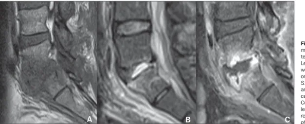
Figure 1. Segmental involve-ment of L5-S1 and of the in-terposed intervertebral disc. Lesion with hyposignal on T1-weighted (A) and hypersignal on STIR (B) images of the L5-S1 disc, with enhancement and irregularities in the adja-cent vertebral plateaus (C). Culture of the material col-lected from the vertebral body revealed the presence of Sta-phylococcus aureus.
are observed on T1- and T2-weighted im-ages, besides focal enhancement of part of medullary spaces of the vertebral body. Such findings allow the demonstration of discrepancy in the involvement between discs and adjacent structures such as ver-tebral bodies and paraverver-tebral region. Thus, disc involvement is noticeable in
later phases of the disease. At early phases, the disc is usually intact, although it is al-ready possible to identify the presence of extensive bone involvement or large paravertebral abscesses/masses which, typically, tend to present well defined con-tours. At early phases of the disease, the differential diagnosis with, for example,
neoplastic (Figure 6), degenerative (Figure 7) or inflammatory etiologies may be hardly made. In such cases, morphological crite-ria, presence of signal from affected regions on T2-weighted images, and the character-istics of the gadolinium enhancement may be useful to shorten the list of diagnostic hypotheses(22).
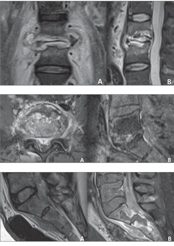
Figure 2. Segmental involve-ment of L3-L4 and of the inter-posed intervertebral disc in the coronal (A) and sagittal planes (B). Remarkable irregularity of the vertebral plateaus in associa-tion with homogeneous hyper-signal of the vertebral body on the STIR image. Culture of the material collected from the inter-vertebral disc revealed the
pres-ence of Enterococcus sp.
A
B
Figure 3. Segmental involve-ment of L5-S1 and of the inter-posed intervertebral disc. Poorly defined paravertebral collection, with intense gadolinium en-hancement on axial (A) and sag-ittal (B) T1-weighted image with fat suppression. Note the early disc involvement. Culture of the collected material revealed the
presence of Pneumococcus sp.
A
B
Figure 4. Multisegmental in-volvement. Paravertebral mass with subligamentous extension on STIR image (A) and intense and heterogeneous contrast enhancement on L5-S1 to S2-S3, with development of intraos-seous abscesses at S1 and S2 on post-gadolinium T1-weighted sequence with fat suppression (B). Note that the corresponding intervertebral disc is apparently intact. PCR of the material was positive for Mycobacterium
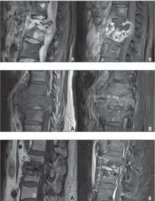
Figure 5. Multisegmental in-volvement. Total collapse of L1 and partial collapse of L2 in as-sociation with heterogeneous medullary space enhancement following intravenous gadolinium injection, demonstrating the presence of intraosseous ab-scess at L2 and heterogeneous, expansile paravertebral mass at the level of L1 to L3 with hyper-signal on STIR (A) and peripheral enhancement (B). The lesion extends toward the paravertebral region, posteriorly compressing the dural sac. Culture of the material revealed the presence of Mycobacterium tuberculosis.
A
B
Figure 6. Large, poorly defined paravertebral mass on T1-weighted image (A), with hetero-geneous contrast-enhancement on T1-weighted image with fat suppression (B), affecting verte-bral bodies T8 and T9, in asso-ciation with intact intervertebral disc. Vertebral body biopsy re-vealed the neoplastic etiology of the lesion (implant).
A
B
Figure 7. Remarkable irregular-ity and hyposignal of vertebral plateaus L2-L3 on T1-weighted image T1 (A), with intense disc enhancement on gadolinium-enhanced T1-weighted image with fat suppression (B). The ver-tebral plateaus do not present contrast enhancement. Several punctures were performed to evaluate the material. Microor-ganisms growth was not ob-served. Vertebral body biopsy revealed the presence of degen-erative joint disease mimicking infectious spondylodiscitis.
A
B
Inflammatory spondylites such as ankylosing spondylitis, psoriatic spondyli-tis and reactive spondylispondyli-tis, over its natu-ral history, may present alterations similar to the ones of degenerative spondylitis. However, the vertebral involvement is typi-cally associated with, and sometimes pre-ceded by either symmetrical or asymmetri-cal, bilateral sacroiliitis, depending on each etiology(23).
Usually, neoplastic lytic lesions with metastasis to the axial skeleton are not as-sociated with reactive sclerosis or peri-osteal reaction; and, typically, the first sites of involvement are the vertebral pedicles(23). Degenerative spondylites, like infec-tious cases, may present enhancement of the intervertebral disc on the post-gado-linium sequences. However, the affected vertebral plateaus tend to present
hypo-signal on T2-weighted images and no con-trast-enhancement after intravenous gado-linium injections(23).
classical paradiscal infection where the disc involvement occurs by contiguity with ad-jacent vertebral plateaus, classified accord-ing their initial involvement(23). Thus the central, anterior subperiosteal and appen-dicular patterns of dissemination are ob-served, all of them corresponding to the findings focused by the present essay, but their description is not included in the scope of this study.
The MRI findings suspicious for tuber-culous spondylodiscitis include multiseg-mental subligamentous involvement (Fig-ure 4); presence of well defined paraverte-bral mass/abscess (Figures 5 and 8); rela-tively spared disc at early phases of the dis-ease (Figure 4); and heterogeneous en-hancement/change in signal of vertebral bodies (Figures 5 and 8).
etiological diagnosis of spondylodiscitis is hardly established on the grounds of iso-lated imaging findings criteria since many times they are nonspecific. However, the disproportion of the degree of involvement of the intervertebral disc in relation to the corresponding vertebral plateaus, the ho-mogeneity of the vertebral bodies signal and enhancement, as well as the paraverte-bral masses volume and contours(19) should be appropriately taken into consideration as useful – although not definitive – tool in the investigation of a possible etiologic agent (Table 1). Thus, the MRI specificity for the diagnosis of spondylodiscitis is directly dependent on the characteristics of the sig-nal, on the anatomic distribution, on the proportionality between vertebral bodies and intervertebral discs involvement, on the homogeneity of enhancement of the med-ullary spaces of vertebral bodies and, in-variably, on the clinical history of the pa-tient, although it is not always available at the moment of the imaging reporting(17).
REFERENCES
1. Ribeiro DS, Araújo Neto C, D’Almeida F, et al. Achados de imagem das alterações musculoes-queléticas associadas ao lúpus eritematoso sistê-mico. Radiol Bras. 2011;44:52–8.
2. Simão MN, Nogueira-Barbosa MH. Ressonância magnética na avaliação das variações anatômicas meniscais e da anatomia ligamentar perimeniscal: potenciais causas de erro de interpretação. Radiol Bras. 2011;44:117–22.
3. Nunes RB, Amaral DT, Oliveira VS. Propedêutica radiológica do impacto femoroacetabular em tem-pos de tomografia computadorizada e ressonân-cia magnética: o que o radiologista precisa saber. Radiol Bras. 2011;44:249–55.
4. Cotta AC, Melo RT, Castro RCR, et al. Dificul-dades diagnósticas no osteoma osteoide do coto-velo: estudo clínico, radiológico e histopatológico. Radiol Bras. 2012;45:13–9.
5. Moura MVT. Interposição de fragmento perios-teal na fratura da placa epifisária femoral distal: es-tudo por ressonância magnética. Radiol Bras. 2012;45:184–6.
6. Chojniak R, Grigio HR, Bitencourt AGV, et al. Biópsia percutânea por agulha grossa de
tumo-res de partes moles guiada por tomografia com-putadorizada: resultados e correlação com análise da peça cirúrgica. Radiol Bras. 2012;45:259–62. 7. Tavares Júnior WC, Faria FM, Figueiredo R, et al. Fadiga óssea: causa de dor em joelhos na os-teoartrite. Radiol Bras. 2012;45:273–8. 8. Grassi CG, Diniz FV, Garcia MRT, et al.
Aspec-tos de imagem na tendinite calcária pré-vertebral. Radiol Bras. 2011;44:327–30.
9. Jacob Jr C, Barbosa DM, Batista PR, et al. Fratura toracolombar do tipo explosão: o que o radiolo-gista deve conhecer. Radiol Bras. 2012;45:101–4. 10. Simão MN, Helms CA, Richardson WJ. Achados de ressonância magnética em cistos epidurais de origem discal em pacientes não operados e após microdiscectomia. Radiol Bras. 2012;45:205–9. 11. Maiuri F, Iaconetta G, Gallicchio B, et al. Spondy-lodiscitis. Clinical and magnetic resonance diag-nosis. Spine (Phila Pa 1976). 1997;22:1741–6. 12. Sharif HS. Role of MR imaging in the
manage-ment of spinal infections. AJR Am J Roentgenol. 1992;158:1333–45.
13. Malawski SK, Lukawski S. Pyogenic infection of the spine. Clin Orthop. 1991;(272):58–66. 14. Varma R, Lander P, Assaf A. Imaging of pyogenic
infectious spondylodiskitis. Radiol Clin North Am. 2001;39:203–13.
15. Prasad A, Manchanda S, Sachdev N, et al. Imag-ing features of pediatric musculoskeletal tubercu-losis. Pediatr Radiol. 2012;42:1235–49. 16. Daher S, Pimenta Jr WE, Souza Jr ZA, et al. Qual
o seu diagnóstico? Radiol Bras. 2004;37:v–vii. 17. Hong SH, Choi JY, Lee JW, et al. MR imaging
assessment of the spine: infection or an imitation? Radiographics. 2009;29:599–612.
18. Resnik D. Osteomyelitis, septic arthritis and soft tissue infection: axial skeleton. In: Resnick D, edi-tor. Diagnosis of bone and joint disorders. 4th ed. Philadelphia, PA: Saunders; 2002. p. 2481–509. 19. Ledermann HP, Schweitzer ME, Morrison WB, et al. MR imaging findings in spinal infections: rules or myths? Radiology. 2003;228:506–14. 20. Moon MS. Tuberculosis of the spine.
Controver-sies and a new challenge. Spine (Phila Pa 1976). 1997;22:1791–7.
21. Gouliamos AD, Kehagias DT, Lahanis S, et al. MR imaging of tuberculous vertebral osteomyeli-tis: pictorial review. Eur Radiol. 2001;11:575–9. 22. Bron JL, de Vries MK, Snieders MN, et al. Disco-vertebral (Andersson) lesions of the spine in ankylosing spondylitis revisited. Clin Rheumatol. 2009;28:883–92.
23. Jacobson JA, Girish G, Jiang Y, et al. Radio-graphic evaluation of arthritis: inflammatory con-ditions. Radiology. 2008;248:378–89.
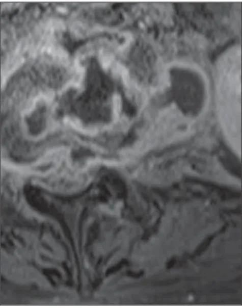
Figure 8. Paravertebral, well-defined, thin-walled collection on post-gadolinium T1-weighted se-quence. Note the presence of exuberant, periph-eral contrast enhancement, typical of collections. Culture of the collected material revealed Mycobac-terium tuberculosis.
Table 1 Main findings in the differential diagnosis of spondylodiscitis.
Vertebral body enhancement Intervertebral disc Vertebral involvement Pyogenic Homogeneous Early involvement Segmental Tuberculous Heterogeneous Relatively spared Multisegmental CONCLUSION