139
Tyng CJ et al. Percutaneous biopsy of the pancreas with pneumodissection
Radiol Bras. 2013 Mai/Jun;46(3):139–142
Computed tomography-guided percutaneous biopsy
of pancreatic masses using pneumodissection
*
Biópsia percutânea de massas pancreáticas guiada por tomografia computadorizada com pneumodissecção
Chiang Jeng Tyng1, Almir Galvão Vieira Bitencourt2, Maria Fernanda Arruda Almeida3, Paula Nicole Vieira Barbosa1, Eduardo Bruno Lobato Martins2, João Paulo Kawaoka Matushita Junior4, Rubens Chojniak5, Felipe José Fernandez Coimbra6
Objective: To describe the technique of computed tomography-guided percutaneous biopsy of pancreatic tumors with pneumodissection. Materials and Methods: In the period from June 2011 to May 2012, seven computed tomography-guided percutaneous biopsies of pancreatic tumors utilizing pneumodissection were performed in the authors’ institution. All the procedures were performed with an automatic biopsy gun and coaxial system with Tru-core needles. The biopsy specimens were histologically assessed. Results: In all the cases the pancreatic mass could not be directly approached by computed tomography without passing through major organs and structures. The injection of air allowed the displacement of adjacent structures and creation of a safe coaxial needle pathway toward the lesion. Biopsy was successfully performed in all the cases, yielding appropriate specimens for pathological analysis. Conclusion: Pneumodissection is a safe, inexpensive and technically easy approach to perform percutaneous biopsy in selected cases where direct access to the pancreatic tumor is not feasible.
Keywords: Pancreatic neoplasms; Needle biopsy; Computed tomography.
Objetivo: Descrever a técnica de biópsia percutânea de tumores pancreáticos guiada por tomografia computadorizada com pneumodissecção. Materiais e Métodos: No período de junho de 2011 a maio de 2012, foram realizadas sete biópsias percutâneas de tumores pancreáticos guiadas por tomografia computadorizada utilizando a manobra de pneumo-dissecção em nossa instituição. Todas as biópsias foram realizadas utilizando pistola de disparo automático e sistema coaxial, com agulhas Tru-core. As amostras colhidas foram submetidas a avaliação histológica. Resultados: Para todos os casos, não havia um acesso direto seguro pela tomografia computadorizada para atingir o tumor pancreático sem atravessar órgãos e estruturas importantes. A injeção de ar foi capaz de deslocar as estruturas adjacentes e criar uma nova rota de acesso, permitindo um trajeto seguro da agulha coaxial até a lesão, e a biópsia foi realizada com sucesso em todos os casos. Todas as biópsias forneceram material suficiente para análise histológica. Conclusão: Esta técnica é segura, barata e tecnicamente fácil, podendo auxiliar na realização de biópsias percutâneas de tumores pancreáticos guiadas por tomografia computadorizada de casos selecionados em que não existe acesso direto à lesão.
Unitermos: Neoplasias pancreáticas; Biópsia por agulha; Tomografia computadorizada.
Abstract
Resumo
* Study developed at Hospital A. C. Camargo, São Paulo, SP, Brazil.
1. Masters and Fellows PhD degree, Full Professors, Depart-ment of Imaging, Hospital A. C. Camargo, São Paulo, SP, Brazil. 2. PhDs, Full Professors, Department of Imaging, Hospital A. C. Camargo, São Paulo, SP, Brazil.
3. Fellow PhD degree, Department of Imaging, Hospital A. C. Camargo, São Paulo, SP, Brazil.
4. Master, Department of Imaging, Hospital A. C. Camargo, São Paulo, SP, Brazil.
5. PhD, Director, Department of Imaging, Hospital A. C. Camargo, São Paulo, SP, Brazil.
6. PhD, Director, Department of Abdominal Surgery, Hospi-tal A. C. Camargo, São Paulo, SP, Brazil.
Tyng CJ, Bitencourt AGV, Almeida MFA, Barbosa PNV, Martins EBL, Matushita Junior JPK, Chojniak R, Coimbra FJF. Computed tomography-guided percutaneous biopsy of pancreatic masses using pneumodissection. Radiol Bras. 2013 Mai/Jun;46(3):139–142.
0100-3984 © Colégio Brasileiro de Radiologia e Diagnóstico por Imagem ORIGINAL ARTICLE
lesions such as imaging-guided percutane-ous biopsy, and endoscopic or surgical bi-opsies(2).
Recently, a series of studies published in Brazil have highlighted the relevance of interventional radiology in the diagnosis and treatment of diseases in different com-partments of the body(3–10). Computed
to-mography (CT)-guided percutaneous bi-opsy is a safe and well-established tech-nique, with high accuracy in the diagnosis of focal pancreatic lesions and low rate of complications(11,12).
Most pancreatic biopsies involve direct approach to the organ. However, different overlying anatomic structures, such as the vances of imaging methods, including
functional evaluation by positron emission computed tomography and by magnetic resonance imaging(1), the diagnosis can be
suggested with no need for invasive proce-dures. In some cases, the non-invasive im-aging evaluation cannot establish an accu-rate diagnosis, and a histological analysis is required before the treatment. There are several techniques of collection of speci-mens for histological analysis of pancreatic
Mailing Address: Dr. Chiang Jeng Tyng. Rua Professor An-tônio Prudente, 211, Liberdade. São Paulo, SP, Brazil, 01509-010. E-mail: [email protected].
Received November 11, 2012. Accepted after revision Feb-ruary 7, 2013.
INTRODUCTION
Focal pancreatic lesions may be related to several pathological conditions, with dif-ferent therapeutic options. With the
140
Tyng CJ et al. Percutaneous biopsy of the pancreas with pneumodissection
Radiol Bras. 2013 Mai/Jun;46(3):139–142 stomach, bowel loops, liver, kidneys,
spleen and vessels may preclude a safe and direct access to the lesion. In such cases, alternative techniques can be utilized to access the lesion.
The present article is aimed at describ-ing the technique of CT-guided percutane-ous biopsy of pancreatic tumors with pneumodissection.
MATERIALS AND METHODS
In the period from June 2011 to May 2012, seven CT-guided percutaneous biop-sies of pancreatic tumors utilizing pneumo-dissection were undertaken in the authors’ institution. All the procedures were per-formed with an automatic biopsy gun and coaxial system with Tru-core needles (Angiotech; Vancouver, Canada) measur-ing 10 cm or 15 cm, dependmeasur-ing on the dis-tance between the skin and the lesion. Co-agulation tests were performed as a prepro-cedural routine. The collected specimens were submitted to histological analysis.
Biopsy technique
The preferential position for biopsy is that most comfortable for the patient, which most frequently is the prone position. Af-ter asepsis and local anesthesia with lidocaine 2%, a 17-gauge coaxial needle is inserted into the intra-abdominal fatty tis-sue and new CT images are acquired for access route planning. After correction of the needle tip location, 50 to 100 ml of air are injected to displace the adjacent organs and structures, while the needle is carefully advanced through the newly created access route under CT guidance. Once the coaxial needle is positioned inside or adjacent to the organ, five or six specimens are obtained with an 18-gauge cutting needle. The num-ber of specimens may be higher or lower, depending on the quality of such specimens and on the presence of bleeding or other complications. The whole procedure takes about 30 minutes to be completed.
RESULTS
In this study, most patients were women and the mean age was 69 years, ranging between 39 and 82 years. In five cases, the lesion was located in the pancreatic head, and in two cases, in the body or tail of the
pancreas. The mean diameter of the lesions was 33 mm, ranging between 17 and 47 mm. Table 1 describes all patients’ data.
In all the cases the pancreatic tumor could not be safely approached by CT with-out passing through major organs and struc-tures. The patients were positioned in lat-eral/oblique (Figure 1), dorsal or ventral decubitus (Figure 2), depending on the lo-cation of the lesion and on the planned bi-opsy route. The air injection could displace adjacent structures, creating a new access route to allow a safe advance of the coaxial needle up to the lesion. Biopsy could be successfully performed in all the cases.
No significant bleeding was observed during the procedure and none of the pa-tients complained of important pain during the air injection. One patient presented subtle pneumothorax during the procedure, but drainage was not required. Another pa-tient present slight increase in serum lev-els of pancreatic enzymes, which resolved spontaneously with no specific treatment. No late complication was reported.
All biopsies provided sufficient material for histological analysis. Five lesions were confirmed as primary pancreatic adenocar-cinoma, and two patients presented meta-static lesions (one from melanoma and other from epidermoid carcinoma of the scalp).
DISCUSSION
Several techniques have been described for percutaneous biopsy of pancreatic tu-mors where direct access to the lesion is not feasible, with good results and low rates of complications(12). Such techniques include
transgastric and transhepatic biopsies and hydrodissection maneuvers. Hydrodis-sections maneuver may also be utilized to displace structures such as vessels and
bowel loops, creating a safe access route for biopsy. Such a technique has already been described for other locations such as adre-nal and mediastiadre-nal lesions(13,14).
Pneumodissection may be utilized ei-ther isolatedly or in association with hydrodissection to displace nontarget struc-tures. However, few authors have described the utilization of such maneuver in percu-taneous procedures. In pneumodissection, either ambient air or carbon dioxide (CO2) may be injected with a syringe capable of delivering controlled gas volumes to the area of interest. A relatively great amount of gas may be injected in a way that, even in case the needle cannot be positioned between the lesion and the adjacent organs, such organs can be separated, provided a sufficient amount of gas is injected into the compartment. Both liquids and gas tend to disperse as they are injected into the ab-dominal cavity. Liquids tend to accumulate in the lower regions, while gas tends to accumulate in the superior regions, which may be useful in the selection of the mate-rial to be utilized to displace the nontarget structures in each specific case.
The safety of the intra-abdominal CO2 injection has already been proved in stud-ies about laparoscopy. CO2 pulmonary em-bolism has already described during laparoscopic surgery(15). However, the
amount of CO2 utilized during laparoscopy is much greater than the amount utilized in percutaneous procedures. Kariya et al.(16)
and Buy et al.(17) have described cases of
pneumodissection with CO2 in percutane-ous radioablation of abdominal tumors, utilizing up to 1,500 ml of gas, without complications. The probability of pulmo-nary embolism seems to be minimal with the amount of gas utilized in the presently reported procedures.
Table 1 Data from seven patients submitted to CT-guided percutaneous biopsy of pancreatic lesions with pneumodissection maneuver.
Sex / age
F / 54 years F / 75 years F / 39 years F / 52 years F / 52 years F / 51 years M / 82 years
Location Pancreatic head Pancreatic head Pancreatic head Pancreatic head Pancreatic head Pancreatic body Pancreatic tail Size (mm) 40 17 44 20 25 47 40 Biopsy access Posterior (left) Posterior (right) Posterior (right) Anterior Posterior (right) Lateral (left) Lateral (left) Histological result Adenocarcinoma Adenocarcinoma Adenocarcinoma Metastatic SCC Metastatic melanoma Adenocarcinoma Adenocarcinoma
141
Tyng CJ et al. Percutaneous biopsy of the pancreas with pneumodissection
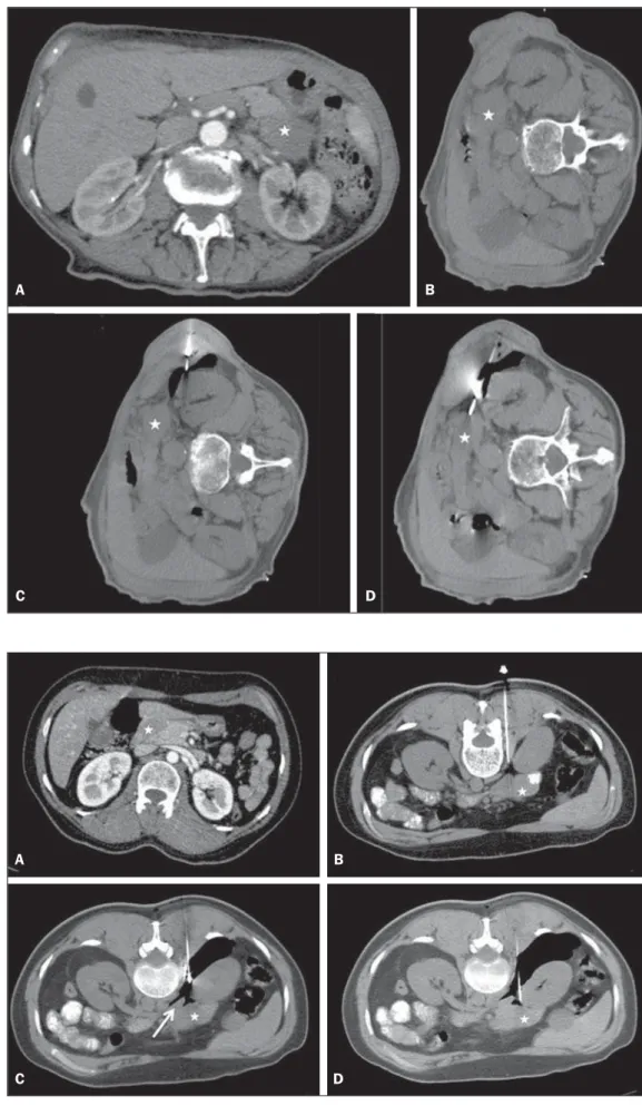
Radiol Bras. 2013 Mai/Jun;46(3):139–142 Figure 2. Female, 52-year-old
pa-tient submitted to CT-guided percu-taneous biopsy of pancreatic tumor, with posterior approach and pneu-modissection maneuver. A: Contrast-enhanced, axial CT section shows hypodense lesion in the pancreatic head (star). B: Computed tomogra-phy image acquired with the patient in ventral decubitus demonstrates the needle positioned in the right paravertebral space. C: Air was in-jected into the paravertebral space, widening the gap between the right kidney and the inferior vena cava (ar-row). D: CT image demonstrates the positioning of the coaxial needle between the right kidney and the inferior vena cava, adjacent to the pancreatic lesion. The histological result corresponded to metastatic melanoma.
A B
C D
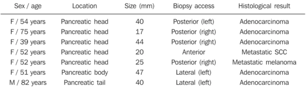
Figure 1. Male, 82-year-old patient submitted to CT-guided percutane-ous biopsy of a pancreatic mass, with lateral approach and pneumodis-section maneuver. A: Contrast-en-hanced, axial CT section demon-strates an ill-defined lesion in the pancreatic tail (star). B: Prepro-cedural planning CT image acquired with the patient positioned in lateral decubitus demonstrates the left kid-ney, spleen and stomach overlying between the lesion and the abdomi-nal wall. C: Air was injected into the perirenal space, widening the gap between the left kidney and the spleen. D: CT image demonstrates the progression of the coaxial needle through the space created by pneumodissection and the needle positioning adjacent to the lesion. The histological result corresponded to adenocarcinoma.
A B
142
Tyng CJ et al. Percutaneous biopsy of the pancreas with pneumodissection
Radiol Bras. 2013 Mai/Jun;46(3):139–142 In the presently described cases, the
pancreatic tumors were involved by other abdominal organs or structures such as bowel loops, kidneys, liver, spleen and great vessels. The approach through the liver, spleen or kidneys should be avoided be-cause of the risk for bleeding. Fine needle puncture through the gastrointestinal tract seems to be safe, however no study about core biopsy is found in the literature, so the possibility of peritonitis should ever been considered. Thus, the pneumodissection maneuver was selected as the best approach for these specific lesions, avoiding the in-volvement of adjacent organs and reducing the risks inherent to the procedure.
Finally, pneumodissection is a safe, low-cost and technically easy-to-perform maneuver that may be useful in selected cases requiring CT-guided percutaneous biopsy of pancreatic tumors where direct access to the lesion is not feasible.
REFERENCES
1. Hernandes MA, Semelka RC, Elias Jr J, et al. Whole-body MRI: comprehensive evaluation on a 48-channel 3T MRI system in less than 40
min-utes. Preliminary results. Radiol Bras. 2012;45: 319–25.
2. Goldin SB, Bradner MW, Zervos EE, et al. Assess-ment of pancreatic neoplasms: review of biopsy techniques. J Gastrointest Surg. 2007;11:783–90. 3. Chojniak R, Pinto PNV, Tyng CJ, et al. Computed tomography-guided transthoracic needle biopsy of pulmonary nodules. Radiol Bras. 2011;44:315– 20.
4. Chojniak R, Grigio HR, Bitencourt AGV, et al. Percutaneous computed tomography-guided core needle biopsy of soft tissue tumors: results and correlation with surgical specimen analysis. Radiol Bras. 2012;45:259–62.
5. Guimarães MD, Fonte AC, Andrade MQ, et al. Computed tomography-guided core-needle bi-opsy of lung lesions: an oncology center experi-ence. Radiol Bras. 2011;44:75–80.
6. Wajnberg E, Rodrigues G, Abud DG. Use of drug-eluting stents for the treatment of vertebral artery stenosis. Radiol Bras. 2011;44:343–8. 7. Queiroz HMC, Costa FA, Campos Jr MM, et al.
Arterial embolization in the treatment of hemo-bilia after hepatic trauma: a case report. Radiol Bras. 2012;45:63–4.
8. Ceratti S, Giannini P, Souza RAS, et al. Ultra-sound-guided fine-needle aspiration of thyroid nodules: assessment of the ideal number of punc-tures. Radiol Bras. 2012;45:145–8.
9. Novero ER, Metzger PB, Obregon J, et al. Endo-vascular treatment of thoracic aortic diseases: a single center result analysis. Radiol Bras. 2012; 45:251–8.
10. Ceratti S, Okano FM, Pontes ABG, et al. Ultra-sound-guided foam sclerotherapy in the treatment of chronic venous insufficiency. Radiol Bras. 2011;44:167–71.
11. Paulsen SD, Nghiem HV, Negussie E, et al. Evalu-ation of imaging-guided core biopsy of pancreatic masses. AJR Am J Roentgenol. 2006;187:769– 72.
12. Tseng HS, Chen CY, Chan WP, et al. Percutane-ous transgastric computed tomography-guided biopsy of the pancreas using large needles. World J Gastroenterol. 2009;15:5972–5.
13. Tyng CJ, Bitencourt AG, Martins EB, et al. Tech-nical note: CT-guided paravertebral adrenal bi-opsy using hydrodissection – a safe and techni-cally easy approach. Br J Radiol. 2012;85:e339– 42.
14. de Bazelaire C, Sabatier F, Pluvinage A, et al. CT-guided percutaneous biopsies. J Radiol. 2011;92: 842–59.
15. Dion YM, Lévesque C, Doillon CJ. Experimen-tal carbon dioxide pulmonary embolization after vena cava laceration under pneumoperitoneum. Surg Endosc. 1995;9:1065–9.
16. Kariya S, Tanigawa N, Kojima H, et al. Radiofre-quency ablation combined with CO2 injection for treatment of retroperitoneal tumor: protecting sur-rounding organs against thermal injury. AJR Am J Roentgenol. 2005;185:890–3.