O
RIGINAL
A
RTICLE
Revista Brasileira de FisioterapiaStrain and slackness of achilles tendon
during passive joint mobilization
via imaging ultrasonography
Deformação relativa e frouxidão do tendão calcanear durante mobilização
articular passiva através de ultra-sonografi a por imagem
Peixinho CC1, Alves DS1, Lacerda RG2, Vieira TMM2, Oliveira LF2
Abstract
Background:In vivo study of the mechanical behavior of tendons may bring advances in evaluating the impact of intervention programs
for fl exibility and strength, in clinical practice and sports. Objective: The aim of this study was to quantify the relative strain and slackness of achilles tendons during passive mobilization, for four ankle joint angles and two knee angles. Methods: The displacement of the muscle-tendon junction was quantifi ed by means of ultrasound images acquired during passive ankle mobilization, with the aid of an electrogoniometer and an electromyograph to ensure the achievement of the required angles and muscle inactivity, respectively.
Results: The strain values ranged from 4.28%±2.37 to -0.94%±1.58 for the fully extended knee, and from 2.38%±1.63 to -2.32%±2.16% for the fl exed knee. Conclusions: The values found in this study confi rm those in the literature and demonstrate how the Achilles tendon participates in length changes in the muscle-tendon unit during passive movement. These results suggest that the mechanical properties of tendinous tissues affect the relationship between the length of muscle fi bers and the joint angle, even during this type of movement.
Key words: calcanear tendon; strain; slackness; ultrasonography.
Resumo
Contextualização: O estudo do comportamento das propriedades mecânicas do tendão in vivo pode trazer avanços na avaliação do
impacto de programas de intervenção para fl exibilidade e força, nas áreas clínica e desportiva. Objetivo: O objetivo deste trabalho foi quantifi car a deformação (strain) e a frouxidão (slackness) relativas do tendão calcanear, durante mobilização passiva para quatro
ângulos articulares do tornozelo e dois do joelho. Materiais e métodos: O deslocamento da junção miotendínea foi quantifi cado
através de imagens ultra-sonográfi cas capturadas durante a mobilização passiva do tornozelo, com o auxílio de um eletrogoniômetro
e um eletromiógrafo, para garantir as angulações requeridas e a inatividade muscular, respectivamente. Resultados: Os valores de
deformação relativa encontrados variaram de 4,28±2,37 a -0,94±1,58% para o joelho estendido e de 2,38±1,63 a -2,32±2,16% para o joelho fl etido. Conclusões: Os valores encontrados ratifi cam os da literatura, demonstrando a participação do tendão calcanear na variação do comprimento da unidade músculo-tendão, durante movimentação passiva. Estes resultados sugerem que as propriedades mecânicas dos tecidos tendinosos afetam a relação entre o comprimento das fi bras e o ângulo articular, até mesmo nesse tipo de movimento.
Palavras-chave: tendão calcanear; deformação; frouxidão; ultra-sonografi a.
Received: 15/09/2007 – Revised: 25/03/2008 – Accepted: 07/07/2008
1 Ultrasound Laboratory, Biomechanics Engineering Program, Alberto Luiz Coimbra Postgraduate and Research Institute of Engineering, Universidade Federal do Rio de Janeiro (PEB/COPPE/UFRJ)
– Rio de Janeiro (RJ), Brazil
2 Biomechanics Laboratory, School of Physical Education and Sports (EEFD), UFRJ
Introduction
The mechanical characteristics of free tendons and aponeuroses in humans have been investigated mainly through in vivo research1-6. However, a number of questions remain about the interactions of these structures and their properties during different types of joint movement. It is known that tendinous tissues are not unextendable as considered by some models of muscular contraction. On the contrary, they show elastic properties and play the role of biological coils that allow a dynamic mechanical interaction between muscles and tendons1-3.
Recent evidence has demonstrated that tendinous tissues display increased compliance within an initial range of deformation under low overload (known as toe region), thus the application of a low intensity passive force produced by the muscle fibers may deform the tissues4. Because the passive force of the muscle-tendon unit is a function of the muscular length, the degree of deformation of tendinous tissues can be modified according to the joint angle. Also, because the force transmitted to the bone segment must be preserved, this deformation may require additional shortening of the muscular fibers during the contraction and according to the joint angle4. Therefore, the study of tendon length variation combined with the joint angle during passive movements is vital to the understanding of the load/length ratio (stiffness) of this tissue and to the understanding of the mechanics in active conditions such as estimations of the speed of shortening and the length of the muscle fibers4.
The tendon length/joint angle ratio in passive conditions is especially important in muscles that have short fibers when compared to the size of the tendon, such as the gastrocnemius medialis (GM). In this case, a certain relative deformation corresponds to a greater absolute change in muscular fiber length4.
In vivo ultrasound imaging has been frequently used to determine the mechanical properties of tendons and of aponeuroses in humans5-9. Because it is a non-invasive, easy-to-use and relatively low-cost method of obtaining high resolution images of structures of different sizes and depths in the human body. This technique allows real time monitoring of moving structures and the post-processing of the generated images, avoiding limitations imposed by other methods1. The GM has been extensively studied because it is superficial and easy to view with current high resolution image techniques5,10, in addition to being part of a muscle group of great functional importance to human locomotion.
One parameter that is often used to characterize these tissues is the relative deformation or strain, which is determined in vivo through image analysis during
passive movements and maximum voluntary isometric contractions3-5,9,11,12. However, most studies focus on intense isometric contractions and give little attention to the direct measurement of muscle and tendon length increase at low levels of tension, typically related to relaxed muscles4,5,7. Another equally important but less investigated parameter is tendon slackness, evidenced by negative strain values and present in short muscle lengths4 where tendons are “loose”. Herbert and Gandevia13 suggested that a greater internal shortening of muscle fibers and/or a greater pennation angle in a short muscle length can be attributed to tendon slackness.
The structural and functional characteristics of tendinous tissues change with injury7,9,14 or during the recovery process in a rehabilitation program7,11,14-17. This methodology has been important in studies on the effects of stretching and fatigue on the viscoelastic properties of the tendon as they require not only passive movement but also muscular activation11,14-16. We can cite the articles of Kubo, Kanehisa and Fukunaga11 and Kubo et al.15 who studied the acute and chronic effects of passive stretching, commonly used in injury prevention and performance improvement programs, through joint range of motion gain. They reported that the potential mechanism to reduce risk of injury combined with flexibility gain is caused by variations in the viscoelastic properties of the muscle-tendon units. Mademli, Arampatzis and Walsh14 and Kubo et al.16 used a similar methodology to study the influence of repeated muscle contractions and fatigue on tendon elasticity, which has an important application in the prescription of physical training regimes. The authors also tried to underline the influence of variables such as type and duration of contractions on the magnitude of tendinous tissue adaptation.
h e study of the mechanical properties of this tissue in vivo allows the examination of the adaptation of tendons and aponeuroses to physical activity and the impact of interventions such as stretching programs or resisted exercise on these structures. It also allows the understanding of the function and performance capability of muscle-tendon units and provides relevant information concerning the input parameters for the simulation of human system models.
The objectives of this study were to quantify the strain and the slackness of the free tendon of the GM during passive movements of the ankle joint, and to investigate the variations in the analyzed parameters for different knee joint angles.
Strain and slackness of calcanear tendon measured via ultrasonography
Eleven subjects ( five men and six women) aged (mean±standard deviation) 23.64±3.56 years, 170.36±7.45cm tall and weighting 70.36±14.45kg took part in this study. Subjects did not report any history of bone, muscle or joint injury to the lower limbs and signed a free and informed consent form. The present study was approved by the Ethics in Research Committee of Universidade Federal do Rio de Janeiro (UFRJ) (approval no. 03107).
The images were acquired using an ultrasound system (EUB-405 by Hitachi Medical Corporation, Tokyo, Japan) with a linear transducer (7.5MHz frequency). Gel (Ultrex-gel, Farmativa Indústria e Comércio, Rio de Janeiro) was also used for the acoustic coupling and to prevent skin surface depression. A single researcher handled the device for the entire period of data collection. He was trained by means of data collection on ultrasound phantoms for inter- and intra-examiner reproducibility tests and other previous experimental tests with humans. In addition, a four-channel, 2kHz sampling frequency electromyograph was used (Miotec, Equipamentos Biomédicos Porto Alegre, RS, Brazil) with Ag-AgCl surface electrodes (Meditrace Kendall, CA, USA) and an electrogoniometer (Miotec, Equipamentos Biomédicos Porto Alegre, RS, Brazil)
the ankle joint from 75° (dorsil exion) to 120° (plantar l exion), at 15° intervals (75 to 90°, 90 to 105°, 105 to 120°), with the knee in two positions ( full extension and 90° l exion). h e speed of approximately 2°/seconds was set by a timer, and the tests were conducted by the same examiner who was trained for three months. h e subject remained in the prone position on a stretcher with feet free, however a plate was attached to the right foot with Velcro strips and the electrogoniometer was coaxially positioned on the ankle joint. Each subject performed the tests twice with a minimum interval of 48 hours.
Initially, the longitudinal mean axis of the GM muscle-tendon unit was determined by the methodology described by Narici et al.8, Maganaris6,9 and Maganaris and Paul12. This protocol consisted of generating ultrasound images in the axial plan with a 2cm interval. The images were used to identify the lateral and medial edges, and the midpoint between the edges was later marked on the skin. The mid-longitudinal axis of the GM is the straight line that links the midpoint marked on the skin to the distal insertion point of the calcaneal tendon, which was also identified by the ultrasound images. The transducer was then positioned longitudinally along this axis in order to locate the myotendinous junction (MTJ).
The length of the calcaneal tendon was defined as the distance between its most distal insertion point and the MTJ of the right GM (extramuscular portion), identified with the ultrasound. The resting length of the calcaneal tendon was measured with the ankle in a relaxed position, and the joint angle was recorded. At every passive variation of the joint range of motion, a new location of the MTJ was considered for measurement of the length of the corresponding tendon. Figure 1 shows ultrasound images that were obtained during the procedure and that allowed the measurement of the length of the calcaneal tendon and the calculation of its strain.
Fully extended knee Flexed Knee (90º) Θr 195.72±20.77 196.27±21.37 75º# 204.09±21.81* 201.09±20.64* 90º 190.09±22.02* 197.72±21.20 105º 195.63±20.81 196.27±20.44 120º 193.81±20.19 191.90±18.90*
Θr(º)=joint angle adopted for measurement of resting tendon length; *=statistical
difference compared to resting length (p<0.05); #=statistical difference between knee positions (p<0.05).
Table 1: Gastrocnemius medialis tendon length (mm) at each ankle angle for both knee positions.
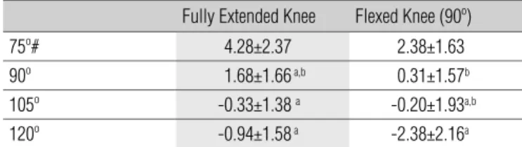
Fully Extended Knee Flexed Knee (90º)
75º# 4.28±2.37 2.38±1.63
90º 1.68±1.66 a,b 0.31±1.57b
105º -0.33±1.38 a -0.20±1.93a,b
120º -0.94±1.58 a -2.38±2.16a
Table 2. Strain (%) of the gastrocnemius medialis tendon at each ankle angle for both knee positions.
a=statistical difference compared to 75º (p<0.05); b=statistical difference compared to 120º(p<0.05);# =statistical difference between knee positions(p<0.05).
analysis and the hypothesis tests. The adherence of the data to the normal distribution was confirmed by the Kolmogorov-Smirnov test. Student´s t-test was applied to analyze the differences between the length of the tendon in each of the four angles of the ankle and with the rest angle, as well as for the differences between the values for all parameters measured with the flexed and extended knee. The same procedure was used to compare the results between the two days of the test. The ANOVA and the Tukey post-hoc test were used for the statistical analysis of the differences between the relative strains of the four ranges of passive mobilization. The level of significance adopted for these tests was p<0.05.
The reliability of this methodology was previously assured in other similar studies1,3,4,9. The inter- and intra-examiner variations for ultrasound measurements of the resting length of the connective tissue and during isometric contraction were confirmed at 2 to 4%19. The reproducibility of the measurements was verified through the coefficients of variation which were 1.59, 1.02, 10.39 and 10.06% for the rest angle with extended and flexed knee, and the tendon lengths with extended and flexed knee, respectively.
Results
The results obtained in the first and second day did not show any significant differences. There was no myoelectrical activity in the LG during passive mobilization of the ankle.
The values of resting tendon length for the extended and flexed knee were 195.72±20.77mm for an angle of 109.09±4.15° and 196.27±21.37mm for an angle of 106.63±3.90° respectively, with a significant difference for the angle, but not for the length. Table 1 shows the lengths of the tendon at the analyzed ankle angles for both knee positions.
Table 2 demonstrates the tendon strain with flexed and extended knee and Figure 2 shows the dispersions of the relative strain values in the ranges of ankle mobilization for the extended and flexed knee, respectively. It can be highlighted that positive values of relative strain indicate tendon strain, and negative values indicate relative slackness.
Discussion
The values of the present study for resting tendon length (195.7±20.7mm) were within the range of mean values found in the literature with similar measurement methodology: 178±24mm4, 190±30mm10, 225± 20mm6 and 240.3±39.9mm20.
Some of these variations can be explained by the differences in height of the analyzed groups, which suggests different leg lengths given that the other characteristics of the subjects were similar. As an example, the subjects analyzed by Arampatzis et al.20 had a mean height of 185±6cm, greater than the values in the present study, and resting tendon lengths were also greater. Herbert et al.5 reported far greater values of resting length (302±28mm). In this case, the difference may be due to the methodology because the resting tendon length of the GM was estimated by subtracting the variation in fasciae length (measured by ultrasound) from the total variation of the myotendineous unit, based on anthropometric data of cadavers. These differences point to the importance of analyzing the methodology used to calculate this parameter in comparative studies on relative tendon strain.
Values for relative strain of tendinous tissues are reported in the literature for animals and humans in vitro
which vary between 2 and 12%1. Some examples are: 2% for the gastrocnemius of frogs21 and 3.68±0.31% for the wrist tendons of human cadavers22. For in vivo studies, the relative strain of the calcaneal tendon has been mainly quantified during maximum voluntary isometric contractions. As an example, Arampatzis et al.20 reported a relative strain of 4.72±1.85%, Magnusson et al.23 of 4.4±5.6%, Muramatsu et al.3 of 5.1±1.1%, Kubo, Kanehisa, Fukunaga et al.7 of 5.2±1.4% and Muraoka et al.24 of 5.3±1.6%. Despite differences in Strain and slackness of calcanear tendon measured via ultrasonography
methodology, these studies report similar values for relative strain (around 5%), which are greater than those found in the present study.
These lower values may be attributed to the different type of muscular activity because, during maximum voluntary contraction, the muscle shortens as it produces force, applying traction to the tendon. Furthermore, Herbert et al.5 suggest that, when resting muscles are stretched, most of the increase in total length occurs in the tendon (even without muscle traction), although the mean values of relative strain are lower than in active movements. Some authors also suggest that the extramuscular part of the calcaneal tendon becomes slack or very compliant when the ankle receives passive dorsiflexion. This does not occur during passive plantar flexion, as suggested by the results for relative strain and slackness of the present study.
Little data was found in the literature with measurements during passive movement. The results for relative strain of the present study (4.28, 1.68, -0.33 and -0.94% for 75°, 90°, 105° and 120° angles) were in agreement with those of Muraoka et al.10, who found relative strain values of -2.6, -0.5, 0.6, 1.4 and 1.8% for ankle angles of 120°, 110°, 100°, 90° and 85° angles, respectively. These tests were done with the knee extended, and the resting tendon length was measured with the ankle joint at a mean angle of 106±5°, similar to the present study. The values shown in this study confirm the findings of another study on the same group4, in which relative strain values during passive movements for 83 and 126° angles were 2.4±1.0% and -3.5±1.6%, respectively. The difference in the maximum value for relative strain may be
explained by the use of a greater angle of dorsiflexion than the one used by other authors, which would cause greater relative strain not only of the tendinous structure, but of the entire muscle-tendon unit. Muraoka et al.4 suggest that this strain feature, confirmed by the present study, makes the compliant calcaneal tendon act as a shock absorber in high impact movements because it allows adaptation to changes in length during this type of movement. However, another important feature is the relative slackness of the tendon without which the shock-absorbing property would be impaired in short muscle lengths due to their low potential for strength production4.
The mean value for relative slackness found in this study with the ankle at 120° and extended knee (-0.94±1.58%) was lower than the one reported in the two studies carried out under the same conditions (approximately -2.6±-3.5%)4,10. This fact might be derived from methodological differences between the studies. Muraoka et al.4,10 quantified the relative slackness by processing scanned ultrasound images then subtracting the displacements of the distal points of insertion of the tendon from those of the MTJ, which could increase the total strain. Another methodological difference is the cyclical mobilization of the ankle joint during warm-up, which could also increase the relative tendon slackness.
The mean value for relative slackness with the ankle at 120° and flexed knee was 2.32±2.16%, higher than with the extended knee but with marginal statistical results (p=0.07). Although no data under similar conditions were found in the literature, this result was expected as the gastrocnemius in this knee position has reduced length and no passive
Figure 2. Boxplots of relative strain of the gastrocnemius medialis (GM) tendon at each ankle angle for extended knee (A) and fl exed knee -90o (B).
The fi gure clearly shows the pattern of strain (%) reduction as joint angles increase, for both knee positions
Strain (5)
-2
2
4
6
8
10
075 090 105 120
Angles
Strain (5)
-4
-2
0
2
4
075 090 105 120
Angles
tension. This is confirmed by the fact that the resting length was reached at different joint angles with the knee extended and flexed: 109.09±4.15° and 106.63±3.90°, respectively. This result was also corroborated by the values for relative strain and tendon length with the ankle at 75° where an inverse relation was found, i.e. significantly higher values with extended knee (4.28±2.37% and 204.09±21.81mm for strain and length, respectively) in comparison with flexed knee (2.38±1.63% and 201.09±20.64mm). Riener and Edrich26 confirm these results as they observed the zero moment in the ankle at a lower plantar flexion position, when the knee was flexed at 60o. This can be explained by the fact that passive joint torque depends on the properties of the surrounding tissues4,10.
These results support the suggestion of previous studies2,3 regarding the importance of standardizing the joint angle considered as reference of resting tendon length, which could guarantee the absence of strain. The previous studies used 0% relative strain for tendon length when the passive moment is zero. Deviations from this standard would result in a different load-strain ratio of tendinous tissues and would hinder adequate comparisons of results between different studies. The adoption of the neutral anatomical position (90º) as a reference point may cause errors in the measurement of relative strain because the tendon already displays a degree of strain when the knee is extended, as shown in the present study.
The use of the methodology of the present study will allow the follow-up of rehabilitation programs that involve muscle stretching and strengthening exercises, as already reported by Kubo, Kanehisa and Fukunaga11 and Kubo et al.15-17. The authors reported a reduction in viscosity and an increase in tendon elasticity immediately after passive stretching sustained for ten minutes, with a significant difference in relative strain values before (8.1±1.6%) and after (8.6±1.7%) stretching15. In another study conducted by the same group, researchers observed that repeated muscle contractions led to changes in tendon compliance and strain, suggesting that
elasticity could be modified by the duration of the action and not by the level of strength or type of muscular action16. Finally, Muraoka et al.24, using a similar methodology, found a positive correlation (r=0.39) between the muscle strength and the relative strain (5.3±1.6%) of the calcaneal tendon, which suggests that individuals with higher levels of strength are able to store more elastic energy in the tendon and indicates the possibility and the need to investigate the adaptation of tendon properties to strength training. However, the plastic changes resulting from programs that seek long-term effects and from passive joint mobilization have yet to be investigated, confirming the potential for the application of the described technique.
Conclusions
The present study described the values of relative calcanear tendon strain and slackness by means of ultrasound image analysis technique during passive ankle joint mobilization. The values found in the present study confirmed those described in the literature and demonstrated the participation of the calcanear tendon in the length variation of the muscle-tendon unit during passive movement. Considering that the length of the GM tendon changes during passive joint movements, it can be concluded that the mechanical properties of tendinous tissues quantitatively change the fiber length/joint angle ratio, thus affecting the mechanical properties of the muscle, such as strength production potential, shortening speed and load/length ratio (rigidity) even in this type of movement.
The employed method of analysis showed similar results to those found in the literature, as reference for the same test conditions. It is fair to assume that the method can be used to follow-up the impact of stretching and resisted exercise programs on tendinous structures of the human body, as prescribed in research and clinical follow-ups in the clinical and sport fields.
Magnusson SP, Hansen P, Aagaard P, Brønd J, Dyhre-Poulsen 1.
P, Bojsen-Moller J et al. Differential strain patterns of the human gastrocnemius aponeurosis and free tendon, in vivo. Acta Physiol Scand. 2003;177(2):185-95.
Maganaris CN, Paul JP. In vivo human tendon mechanical properties. J 2.
Physiol. 1999;521 (Pt 1):307-13.
Muramatsu T, Muraoka T, Takeshita D, Kawakami Y, Hirano Y, 3.
Fukunaga T. Mechanical properties of tendon and aponeurosis of
human gastrocnemius muscle in vivo. J Appl Physiol. 2001;90(5): 1671-8.
Muraoka T, Muramatsu T, Takeshita D, Kawakami Y, Fukunaga T. Length 4.
change of human gastrocnemius aponeurosis and tendon during passive joint motion. Cells Tissues Organs. 2002;171(4):260-8.
Herbert RD, Moseley AM, Butler JE, Gandevia SC. Change in length of 5.
relaxed muscle fascicles and tendons with knee and ankle movement in humans. J Physiol. 2002;539(Pt 2):637-45.
References
Strain and slackness of calcanear tendon measured via ultrasonography
Maganaris CN, Paul JP. Tensile properties of the in vivo human 6.
gastrocnemius tendon. J Biomech. 2002;35(12):1639-46.
Kubo K, Kanehisa H, Fukunaga T. Effects of cold and hot water immersion 7.
on the mechanical properties of human muscle and tendon in vivo. Clin Biomech. 2005;20(3):291-300.
Narici MV, Binzoni T, Hiltbrand E, Fasel J, Terrier F, Cerretelli P. In vivo 8.
human gastrocnemius architecture with changing joint angle at rest and during graded isometric contraction. J Physiol. 1996;496(pt 1): 287-97.
Maganaris CN. Tensile properties of the in vivo human tendinous tissue. 9.
J Biomech. 2002;35(8):1019-27.
Muraoka T, Muramatsu T, Fukunaga T, Kanehisa H. Infl uence of tendon 10.
slack on electromechanical delay in the human medial gastrocnemius in vivo. J Appl Physiol. 2004;96(2):540-4.
Kubo K, Kanehisa H, Fukunaga T. Effect of stretching training on the 11.
viscoelastic properties of human tendon structures in vivo. J Appl Physiol. 2002;92(2):595-601.
Maganaris CN, Paul JP. Load-elongation characteristics of in vivo human 12.
tendon and aponeurosis. J Exp Biol. 2000;203(Pt 4):751-6.
Herbert RD, Gandevia SC. Changes in pennation with joint angle and 13.
muscle torque: in vivo measurements in human brachialis muscle. J Physiol. 1995;484(Pt 2):523-32.
Mademli L, Arampatzis A, Walsh M. Effect of muscle fatigue in the 14.
compliance of the gastrocnemius medialis tendon and aponeurosis. J Biomech. 2006;39(3):426-34.
Kubo K, Kanehisa H, Kawakami Y, Fukunaga T. Infl uence of static 15.
stretching on viscoelastic properties of human tendon structures in vivo. J Appl Physiol. 2001;90(2):520-7.
Kubo K, Kanehisa H, Kawakami Y, Fukunaga T. Infl uences of repetitive 16.
muscle contractions with different modes on tendon elasticity in vivo. J Appl Physiol. 2001;91(1):277-82.
Kubo K, Akima H, Kouzaki M, Ito M, Kawakami Y, Kanehisa KH et 17.
al. Changes in the elastic properties of tendon structures following 20 days bed-rest in humans. Eur J Appl Physiol. 2000;83(6): 463-8.
Freriks B, Hermens HJ, Disselhorst-Klug C, Rau G
18. . Development of
recommendations for SEMG sensors and sensor placement procedures. J Electromyogr Kinesiol. 2000;10(5):361-74.
Maganaris CN, Kawakami Y, Fukunaga T. Changes in aponeurotic 19.
dimensions upon muscle shortening: in vivo observations in man. J Anat. 2001;199(Pt 4):449-56.
Arampatzis A, Stafilidis S, DeMonte G, Karaminidis K, Morey-20.
Klapsing G, Brüggemann GP. Strain and elongation of the human gastrocnemius tendon and aponeurosis during maximal plantarflexion effort. J Biomech. 2005;38(4):833-41.
Trestik CL, Lieber RL.
21. Relationship between Achilles tendon mechanical properties and gastrocnemius muscle function. J Biomech Eng. 1993;115(3):225-30.
Loren GJ, Lieber RL. Tendon biomechanical properties enhance human 22.
wrist muscle specialization. J Biomech. 1995;28(7):791-9.
Magnusson SP, Aagaard P, Dyhre-Poulsen P, Kjaer M. Load-displacement 23.
properties of the human triceps surae aponeurosis in vivo. J Physiol. 2001;531(Pt 1):277-88.
Muraoka T, Muramatsu T, Fukunaga T, Kanehisa H. Elastic properties of 24.
human Achilles tendon are correlated to muscle strength. J Appl Physiol. 2005;99(2):665-9.
Kawakami Y, Abe T, Fukunaga T. Muscle-fi ber pennation angles are 25.
greater in hypertrophied than in normal muscles. J Appl Physiol. 1993;74(6):2740-4.
Riener R, Edrich T. Identifi cation of passive elastic joint moments in the 26.