ultrasonography as a supplementary
tool in the differential diagnosis of
fetal cleft lip and palate
Myoung Seok Lee1, Jeong Yeon Cho2,3, Sang Youn Kim2, Seung Hyup Kim2,3, Joong Shin Park3,4, Jong Kwan Jun3,4
1
Department of Radiology, Seoul Metropolitan Government-Seoul National University Boramae Medical Center, Seoul; 2Department of Radiology, Seoul National University Hospital, Seoul; 3College of Medicine, Seoul National University, Seoul; 4Department of Obstetrics and Gynecology, Seoul National University Hospital, Seoul, Korea
https://doi.org/10.14366/usg.16025 pISSN: 2288-5919 • eISSN: 2288-5943 Ultrasonography 2017;36:53-59
Purpose: The purpose of this study was to evaluate the feasibility and usefulness of sagittal color Doppler ultrasonography (CDUS) for the diagnosis of fetal cleft lip (CL) and cleft palate (CP). Methods: We performed targeted ultrasonography on 25 fetuses with CL and CP, taking coronal and axial images of the upper lip and maxillary alveolar arch in each case. The existence of defects in and malalignment of the alveolus on the axial image, hard palate defects on the mid-sagittal image, and low-through defects on CDUS taken during fetal breathing or swallowing were assessed. We compared the ultrasonography indings with postnatal indings in all fetuses. Results: Alveolar defects were detected in 16 out of 17 cases with CP and four out of eight cases with CL. Alveolar malalignment and hard palate defects were detected in 11 out of 17 cases and 14 out of 17 cases with CP, respectively, but not detected in any cases with CL. Communicating low through the palate defect was detected in 11 out of 17 cases of CL with CP. The accuracy of detection in axial scans of an alveolar defect and malalignment was 80% and 76%, respectively. Accuracy of detection of in mid-sagittal images of hard palate defect and low was 80% and 86%, respectively. The overall diagnostic accuracy of combined axial and sagittal images with sagittal CDUS was 92%.
Conclusion: Sagittal CDUS of the fetal hard palate is a feasible method to directly reveal hard palate bony defects and low through defects, which may have additional value in the differential diagnosis of fetal CL and CP.
Keywords: Ultrasonography, prenatal; Cleft lip; Cleft palate; Ultrasonography, Doppler, color; Congenital abnormalities
Received: May 18, 2016 Revised: July 28, 2016 Accepted: August 6, 2016
Correspondence to:
Jeong Yeon Cho, MD, Department of Radiology, Seoul National University Hospital, 101 Daehak-ro, Jongno-gu, Seoul 03080, Korea
Tel. +82-2-2072-3074 Fax. +82-2-747-7418 E-mail: radjycho@snu.ac.kr
ORIGINAL ARTICLE
This is an Open Access article distributed under the terms of the Creative Commons Attribution Non-Commercial License (http://creativecommons.org/ licenses/by-nc/3.0/) which permits unrestricted non-commercial use, distribution, and reproduction in any medium, provided the original work is properly cited.
Copyright © 2017 Korean Society of Ultrasound in Medicine (KSUM)
How to cite this article:
Lee MS, Cho JY, Kim SY, Kim SH, Park JS, Jun JK. Value of sagittal color Doppler ultrasonography as a supplementary tool in the differential diagnosis of fetal cleft lip and palate. Ultrasonography. 2017 Jan;36(1):53-59.
Introduction
congenital anomalies, reported in approximately one in 700 live births [1]. The rates of CL with or without CP are higher in parts of Latin America and Eastern Asia and lower in South Africa and Southern Europe [2]. The accurate prenatal detection and differential diagnosis of CL and CP is of clinical importance because the type and extension of clefts are correlated with fetal outcomes and associated anatomical and chromosomal abnormalities [3,4]. CL without CP is generally known to show favorable prognosis compared to CL with CP. Reconstruction techniques, surgical implications, and risks for chronic otitis media, hearing loss, and abnormal speech are very different when the palate is involved. CL with CP includes greater morbidity and reparations resulting in more extensive surgeries.
Transabdominal ultrasonography (TA-US), performed in the second trimester of pregnancy, is the first choice of imaging modalities to screen for orofacial clefts due to its easy accessibility and the fact that it does not result in radiation exposure. In low-risk groups (unselected population-based groups of pregnant women), the overall detection rate for all types of clefts was reported as 0%-70%, for CL with or without CP it was 33%-88%, and for isolated CP it was 0%-22% [5]. There have been some prospective studies for the detection and identiication of CL and CP in high-risk groups (i.e., referral cases because of abnormal findings during routine prenatal screening ultrasonography [US], family history of CL or CP). The reported detection rate in these cases using 2-dimensional (2-D) TA-US was 91%-96% for CL with or without CP and 41%-49% for CL with CP [6-8]. For the high-risk group in these studies, using 3-dimensional (3-D) TA-US improved the detection rate to 100% for CL with or without CP and 41%-46% of CL with CP [6-8]. Some additional scan techniques have been introduced for 3-D US, such as the reverse face view [9] or lipped face view [10], to improve the diagnostic accuracy of detection and identiication of CL and CP.
However, 3-D US takes a longer time to execute a scan and, in the majority of cases, needs a customized volume transducer. Hence, conventional 2-D US is still important as the first-line imaging modality for the prenatal diagnosis of CL and CP. Efforts to enhance the accuracy of 2-D US, including the application of color or power Doppler have been reported [11-13]. A conventional grayscale ultrasound may miss the existence of CP, because acoustic shadowing from the bony alveolar ridge often obscures the palate defect. Previous communications reported that the color or power Doppler on the sagittal plane can improve the diagnostic accuracy of CP by detecting the slow amniotic luid lows between the buccal space and the nasal fossa during breathing or swallowing, with representative cases at the level of case reports [11-13]. However, to our knowledge, there have been no previous studies validating the use of color Doppler ultrasonography (CDUS) on the sagittal
plane in the diagnosis of CL and CP.
Thus, the aim of our study is to evaluate the feasibility and usefulness of sagittal CDUS for the diagnosis of fetal CL and CP.
Materials and Methods
This prospective study was approved by our Institutional Review Board and Ethics Committee. Informed consent was carefully obtained from all study participants.
Study Population
From January 2007 to January 2010, 25 pregnant women were consecutively enrolled in our study. The study participants were those patients whose fetuses were suspected to have CL or CP as a result of routine screening US scans in the obstetrics clinic of our center, followed by a referral to the radiology department for targeted US scans. There were no multiple gestation cases, so a total of 25 fetuses were evaluated in this study. The mean maternal age was 31.2 years old (range, 26 to 39 years). The mean gestational age at the time of the targeted US was 25.6 weeks old (range, 18.5 to 36.0 weeks). The characteristics of study participants are summarized in Table 1.
Targeted Ultrasound
have taken approximately 5 minutes.
Image Analysis
All targeted US findings were compared to postnatal findings based on hospital records made by experienced obstetricians. Clefts were categorized by location and extent (involving lips, alveolus, and palate) and by laterality (unilateral, bilateral, or midfacial) in both the targeted US and postnatal indings. Four imaging indings (i.e., alveolar defect in an axial scan, alveolar malalignment in an axial scan, palatal echo defect in a sagittal scan, and low-through defect in a sagittal CDUS image) were documented in each case. The existence rates of each inding for each fetus with CL with CP or isolated CL were calculated. Finally, the accuracy of detecting CL with or without CP using a combined scan of axial and sagittal images, including CDUS images, was also calculated.
Results
All of the 25 fetuses were conirmed postnatally to have an orofacial cleft. Among them, 17 had CL with CP and eight had isolated CL only (CL without CP). Four of the eight isolated CL cases had a cleft alveolus. Due to weak fetal swallowing, a sagittal CDUS could not be obtained in four out of the 25 cases (three out of 17 CL with CP cases and one out of eight isolated CL case).
An alveolar defect in an axial image was detected in 16 out of 17 cases of CL with CP (94.1%), and in four out of eight (50%) isolated CL cases. The overall accuracy for diagnosing CL with or without CP using the inding of an alveolar defect in an axial image was 80% (20 out of 25 cases). Alveolar malalignment was detected in 11 out of 17 cases of CL with CP (57.8%); however, it was not detected in any cases of isolated CL. Palate echo defects in sagittal scans were detected in 14 of 17 cases (82.4%). Communicating
Table 1. Summary of study populations
Case No.
Maternal age (yr)
GA at US (wk)
Sagittal US
grayscale Sagittal CDUS
Arch defect
Arch
mal-alignment US diagnosis Postnatal diagnosis
1 34 27.5 Nonspeciic No transpalatal low - - Cleft lip Cleft lip
2 33 21.4 Nonspeciic No transpalatal low + + Cleft lip and palate Cleft lip and palate
3 28 36.0 Hard palate defect (+), low through defect + - Cleft lip and palate Cleft lip and alveolus
4 31 22.0 Hard palate defect Could not acquired + - Cleft lip and palate Cleft lip and palate
5 34 20.0 Hard palate defect Could not acquired + + Cleft lip and palate Cleft lip and palate
6 33 34.4 Hard palate defect Could not acquired + + Cleft lip and palate Cleft lip and palate
7 33 30.2 Hard palate defect (+), low through defect + + Cleft lip and palate Cleft lip and palate 8 30 30.1 Hard palate defect (+), low through defect + + Cleft lip and palate Cleft lip and palate 9 32 26.4 Hard palate defect No transpalatal low + Equivocal Cleft lip and alveolus Cleft lip and alveolus
10 38 21.3 Hard palate defect (+), low through defect + + Cleft lip and palate Cleft lip and palate 11 34 31.5 Hard palate defect (+), low through defect + - Cleft lip and palate Cleft lip and palate 12 31 25.0 Hard palate defect (+), low through defect + + Cleft lip and palate Cleft lip and palate 13 29 28.2 Hard palate defect (+), low through defect + - Cleft lip and palate Cleft lip and palate
14 32 18.5 Hard palate defect (+), low through defect + - Cleft lip and palate Cleft lip and palate
15 28 37.0 Nonspeciic No transpalatal low - - Cleft lip Cleft lip and alveolus
16 26 21.3 Nonspeciic No transpalatal low - - Cleft lip Cleft lip
17 30 21.6 Nonspeciic No transpalatal low + - Cleft lip Cleft lip and alveolus
18 39 27.3 Nonspeciic No transpalatal low - - Cleft lip Cleft lip
19 24 34.4 Nonspeciic No transpalatal low + +, mild Cleft lip and alveolus Cleft lip and palate
20 26 26.2 Nonspeciic (+), low through defect - - Cleft lip and palate Cleft lip and palate
21 37 25.1 Nonspeciic Could not acquired + - Cleft lip and alveolus Cleft lip and alveolus
22 32 30.0 Hard palate defect (+), low through defect + + Cleft lip and palate Cleft lip and palate 23 30 22.5 Hard palate defect (+), low through defect + + Cleft lip and palate Cleft lip and palate 24 28 22.0 Hard palate defect (+), low through defect + - Cleft lip and palate Cleft lip and palate
25 27 35.0 Hard palate defect (+), low through defect + + Cleft lip and palate Cleft lip and palate
identiied as transpalatal low in the CDUS.
The sensitivity, speciicity, positive predictive values (PPV), negative predictive values (NPV), and accuracy of each of the parameters (i.e., alveolar arch defect in an axial scan, alveolar malalignment in an axial scan, hard palate defect in a sagittal scan, transpalatal low in a CDUS, and combined US interpretation using all parameters) for differentiating CL with CP from isolated CL are summarized in Table 2. Sagittal CDUS presented slightly improved sensitivity, accuracy, and PPV compared to sagittal grayscale scan (85.71% vs. 82.35% sensitivity, 85.71% vs. 80.00% PPV, and 92.31% vs. 87.50% accuracy, respectively). The combined US interpretation of all parameters showed good diagnostic performance, with 94.12% sensitivity, 94.12% PPV, and 91.67% accuracy.
Discussion
The standard US evaluation method of fetal upper lip and anterior low-through defects in the sagittal CDUS were identiied in 12 of
17 cases (70.6%) of CL with CP (Fig. 1).
Case 20 showed no definite defect in the hard palate, but a transpalatal communicating low was detected in the sagittal CDUS. Therefore, we suggested the possibility of a diagnosis of CL with CP based on the inding in the sagittal CDUS, conirmed by postnatal neonate examination.
Case 9 showed a suspicious hard palate defect on the grayscale scan and was initially considered to be CL with CP. However, there was no definite transpalatal communicating flow on consecutive sagittal CDUS. The US diagnosis was reported as CL with alveolar involvement and conirmed by postnatal neonate examination (Fig. 2).
Case 3 showed a hard palate defect in the sagittal grayscale ultrasound and a transpalatal flow in the CDUS; these were conirmed as an isolated CL with alveolar defect. It was suggested that the flow through the alveolar cleft had been improperly
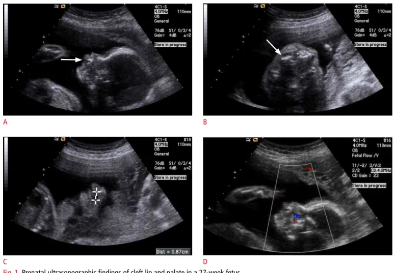
Fig. 1. Prenatal ultrasonographic indings of cleft lip and palate in a 27-week fetus.
A. Sagittal image of upper lip shows narrow defect of upper lip (arrow). B. Axial image of upper alveolar shows a small alveolar defect and malalignment (arrow). C. Coronal image of the hard palate reveals approximately 0.9 cm long defect on the hard palate. D. Color Doppler sonogram shows low through the hard palate defect.
A B
Fig. 2. Prenatal ultrasonographic indings of cleft lip with alveolar involvement in a 26.4 week fetus.
A. Coronal image of the upper lip shows a defect of upper lip (arrow). B. Axial image of the alveolar arch shows a small alveolar arch defect (annotated). Arch malalignment is equivocal. C. Sagittal image of the hard palate reveals suspicious focal defect on the hard palate (arrow). D. Color Doppler sonogram shows no deinite low through the hard palate defect.
A B
C D
Table 2. Sensitivity, speciicity, positive and negative predictive values, and accuracy of each parameter
Arch defect on axial grayscale scan
Arch malalignment on axial grayscale scan
Hard palate defect on sagittal grayscale scan
Transpalatal low on sagittal CDUS
Combined US interpretation using
all parameters
Sensitivity (%) 94.12 64.71 82.35 85.71 94.12
Speciicity (%) 50.00 100.00 75.00 85.71 85.70
Accuracy (%) 80.00 76.00 80.00 85.71 91.67
Positive predictive value (%) 80.00 100.00 87.50 92.31 94.12
Negative predictive value (%) 80.00 57.14 66.67 75.00 85.71
Odds ratio 16.00 N/A 14.00 36.00 96.00
Relative risk 4.00 2.33 2.63 3.69 6.59
palate for cleft consists of coronal and axial plane assessment of midfacial anatomy [14]. Although adding fetal facial scans to routine screening 2-D US has been considered optimal [15], the accuracy for detecting CL with or without CP is variable, especially in low-risk patients [5]. Hence, some efforts to improve the detection rate of CL with or without CP have been attempted by adding the sagittal view to the evaluation.
However, detection of a defected palate in a grayscale sonogram is dificult due to acoustic shadowing from the bony alveolar ridge and maxilla [13]. The importance of a sagittal scan, therefore, has been described with a limited view toward detecting “premaxillary protrusion(s)” when bilateral CL with or without CP was suspected in coronal or axial scans. Babcook et al. [16] reported that the echogenic fusion line of the secondary palate was seen in only 26% of in utero fetuses in sagittal sonograms and in 100% of normal fetal specimens.
Delineating the palatine defect by visualization of the slow amniotic flow through the defect using sagittal CDUS has been tried in some case reports [11-13] to increase the detection rate of CL with or without CP by collecting additional information through sagittal US scans. In this study, we extended the methods to a larger group of high-risk patients.
In our study, the accuracy of observing flow in sagittal CDUS to detect CL with CP was 85.7% (CL with CP, 12 out of 14 cases; CL without CP, one out of seven cases showed transpalatal flow in sagittal CDUS). This result is a high detection rate compared to previous studies showing 41%-46% for detection of CL with CP [6,7], which highlights the diagnostic value of adding sagittal CDUS to fetal facial US. For these previous studies, the CL with CP detection rate of 3-D US was 86%-90%. Thus, the results of our study suggest that by only adding sagittal CDUS to fetal facial US, are detection rates similar those achieved using 3-D US to detect CL with CP possible. Detection of palate defects directly in sagittal images or flow in sagittal CDUS showed comparable, or better, accuracy than axial scans for CL with CP (detecting alveolar defects or alveolar malalignment). We infer from this that the sagittal scan with CDUS is a feasible and useful scanning method to detect CL with CP; our high detection rate compared to previous studies can be explained by the use of sagittal CDUS.
We found that the accuracy of detecting CL with CP by delineating a hard palate defect on the grayscale sagittal US was successful (accuracy of 80%). Rotten and Levaillant [17] described sonographic characteristics of secondary palate clefts in sagittal US scans. They reported that viewing defects in hard palate continuity was possible and that the defects were asymmetric. However, they did not report the detection accuracy of a sagittal scan alone, and concluded that the secondary palate was best seen in coronal scans with 87%
concordance between the US report and postnatal evaluation. In our study, we reported comparable accuracy as reported by Rotten et al. [17], supporting our hypothesis that sagittal US scans may have additional value in the diagnosis of CL with or without CP.
Though similar in accuracy, sagittal CDUS had the slight advantage of increasing specificity, PPV, and NPV compared to detecting hard palate defects in sagittal grayscale scans. A combined US interpretation of all parameters could show good diagnostic performance, with up to 94% of sensitivity, 94% of PPV, and 92% of accuracy. The odds ratio and relative risk of sagittal CDUS were better than that of sagittal grayscale scans; positive indings of sagittal CDUS were better correlated with the existence of actual CPs than those of sagittal grayscale scans. Thus, sagittal CDUS should be added to a routine sagittal grayscale sonogram. Occasionally, there can be cases of CL without CP that show alveolar arch defects or malalignment in sagittal grayscale sonogram. In those cases, performing sagittal CDUS for visualizing transpalatal low can help in distinguishing true CP from artifacts (Fig. 1), strengthening the added value of sagittal CDUS.
By combining axial and sagittal sonograms with CDUS, the diagnostic accuracy was 92% (approximately 10% higher than using axial images alone). Our results had comparable accuracy with previous studies [6-8], in which the accuracy of 2-D US to detect CL with or without CP ranged from 90% to 96%. However, limiting the perspective to CL with CP, the accuracy of previous studies were relatively low (31%-46%) [6,8]. Our study showed 94.1% accuracy; using a combination of axial and sagittal images with CDUS had 16 accurate diagnoses out of 17 total. In a real clinical setting, differentiation from CL with alveolar defect or malalignment without CP and CL with CP is crucial; the former requires a less complex surgical correction (usually a one-stage operation) and shows better clinical outcomes in terms of permanent deformity, phonation, and velopharyngeal insuficiency compared to the latter. Based on our results, we suggest adding sagittal US with CDUS on conventional axial and coronal US of fetal faces to provide the additional beneit of a high NPV (85.7%) compared to grayscale scans, consequently increasing the overall accuracy for differentiating CL with alveolus involvement without CP and CL with CP.
of sagittal CDUS. Furthermore, the yield of sagittal CDUS can be variable based on whether the fetus swallows or breathes during the examination. We note that we were not able to obtain sagittal CDUS from 16% (four out of 25) of our study participants. The yield and average additional examination time of getting sagittal CDUS must be assessed through studies of large, low-risk populations to evaluate the validity of adding sagittal US with CDUS to routine fetal facial scan protocols.
Despite these limitations, we can conclude that sagittal US evaluation of the fetal hard palate is a feasible method to directly reveal bony defects of the hard palate and communicating amniotic fluid flow through the defect using CDUS. Adding sagittal scans with CDUS as a part of a routine sonographic fetal face evaluation protocol may have additional value in the differential diagnosis of fetal CL with, or without, CP.
ORCID: Myoung Seok Lee: http://orcid.org/0000-0001-5285-4874; Jeong Yeon Cho: http://orcid.org/0000-0001-8659-0918; Sang Youn Kim: http://orcid.org/0000-0003-4038-7345; Seung Hyup Kim: http://orcid.org/0000-0002-9339-5907; Joong Shin Park: 0002-5246-0477; Jong Kwan Jun: http://orcid.org/0000-0002-0242-1736
Conlict of Interest
No potential conlict of interest relevant to this article was reported.
Acknowledgments
This research was supported by a grant of the Korea Health Technology R&D Project through the Korea Health Industry Development Institute (KHIDI), funded by the Ministry of Health & Welfare, Republic of Korea (grant number : HI15C1532).
References
1. World Health Organization. Global registry and database on craniofacial anomalies: report of a WHO Registry Meeting on Craniofacial Anomalies. Geneva: World Health Organization, 2003. 2. Mossey PA, Little J, Munger RG, Dixon MJ, Shaw WC. Cleft lip and
palate. Lancet 2009;374:1773-1785.
3. Nyberg DA, Sickler GK, Hegge FN, Kramer DJ, Kropp RJ. Fetal cleft lip with and without cleft palate: US classiication and correlation with outcome. Radiology 1995;195:677-684.
4. Berge SJ, Plath H, Van de Vondel PT, Appel T, Niederhagen B, Von Lindern JJ, et al. Fetal cleft lip and palate: sonographic diagnosis, chromosomal abnormalities, associated anomalies and postnatal
outcome in 70 fetuses. Ultrasound Obstet Gynecol 2001;18:422-431.
5. Maarse W, Berge SJ, Pistorius L, van Barneveld T, Kon M, Breugem C, et al. Diagnostic accuracy of transabdominal ultrasound in detecting prenatal cleft lip and palate: a systematic review. Ultrasound Obstet Gynecol 2010;35:495-502.
6. Johnson DD, Pretorius DH, Budorick NE, Jones MC, Lou KV, James GM, et al. Fetal lip and primary palate: three-dimensional versus two-dimensional US. Radiology 2000;217:236-239.
7. Chmait R, Pretorius D, Jones M, Hull A, James G, Nelson T, et al. Prenatal evaluation of facial clefts with two-dimensional and adjunctive three-dimensional ultrasonography: a prospective trial. Am J Obstet Gynecol 2002;187:946-949.
8. Wang LM, Leung KY, Tang M. Prenatal evaluation of facial clefts by three-dimensional extended imaging. Prenat Diagn 2007;27:722-729.
9. Campbell S, Lees C, Moscoso G, Hall P. Ultrasound antenatal diagnosis of cleft palate by a new technique: the 3D "reverse face" view. Ultrasound Obstet Gynecol 2005;25:12-18.
10. Platt LD, Devore GR, Pretorius DH. Improving cleft palate/cleft lip antenatal diagnosis by 3-dimensional sonography: the "flipped face" view. J Ultrasound Med 2006;25:1423-1430.
11. Aubry MC, Aubry JP. Prenatal diagnosis of cleft palate: contribution of color Doppler ultrasound. Ultrasound Obstet Gynecol 1992;2:221-224.
12. Sherer DM, Abramowicz JS, Jaffe R, Woods JR Jr. Cleft palate: conirmation of prenatal diagnosis by colour Doppler ultrasound. Prenat Diagn 1993;13:953-956.
13. Kennelly MM, Moran P. Directional Power Doppler in the midsagittal plane as an aid to the prenatal diagnosis of cleft lip and palate. Prenat Diagn 2008;28:56-58.
14. Babcook CJ. The fetal face and neck. In: Callen PW, ed. Ultrasonography in obstetrics and gynecology. 4th ed. Philadelphia, PA: W.B.Saunders, 2000;307-330.
15. Royal College of Obstetricians and Gynecologists. Report of the RCOG Working Party on Ultrasound Screening for Fetal Abnormalities. London: Royal College of Obstetricians and Gynaecologists, 1997.
16. Babcook CJ, McGahan JP, Chong BW, Nemzek WR, Salamat MS. Evaluation of fetal midface anatomy related to facial clefts: use of US. Radiology 1996;201:113-118.