Characteristics of ascitic luid from patients with suspected
spontaneous bacterial peritonitis in emergency units at a
tertiary hospital
Características do líquido ascítico de pacientes com suspeita de peritonite
bacteriana espontânea nas unidades de emergência de um hospital terciário
Thiago José Buer Reginato
I, Marcelo José Andrade Oliveira
II, Luiz César Moreira
III, Antonieta Lamanna
III, Milena Marques
Pagliarelli Acencio
IV, Leila Antonangelo
VClinical Laboratory Division, Department of Pathology, LIM 03, Hospital das Clínicas (HC), Faculdade de Medicina da
Universidade de São Paulo (FMUSP), São Paulo, Brazil
ABSTRACT
CONTEXT AND OBJECTIVE: Spontaneous bacterial peritonitis (SBP) is a complication of ascites, especially in cirrhosis. Ascitic fluid with 250 or more neutrophils/mm3 is an acceptable criterion for diagnosis, even when
bacterial fluid cultures are negative. The aims here were to estimate SBP frequency among emergency room patients based on cellular criteria and evaluate the biochemical profile of these fluids.
DESIGN AND SETTING: Retrospective study at a public tertiary hospital.
METHODS: Laboratory records of patients with ascites attended in emergency rooms between Novem-ber 2001 and NovemNovem-ber 2006, from whom ascitic fluid samples were sent to the laboratory due to sus-pected SBP, were evaluated. The 691 samples included were divided into group A (presumed SBP: ≥ 250 neutrophils/mm3; n = 219; 31.7%) and group B (no presumed SBP: < 250 neutrophils/mm3; n = 472; 68.3%).
Patients’ sex and age; ascitic fluid characteristics (numbers of neutrophils, leukocytes and nucleated cells); bacteriological characteristics; and protein, lactate dehydrogenase, adenosine deaminase and glucose concentrations were evaluated.
RESULTS: Among group A cultured samples, 63 (33.8%) had positive bacterial cultures with growth of pathogens commonly associated with SBP. In total, the group A samples showed higher lactate dehydro-genase levels than seen in the group B samples. The latter presented predominance of lymphocytes and macrophages.
CONCLUSION: Among the ascitic fluid samples with clinically suspected SBP, 31.7% fulfilled the cellular di-agnostic criteria. Positive bacterial isolation was found in 33.8% of the cultured samples from the presumed SBP group.
RESUMO
CONTEXTO E OBJETIVO: Peritonite bacteriana espontânea (PBE) é uma complicação da ascite, especial-mente na cirrose. Líquido ascítico com 250 ou mais neutrófilos/mm3 é um critério aceitável para o
diagnós-tico, mesmo com cultura bacteriana negativa. Os objetivos foram estimar a frequência de PBE em pacientes atendidos na sala de emergência, baseando-se no critério celular e avaliar o perfil bioquímico desses líquidos peritoneais.
TIPO DE ESTUDO E LOCAL: Estudo retrospectivo em hospital público terciário.
MÉTODOS: Foram avaliados registros laboratoriais de pacientes com ascite atendidos no setor de emergên-cia entre novembro de 2001 e novembro de 2006, cujas amostras de líquido ascítico foram encaminhadas ao laboratório por suspeita de PBE. As 691 amostras incluídas foram divididas em grupo A (PBE presumi-da: ≥ 250 neutrófilos/mm3; n = 219; 31.7%) e grupo B (Ausência de PBE presumida: < 250 neutrófilos/mm3;
n = 472; 68.3%). Também foram avaliados sexo e idade dos pacientes além de características dos líquidos ascíticos: número de neutrófilos, leucócitos e células nucleadas; bacteriologia; e concentrações de proteínas, desidrogenase láctica, adenosina deaminase e glicose.
RESULTADOS: Das amostras cultivadas do grupo A, 63 (33,8%) tiveram cultura bacteriana positiva com cres-cimento de patógenos comumente associados à PBE. O total de amostras do grupo A exibiu maiores níveis de desidrogenase lática que as do grupo B. Este último demonstrou predomínio de linfócitos e macrófagos.
CONCLUSÃO: Dos líquidos ascíticos com suspeita clínica de PBE, 31.7% preencheram o critério diagnóstico celular. O isolamento bacteriano foi positivo em 33.8% das amostras cultivadas no grupo PBE presumida.
IMedical Student. Faculdade de Medicina da
Universidade de São Paulo (FMUSP), São Paulo, Brazil.
IIMD. Clinical Pathologist, Clinical Laboratory,
Department of Pathology, LIM 03, Hospital das Clínicas (HC), Faculdade de Medicina da Universidade de São Paulo (FMUSP), São Paulo, Brazil.
IIIBSc. Biologist. Clinical Laboratory, Department
of Pathology, LIM 03, Hospital das Clínicas (HC), Faculdade de Medicina da Universidade de São Paulo (FMUSP), São Paulo, Brazil.
IVBSc,PhD. Biologist, Heart Institute, Faculdade
de Medicina da Universidade de São Paulo (FMUSP), São Paulo, Brazil.
VMD, PhD. Clinical Pathologist and Professor,
Clinical Laboratory, Department of Pathology, LIM 03, Hospital das Clínicas (HC), Faculdade de Medicina da Universidade de São Paulo (FMUSP), São Paulo, Brazil.
KEY WORDS:
Ascitic fluid. Infection. Paracentesis. Cytology [subheading]. Peritonitis.
PALAVRAS-CHAVE:
INTRODUCTION
Spontaneous bacterial peritonitis (SBP) is a bacterial infection arising in ascitic luid when there is no evident intra-abdomi-nal surgically treatable source of infection. he irst description of SBP was in 1964.1-3 his common but severe complication in patients with liver disease can develop slowly and insidiously or remain clinically unrecognized until the appearance of symptoms like fever and abdominal pain. he mortality rate ater a single episode ranges from 20 to 40%,4,5 and early diagnosis is required for adequate treatment and prevention of new episodes.
he incidence of spontaneous bacterial peritonitis in cirrhotic patients varies between 7% and 30% per year.6,7 he factors asso-ciated with higher risk are coexistent gastrointestinal bleeding, previous episodes of SBP and low levels of protein in ascitic luid. Possible explanations for its pathogenesis include occurrences of bacterial overgrowth with deterioration of the intestinal barrier, lower intestinal motility, changes in local immune defense and lower activity of bacterial opsonization.8-11 Bacterial overgrowth precedes the key event in the pathogenesis of SBP: the bacterial translocation.2,12-14 his is deined as the passage of viable bacte-ria from the intestinal lumen to mesenteric lymph nodes and/or other extraintestinal sites across the intestinal-mucosal barrier.8 Non-enteric Streptococcus sp and Gram-negative aerobic enter-obacteria like Escherichia coli (present in approximately 70% of cases) and Klebsiella sp are the microorganisms most commonly involved.2,15-17
Early detection of SBP is extremely valuable for patients, since the mortality rate among untreated patients is around 50%.18 he laboratory criterion most used for SBP diagnosis is an ascitic luid neutrophil count ≥ 250 cells/mm3, in the absence of a source of intra-abdominal infection.2 Bacterascites (monomicrobial non-neutrocytic bacterascites) is the term used to describe the coloni-zation of ascitic luid by bacteria, with no evidence of local or sys-temic infection and no inlammatory reaction in the bacterial luid (neutrophil count < 250/mm3 and positive bacterial culture). Cul-ture-negative neutrocytic ascites is the term used to describe the clinical situation in which the ascitic luid contains 250 or more neutrophils /mm3, but luid cultures fail to grow any bacteria. his inding is considered to represent the expected 20% failure rate of cultures to isolate microorganisms.2 Despite the low complex-ity of laboratory tests used for diagnoses, prescriptions for antibi-otic therapy are based on the most commonly involved pathogens and generally precede the bacterial culture results. hus, an early diagnosis is highly desirable in order to avoid indiscriminate use of antibiotics, with potential induction of bacterial resistance or other complications relating to their use.
OBJECTIVES
In this context, the aim of this study was to estimate the fre-quency of presumed cases of spontaneous bacterial peritonitis
in the emergency rooms of a tertiary public university hospital, based on cytological criteria, and to assess the microbiological and biochemical proile of these peritoneal luid samples.
METHODS Subjects
We retrospectively analyzed laboratory data on 691 patients (431 males and 260 females; average age 58.1 years) from whom peri-toneal luid samples were collected in emergency rooms at a ter-tiary public hospital between November 2001 and November 2006. All the samples were received at the cytology laboratory containing a written diagnostic hypothesis of SBP on the labora-tory test order. If more than one peritoneal luid sample from any patient included was processed during the study period, only the irst sample was taken into consideration in the study analysis, thus resulting in one sample for each patient.
he 691 samples were divided in two groups, based on their neutrophil count: group A (presumed spontaneous bacterial peri-tonitis: ≥ 250 neutrophils/mm3) and group B (no presumed spon-taneous bacterial peritonitis: < 250 neutrophils/mm3). he study protocol was approved by the Institutional Ethics Committee.
Methods
he following variables were evaluated: (1) patients’ sex and age; and (2) ascitic luid characteristics such as: total number of nucleated cells and total and diferential leukocyte count; pres-ence of bacteria on Gram-stained slides and aerobic and anaer-obic bacterial cultures; and total protein, albumin, adenosine deaminase (ADA), lactate dehydrogenase (LD) and glucose concentrations, when requested.
Ascites samples were collected by paracentesis using a sterile technique and the samples were immediately sent to the labora-tory for analysis. For samples collected into EDTA (ethylenedi-aminetetraacetic acid) coated tubes,19 cells were counted man-ually in a Neubauer chamber20 and the cytological examination was performed on Leishman-stained smear slides. In cases with hemorrhagic luid (red cells ≥ 10,000/ml), the neutrophil count was corrected by subtracting one neutrophil per 250 counted erythrocytes. For biochemical analysis, luid samples collected into tubes containing gel separator plus clot activator were cen-trifuged and the supernatant was tested for total protein, albu-min, globulin, lactate dehydrogenase and glucose concentrations, using a Roche modular analyzer (Roche Diagnostics, Roche, Somerville, United States).
ADA is an enzyme that is produced by lymphocytes and mac-rophages in response to T cell stimuli and is frequently increased in cases of peritoneal tuberculosis. he ADA level was measured using the Giusti modiied manual method.21
Diagnostic Systems, Sparks, United States) and bacterial cation was performed by means of the Vitek automated identii-cation system (BioMérieux Clinical Diagnostics, France).
Statistical analysis
Diferences between groups were evaluated using the Mann-Whit-ney test for non-categorical data and the chi-square test for cate-gorical data, by means of the Statistical Package for the Social Sci-ences (SPSS) sotware (version 11.0, Chicago, United States). he data were presented as means ± standard deviations, unless other-wise indicated. Diferences were considered signiicant if P < 0.05.
RESULTS
Only 219 (31.7%) samples contained 250 or more neutrophils/mm3 (Group A, presumed SBP), while 472 samples (68.3%) had < 250 neutrophils/mm3 (Group B, no presumed SBP). We did not observe any statistically signiicant diference in relation to sex and age distribution between groups A and B. However, there was predominance of males in both groups (Table 1).
Bacterioscopy, or Gram staining, is not obligatory in the rou-tine workup for SBP, since its sensitivity is too low. Nevertheless, it was performed on 135 samples (61.6%) in group A and on 282 samples (59.7%) in group B, which yielded rates of positive ind-ings of 12.6% and 1.1%, respectively (Table 2).
Among the samples in group A, 33 (15%) were not subjected to bacterial culture. Among the cultured group A samples, 123 (66.2%) presented negative cultures and 63 (33.8%) had positive results
(Table 2). he most prevalent agents were Escherichia coli (31.7%), followed by Streptococcus pneumoniae (7.9%), Staphylococcus aureus (7.9%), and Klebsiella pneumoniae (7.9%). he overall rate of posi-tive indings of the genus Streptococcus sp was 23.8%. In group B, 75 samples (15.9%) were not subjected to bacterial culture. Among the cultured group B samples, 373 (94%) presented negative cultures and 24 (6%) had positive results that could be classiied as potential bacterascites. he most prevalent agents in these cases were E. coli (20.8%), S. epidermidis (16.7%), K. pneumoniae (12.5%) and Coryne-bacterium sp (12.5%). A small number of cases displayed growth of atypical agents that are not usually associated with SBP, such as Staphylococcus simulans, Staphylococcus hominis, Providencia stu-artii, Citrobacter braakii and Streptococcus salivarus.
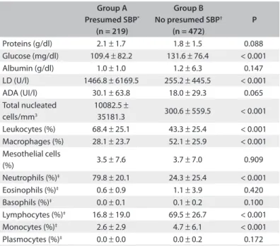
Statistically signiicant diferences were observed between groups A and B regarding the concentrations of glucose (109.4 ± 82.2 mg/dl versus 131.6 ± 76.4 mg/dl; P < 0.001) and LD (1466.8 ± 6169.5 U/l versus 255.2 ± 445.5 U/l; P < 0.001), and in relation to the percentages of total nucleated cells and some cell types.
he concentration of ADA, did not show any signiicant dif-ference between the groups. On cytological examination, the number of nucleated cells was signiicantly higher in group A, mainly due to neutrophil predominance (Table 3).
Group A Presumed SBP*
(n = 219)
Group B No presumed SBP†
(n = 472)
P
Age (mean ± SD) 58.3 ± 13.1 58.0 ± 14.5 0.911 Sex
Males 139 292
0.749
Females 80 180
*Ascitic fluid with ≥ 250 neutrophils/mm3; †ascitic fluid with < 250 neutrophils/
mm3; SD = standard deviation; SBP = spontaneous bacterial peritonitis. Statistical
tests: Mann-Whitney or chi-square;significant if P< 0.05. Table 1. Patients’ demographic data
Group A Presumed SBP*
(n = 219)
Group B No presumed SBP†
(n = 472)
P
Bacterioscopy (Gram stain)
Positive 17 3 < 0.001
Negative 118 279 0.226
Not performed 84 190 0.695 Bacterial culture
Positive 63 24‡ < 0.001
Negative 123§ 373 < 0.001
Not performed 33 75 0.870
*Ascitic fluid with ≥ 250 neutrophils/mm3; †ascitic fluid with < 250 neutrophils/mm3; ‡presumed bacterascites; §culture-negative neutrocytic ascites; SBP = spontaneous
bacterial peritonitis. Statistical tests: Mann-Whitney or chi-square; significant if P< 0.05. Table 2. Microbiological characteristics of ascitic fluids
Table 3. Biochemical and cytological characteristics of ascitic fluids
Group A Presumed SBP*
(n = 219)
Group B No presumed SBP†
(n = 472)
P
Proteins (g/dl) 2.1 ± 1.7 1.8 ± 1.5 0.088 Glucose (mg/dl) 109.4 ± 82.2 131.6 ± 76.4 < 0.001 Albumin (g/dl) 1.0 ± 1.0 1.2 ± 6.3 0.147 LD (U/l) 1466.8 ± 6169.5 255.2 ± 445.5 < 0.001 ADA (UI/l) 30.1 ± 63.8 18.0 ± 29.3 0.065 Total nucleated
cells/mm3
10082.5 ±
35181.3 300.6 ± 559.5 < 0.001 Leukocytes (%) 68.4 ± 25.1 43.3 ± 25.4 < 0.001 Macrophages (%) 28.1 ± 23.7 52.1 ± 25.9 < 0.001 Mesothelial cells
(%) 3.5 ± 7.6 3.7 ± 7.0 0.909 Neutrophils (%)‡ 79.8 ± 20.1 24.3 ± 25.4 < 0.001
Eosinophils (%)‡ 0.6 ± 0.9 1.1 ± 3.9 0.420
Basophils (%)‡ 0.0 ± 0.1 0.1 ± 0.2 0.100
Lymphocytes (%)‡ 16.8 ± 19.0 69.5 ± 26.7 < 0.001
Monocytes (%)‡ 2.6 ± 2.9 4.7 ± 6.1 < 0.001
Plasmocytes (%)‡ 0.0 ± 0.0 0.0 ± 0.2 0.172
*Ascitic fluid with ≥ 250 neutrophils/mm3; †ascitic fluid with < 250 neutrophils/mm3; ‡leukocyte differential; LD = lactate dehydrogenase; SBP: spontaneous bacterial
DISCUSSION
In the present study, application of the cytological criterion for presumed SBP in clinically suspected patients resulted in positive indings in 219 (31.7%) of all the study samples. Another interest-ing indinterest-ing was that 15.6% of the peritoneal luid samples were not sent for bacterial culture, even though paracentesis had been per-formed due to clinically suspected infection. We found that 33.9% (63/186) of the cultured samples in group A presented positive cul-tures. his rate of positive cultures was lower than in the literature, in which rates ranging from 40 to 80% in conirmed SBP cases have been reported.2,22 In fact, cytological examination and bedside luid inocu-lation into bacterial culture bottles are the two most accepted labora-tory tests for investigation of SBP.2,6 For this reason, we emphasize the importance of ordering at least these two laboratory tests (cytology and cultures), in order to establish the diagnosis in suspected cases.
Regarding the microbial agents identiied, our results were sim-ilar to those reported in literature,2 with Escherichia coli as the most prevalent agent.17,23 In a small number of cases with cellular crite-ria for SBP, we observed growth of some bactecrite-ria that are not usu-ally related to this diagnosis. In such cases, it is important to rule out possible sample contamination during paracentesis or sample handling. False-positive results can lead to unnecessary antibiotic therapy, which could increase bacterial resistance to the antibiot-ics most used for treating SBP. In group B, only a small percentage of cases showed positive bacterial cultures, and most of them were for microbial agents that are not usually associated with SBP, which suggests that the peritonitis had non-spontaneous etiology.
We observed predominance of men over women among the study subjects, with an average age of around 60 years. his pattern was similar to what was observed in other reports,24-26 and probably relects the classical natural history of patients with liver diseases who seek emergency care centers due to development of ascites. Most of these patients have cirrhosis with portal hypertension as a complica-tion of a history of alcoholism or chronic hepatitis C virus infeccomplica-tion, and both of these conditions are more prevalent among men.25,26
he inding of lower levels of glucose in the peritoneal luid of patients with a presumed diagnosis of SBP probably relects the con-sumption of this substance by bacteria, whereas the high concentra-tion of LD relects a high degree of peritoneal inlammaconcentra-tion. An anal-ogy can be made with parapneumonic pleural efusions, in which the high concentration of lactate dehydrogenase is one of the criteria used for classifying an efusion as complicated. In these cases, pleural luid LD levels higher than 1,000 U/l, in association with decreased pH and glucose suggests clinical worsening and may be an indica-tion for thoracic drainage.27,28 High LD activity (> 500 U/l) has been widely reported in cases of malignancy and tuberculous and pancre-atic ascites but without enough sensitivity to distinguish it from liver disease. his makes low LD values unsuitable for ruling out malig-nancy, but indicates that elevated LD in luid samples point towards causes other than liver disease.29 High levels of LD can also occur
on SBP, as seen in the group A ascitic luids, but can also occur in secondary bacterial peritonitis, which is frequently associated with intra-abdominal surgically treatable sources of infection,30 such as intestinal perforation. A study conducted by Boyer et al. found that ascitic luids with two out of three of the characteristics of an exu-date (LD > 400 U/l; luid/serum LD ratio > 0.6; and luid/serum total protein ratio > 0.5) tended to indicate a non-hepatic cause for the ascites.31 Since we did not review all the medical records, we could not identify possible cases of secondary peritoneal infection.
he management of ascitic patients is to a great extent inlu-enced by laboratory test results. In clinical practice, since the collec-tion of peritoneal luid samples can be a time-consuming and cum-bersome procedure, the use of this biological material needs to be optimized by ordering relevant tests and paying special attention to pre-analytical best-practice procedures in order to increase the reli-ability of test results. Some of these recommended procedures are: (1) bedside inoculation of ascitic luid into culture bottles and refer-ral to a quality-certiied microbiology laboratory; (2) for adequate cell counting analysis, collection of ascitic luid into EDTA-coated tubes, in order to avoid ibrin formation and cell clumping, and (3) immediate transportation of samples to the laboratory, in order to avoid time and temperature-related pre-analytical errors, especially in biochemical tests. In our laboratory, we routinely perform body cavity luid cell counting in Neubauer chambers (manual technique), instead of using automated counting devices. he latter could be an alternative,19 but these devices show poorer accuracy, particularly for luid samples with low cell counts.20
Among the limitations of our study, it should be noted that we did not review the patients’ clinical records to check for any underlying clinical conditions such as recent gastrointestinal bleeding or abdominal surgery, or to investigate any second-ary sources of peritoneal infection, cirrhosis and other causes of ascites. Furthermore, since we did not check for concomitant or recent use of antibiotics, we were unable to estimate the impact of antibiotic use on negative results from cultures. However, because the samples were sent to the laboratory as probable SBP cases, we supposed they represented a heterogeneous group of patients, mostly with cirrhosis, which is the major underlying condition that raises the suspicion of SBP in patients with ascites.
In any event, it is worth emphasizing to clinicians the impor-tance of proper sample collection and management, as well as correct ordering of relevant laboratory tests for investigating sus-pected SBP cases, not only to achieve early diagnosis, but also to avoid unnecessary antibiotic administration.
CONCLUSIONS
REFERENCES
1. Guarner C, Soriano G. Spontaneous bacterial peritonitis. Semin Liver Dis.
1997;17(3):203-17.
2. Koulaouzidis A, Bhat S, Saeed AA. Spontaneous bacterial peritonitis. World
J Gastroenterol. 2009;15(9):1042-49.
3. Conn HO, Fessel JM. Spontaneous bacterial peritonitis in cirrhosis: variations
on a theme. Medicine (Baltimore). 1971;50(3):161-97.
4. Rerknimitr R, Limmathurotsakul D, Bhokaisawan N, et al. A comparison
of diagnostic efficacies among different reagent strips and automated
cell count in spontaneous bacterial peritonitis. J Gastroenterol Hepatol.
2010;25(5):946-50.
5. Such J, Runyon BA. Spontaneous bacterial peritonitis. Clin Infect Dis.
1998;27(4):669-74; quiz 675-6.
6. Rimola A, García-Tsao G, Navasa M, et al. Diagnosis, treatment and
prophylaxis of spontaneous bacterial peritonitis: a consensus document.
International Ascites Club. J Hepatol. 2000;32(1):142-53.
7. Sapey T, Kabissa D, Fort E, Laurin C, Mendler MH. Instant diagnosis of
spontaneous bacterial peritonitis using leukocyte esterase reagent strips:
Nephur-Test vs. MultistixSG. Liver Int. 2005;25(2):343-8.
8. Guarner C, Soriano G. Bacterial translocation and its consequences in
patients with cirrhosis. Eur J Gastroenterol Hepatol. 2005;17(1):27-31.
9. Morencos FC, de las Heras Castaño G, Martín Ramos L, et al. Small bowel
bacterial overgrowth in patients with alcoholic cirrhosis. Dig Dis Sci.
1995;40(6):1252-6.
10. Guarner C, Runyon BA, Young S, Heck M, Sheikh MY. Intestinal bacterial
overgrowth and bacterial translocation in cirrhotic rats with ascites. J
Hepatol. 1997;26(6):1372-8.
11. Chang CS, Chen GH, Lien HC, Yeh HZ. Small intestine dysmotility and
bacterial overgrowth in cirrhotic patients with spontaneous bacterial
peritonitis. Hepatology. 1998;28(5):1187-90.
12. Garcia-Tsao G, Lee FY, Barden GE, Cartun R, West AB. Bacterial translocation
to mesenteric lymph nodes is increased in cirrhotic rats with ascites.
Gastroenterology. 1995;108(6):1835-41.
13. Cirera I, Bauer TM, Navasa M, et al. Bacterial translocation of enteric
organisms in patients with cirrhosis. J Hepatol. 2001;34(1):32-7.
14. Runyon BA, Squier S, Borzio M. Translocation of gut bacteria in rats with
cirrhosis to mesenteric lymph nodes partially explains the pathogenesis of
spontaneous bacterial peritonitis. J Hepatol. 1994;21(5):792-6.
15. Garcia-Tsao G. Spontaneous bacterial peritonitis. Gastroenterol Clin North
Am. 1992;21(1):257-75.
16. Hoefs JC, Runyon BA. Spontaneous bacterial peritonitis. Dis Mon.
1985;31(9):1-48.
17. Felisart J, Rimola A, Arroyo V, et al. Cefotaxime is more effective than is
ampicillin-tobramycin in cirrhotics with severe infections. Hepatology.
1985;5(3):457-62.
18. Volk ML, Marrero JA. Advances in critical care hepatology. Minerva
Anestesiol. 2006;72(5):269-81.
19. Riggio O, Angeloni S, Parente A, et al. Accuracy of the automated cell
counters for management of spontaneous bacterial peritonitis. World J
Gastroenterol. 2008;14(37):5689-94.
20. Clinical Laboratory Standard Institute. Body Fluid Analysis for Cellular
Composition; Approved Guidelines. Pennsylvania: Clinical and Laboratory
Standards Institute; 2006. Available from: http://www.clsi.org/source/
orders/free/h56-a.pdf. Accessed in 2011 (Mar 31).
21. Giusti G. Adenosine deaminase. In: Bergmeyer HU, editor. Methods of
enzymatic analysis. 2nd ed. New York: Academic Press; 1974. p. 1092-99.
22. Figueiredo FAF, Coelho HSM, Soares JAS. Peritonite bacteriana espontânea
na cirrose hepática: prevalência, fatores preditivos e prognóstico
[Sponteneous bacterial peritonitis in hepatic cirrhosis: prevalence, predictive
factors and prognosis]. Rev Assoc Med Bras (1992). 1999;45(2):128-36.
23. Arroyo V, Bataller R, Ginès P. Spontaneous bacterial peritonitis. In: O’Grady
JG, Lake JR, editors. Comprehensive clinical hepatology. 1st ed. Barcelona:
Mosby; 2000. p. 7.10-7.14.
24. Rehm J, Mathers C, Popova S, et al. Global burden of disease and injury
and economic cost attributable to alcohol use and alcohol-use disorders.
Lancet. 2009;373(9682):2223-33.
25. Mueller S, Millonig G, Seitz HK. Alcoholic liver disease and hepatitis
C: a frequently underestimated combination. World J Gastroenterol.
2009;15(28):3462-71.
26. Mehta G, Rothstein KD. Health maintenance issues in cirrhosis. Med Clin
North Am. 2009;93(4):901-15, viii-ix.
27. Yataco JC, Dweik RA. Pleural effusions: evaluation and management. Cleve
Clin J Med. 2005;72(10):854-6, 858, 862-4 passim.
28. Colice GL, Curtis A, Deslauriers J, et al. Medical and surgical treatment
of parapneumonic effusions: an evidence-based guideline. Chest.
2000;118(4):1158-71.
29. Tarn AC, Lapworth R. Biochemical analysis of ascitic (peritoneal) fluid: what
should we measure? Ann Clin Biochem. 2010;47(Pt 5):397-407.
30. Akriviadis EA, Runyon BA. Utility of an algorithm in differentiating
spontaneous from secondary bacterial peritonitis. Gastroenterology.
1990;98(1):127-33.
31. Boyer TD, Kahn AM, Reynolds TB. Diagnostic value of ascitic fluid
lactic dehydrogenase, protein, and WBC levels. Arch Intern Med.
1978;138(7):1103-5.
Sources of funding: Research supported by Conselho Nacional de Desenvolvimento Científico e Tecnológico (CNPq), PIBC 116425/2008-3, Brazil
Conflict of interest: None
Date of first submission: October 4, 2010
Last received: January 10, 2011
Accepted: April 15, 2011
Address for correspondence:
Leila Antonangelo
Av. Dr. Enéas de Carvalho Aguiar, 155 — 2o andar — bloco 08
Cerqueira César — São Paulo (SP) — Brasil
CEP 05403-000
Tel. (+55 11) 3069-6158