* Study carried out at the Hospital do Círculo, Caxias do Sul, Brazil. 1. Ph.D. in Pulmonology, Hospital Pompéia, Caxias do Sul, Brazil.
2. Head of the Intensive Care Unit. Unimed Hospital of Caxias do Sul, Caxias do Sul, Brazil.
Correspondence to: André Germano dos Santos Leite. Rua Gal. Arcy da Rocha Nóbrega, 401, Sala 303, Bairro Madureira, CEP 95040-000, Caxias do Sul, RS, Brasil.
Tel 55 54 3028-6040. E-mail: [email protected] Submitted: 19 December 2006. Accepted, after review: 11 April 2007.
Management of recurrent distal tracheal stenosis
using an endoprosthesis: a case report*
André Germano Leite1, Douglas Kussler2
Abstract
The authors report the case of a patient with recurrent distal tracheal stenosis after several tracheal resections. A T-Y tracheobronchial stent was inserted for the management of distal airway obstruction. The prosthesis was adapted based on the specifications obtained through a computed tomography scan of the chest with three-dimensional reconstruction.
Keywords: Tracheal stenosis; Prostheses and implants; Trachea/surgery; Case reports [publication type].
Introduction
Tracheal stenosis is not a new problem. In 1886, Colles described four cases of tracheal stenosis as the cause of tracheal obstruction in 57 individuals who had undergone tracheostomy for the treatment of diphtheria.(1)
Since 1960, the increased use of endotracheal tubes, tracheostomy, and cricothyroidotomy has produced a broad spectrum of airway lesions that can affect an area ranging from the larynx to the lower trachea.(2)
The use of endotracheal tubes with high-volume, low-pressure cuffs was first proposed by Cooper & Grillo.(3) Their
use has significantly decreased the occurrence of postintu-bation tracheal stenosis,(4-7) although this continues to be
the most common postoperative complication affecting the trachea.(2)
Obstructive tracheal lesion following intubation can occur at the following sites: the tracheostomy stoma (opening); the cuff site; the segment between the stoma and the cuff; and the tip of the endotracheal tube (or tracheostomy tube).(7)
Satisfactory palliation of the lesions involving the distal trachea and the carina is difficult to achieve because any attempt to do so presupposes an intervention in a vital system that is anatomically impaired and in highly symp-tomatic patients.
Case report
We report the case of a 37-year-old female who had been experiencing dyspnea at rest and hoarseness for two months. In addition, the patient had undergone surgery for the correction of a cerebral aneurysm four months prior and, as a consequence, had been orotracheally intubated for three weeks.
of the distal tracheal stenosis, at the site of the anastomosis, affecting 60% of the airway lumen.
The patient underwent balloon dilatation (tubes of 2.5 to 7.0 mm), as well as at least six dilatation procedures using rigid bronchoscopy at different time points, and did not remain asymptomatic for periods longer than 15 days.
Nine months after the first surgery, the patient underwent a second tracheal resection via right thoracotomy. Immediately after the surgery, both pulmonary hila were released (the left one, via video-assisted thoracoscopy, and the right one, via thoracotomy) in order to allow resection of the sten-osis and adequate approximation of the bronchial stumps for re-anastomosis. A total of 20 mm of the distal trachea were resected, and the anastomosis between the carina and the proximal trachea was performed next to the apex of the pleural cavity, with a certain degree of tension, and covered with a pedicle flap of intercostal muscle.
The orotracheal tube (OTT) was removed under endoscopic visualization, and the anastomosis remained intact, but with a significant (50%) reduction in the diameter of the tracheal lumen. The patient presented progressive worsening of the respiratory pattern within 24 hours after extubation, tracheal stenosis (15 mm in length) 20 mm above
the tracheal carina.
The patient was submitted to fiberoptic bron-choscopy, which revealed stenosis of 70-80% of the tracheal lumen. It was not possible to advance the device through the stenosis. There was no evidence of inflammatory process. Nor were there any alterations of the tracheal or larynx proximal to the stenosis. Dilatation using a rigid broncho-scope was performed, albeit with no satisfactory results. Tracheoplasty, using a median sternotomy approach, was then performed.
Approximately 20 mm of the distal trachea were dissected, and a primary airway anastomosis, without tension, was performed in apparently undamaged tissue. The patient was extubated in the operating room and was discharged 7 days later.
A control fiberoptic bronchoscopy showed that the anastomosis remained permeable, without granulation or stenosis. The anatomopatholog-ical examination revealed a chronic inflammatory process and tracheal fibrosis, with no signs of gran-uloma or necrosis.
Six months after the surgery, the patient again presented stridor accompanied by progressive dyspnea. Another bronchoscopy revealed recurrence
R L
a
b
c
d
One week after the insertion of the prosthesis, the patient underwent fiberoptic bronchoscopy and a control CT scan (Figure 3), which allowed us to determine the perfect positioning of the prosthesis. The patient was discharged ten days after the inser-tion of the tracheal stent.
The patient remained asymptomatic for a follow-up period of one year. A control bronchos-copy after a follow-up period of one year revealed a tiny granuloma near the proximal border of the tracheal stent (subglottic region). There were no infectious complications in this period, and the prosthesis examined did not present any type of deposition or secretion on its internal wall.
Whether this prosthesis can be safely removed or should be permanently left in place remains unclear.
Discussion
Imaging studies should precede respiratory endoscopy and are also useful for guiding its performance. These studies help to evaluate the airway distal to the stenosis in cases in which the endoscope cannot be advanced past the stenosis. This finding is essential for surgical planning. and it was necessary to reintroduce the
endotra-cheal tube through the anastomosis with the aid of a flexible bronchoscope. Since the anastomosis was located quite near the tracheal carina, the distal edge of the OTT occasionally caused atelectasis of the left lung due to right selective intubation.
Since there was no possibility of re-intervention or dilatation so soon after the surgical procedure, we opted for re-establishing the permeability of the airway through the insertion of an endoprosthesis.
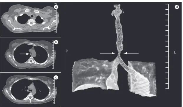
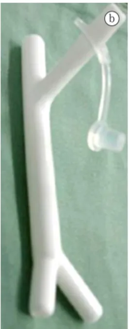
A CT scan of the airway was performed on postoperative day 3 (with the OTT being tempo-rarily removed under endoscopic guidance), and the dimensions of the tracheal stenosis were obtained. The three-dimensional reconstruction of the trachea and the measurements of the trachea were sent to Hood Laboratories (Pembroke, MA, USA). Based on those dimensions, a silicone (T-Y) tracheobron-chial stent compatible with the patient airway was purchased (Figure 2).
On postoperative day 10, the prosthesis was introduced in the patient airway via cervicotomy. The prosthesis was inserted, via tracheotomy, between the third and fourth rings of the trachea. It was positioned in the distal airway with the aid of intra-operative fluoroscopy and fiberoptic bronchoscopy.
a
b
the tracheal carina, thus creating difficulties in the right branch of the T-Y stent.(11,12)
Such T tubes have been used as temporary airway stents, as well as in palliative procedures for the permanent resolution of airway obstruction and in the treatment of postoperative complications following airway reconstruction.
In a study conducted at Massachusetts General Hospital and involving patients who underwent placement of a T tube after unsuccessful tracheal resection,(2) 5 of the 32 patients evaluated evolved
to decannulation, and 5 later underwent another (successful) resection. In 11 cases, the prosthesis was left in place permanently.
In another study,(13) patients presenting
postin-tubation tracheal stenosis underwent surgical resection, the failure rate of which was 3.9% in the initial procedure and 5.6% in the follow-up proce-dure. The low rate of surgical success in patients who had previously undergone unsuccessful postintubation tracheal reconstruction confirms the observation that the chance of achieving favo-rable results is greater in the initial procedure. Therefore, all efforts should be made to approxi-mate the original anatomy as closely as possible, using tension-free tracheal anastomosis in healthy tracheal tissue.
The success in the surgical management of patients with tracheal stenosis requires complete synchrony between the surgery and anesthesiology teams. Ventilation can be obtained using high-frequency ventilation, jet ventilation, distal tracheal intubation, spontaneous ventilation, or extracor-poreal circulation.(14)
In the case described here, we opted to use superficial intravenous anesthesia and spontaneous ventilation through the open airway in the surgical field.(14,15) An alternative means of maintaining the
patient oxygenated would be to catheterize the distal airway and provide a continuous supply of oxygen.
A second attempt at reconstruction of the trachea is always a great challenge, not only due to the reduced length of normal trachea available and the surgical scar (which makes dissection and displacement of the trachea difficult) but also to the danger of cutting off the blood supply to the trachea and to the recurrent laryngeal nerves.
Finally, a permanent tracheal stent might be the best option for patients with extensive tracheal lesion.
Imaging studies also play an important role in the diagnosis of lesions that are sometimes difficult to detect through bronchoscopy, such as malacia.(8)
Three-dimensional CT reconstruction offers optimal accuracy in the diagnosis of tracheobronchial sten-osis, including the identification of the anatomical location, the quantification of the number of lesions present, and the characterization of the status of the airway distal to the lesion. On group of authors found that, in patients with tracheobronchial sten-osis, reconstruction CT has a sensitivity of 100%.(9)
It is essential to rule out the presence of any type of concomitant laryngeal lesion prior to repairing the tracheal lesion.
The access to the distal trachea and the carina can be optimized by using the median sternotomy approach. The principal advantages of this approach are the excellent exposure of the region and the possibility of avoiding the morbidity associated with thoracotomy.(8)
Hilar release is frequently used in complex lower tracheal resections, since it allows the distal trachea to be displaced by 10 to 20 mm. However, laryngeal lowering is not routinely used in such situations.(10)
The use of bifurcated (T-Y) endotracheal stents in patients with tracheobronchial obstruction was first described by Westaby et al.(11)
One limitation of the use of tubes that involve the tracheal carina is that the emergence of the right upper lobe bronchus often occurs very close to
In: Grillo HC, editor. Surgery of the trachea and bronchi. Hamilton: BC Decker; 2004. p. 587-98.
9. Rooney CP, Ferguson JS, Barnhart W, Cook-Granroth J, Ross A, Hoffman EA, et al. Use of 3-dimensional computed tomography reconstruction studies in the preoperative assessment of patients undergoing balloon dilatation for tracheobronchial stenosis. Respiration. 2005;72(6):579-86. 10. Grillo HC, Dignan EF, Miura T. Extensive resection and
reconstruction of mediastinal trachea without prosthesis or graft: an anatomical study in man. J Thorac Cardiovasc Surg. 1964;48:741-9.
11. Westaby S, Jackson JW, Pearson FG. A bifurcated silicone rubber stent for relief of tracheobronchial obstruction. J Thorac Cardiovasc Surg. 1982;83(3):414-7.
12. Ricci F, Puma F, Santoprete S, Urbani M, Vinci D, Sanguinetti A, et al. [Use of the Dynamic Stent in the palliation of carinal and distal tracheal stenosis][Article in Italian]. Ann Ital Chir. 2002;73(2):211-7; discussion 217-8.
13. Grillo HC, Donahue DM, Mathisen DJ, Wain JC, Wright CD. Postintubation tracheal stenosis. Treatment and results. J Thorac Cardiovasc Surg. 1995;109(3):486-92; discussion 492-3.
14. Pinsonneault C, Fortier J, Donati F. Tracheal resection and reconstruction. Can J Anaesth. 1999;46(5 Pt 1):439-55. 15. Grace RF. Spontaneous respiration via an open trachea for
resection of a high tracheal stenosis in a child. Anaesth Intensive Care. 2002;30(4):502-4.
References
1. Colles CJ. I. On Stenosis of the Trachea after Tracheotomy for Croup and Diphtheria. Ann Surg. 1886;3(6):499-507. 2. Grillo HC. Tracheal T Tubes. In: Grillo HC, editor. Surgery of
the trachea and bronchi. Ontario: BC Decker, Inc., Hamilton; 2004. p.749-62.
3. Cooper JD, Grillo HC. Experimental production and prevention of injury due to cuffed tracheal tubes. Surg Gynecol Obstet. 1969;129(6):1235-41.
4. Ching NP, Ayres SM, Paegle RP, Linden JM, Nealon TF Jr. The contribution of cuff volume and pressure in tracheostomy tube damage. J Thorac Cardiovasc Surg. 1971; 62(3): 402-10.
5. Ching NP, Nealon TF Jr. Clinical experience with new low-pressure high volume tracheostomy cuffs. Importance of limiting intracuff pressure. N Y State J Med. 1974; 74(13): 2379-84.
6. Geffin B, Pontoppidan H. Reduction of tracheal damage by the prestretching of inflatable cuffs. Anesthesiology. 1969;31(5):462-3.
7. Grillo HC, Cooper JD, Geffin B, Pontoppidan H. A low-pressure cuff for tracheostomy tubes to minimize tracheal injury. A comparative clinical trial. J Thorac Cardiovasc Surg. 1971;62(6):898-907.