SOTOS SYNDROME (CEREBRAL GIGANTISM)
Analysis of 8 cases
Débora Gusmão Melo
1, Angelina Xavier Acosta
1, Maria Aparecida de Almeida Salles
1,
João Monteiro de Pina-Neto
1, José Daniel Vieira de Castro
2, Antonio Carlos Santos
2ABSTRACT - Sotos syndrome or cerebral gigantism is characterized by macrocephaly, overgrowth, mental retardation and central nervous system abnormalities. Congenital heart defects may be present. We report 8 patients with this syndrome and relate their clinical features, neuroimaging and echocardiographic findings.
KEY WORDS: Sotos syndrome, cerebral gigantism, macrocephaly, mental retardation.
Síndrome de Sotos (gigantismo cerebral): análise de 8 casos
RESUMO - A síndrome de Sotos ou gigantismo cerebral é caracterizada por macrocefalia, hipercrescimento, dismorfias faciais típicas, deficiência mental e alterações do sistema nervoso central. Malformações cardíacas podem estar presentes. Nós relatamos 8 pacientes com esta síndrome e descrevemos seus achados clínicos, de neuroimagem e ecocardiográficos.
PALAVRAS-CHAVE: síndrome de Sotos, gigantismo cerebral, macrocefalia, deficiência mental.
1Departamento de Genética and 2Departamento de Radiologia da Faculdade de Medicina de Ribeirão Preto, Universidade de São Paulo (USP), Riberão Preto SP, Brazil.
Received 2 July 2001, received in final form 11 October 2001. Accepted 24 October 2001.
Dr. João Monteiro de Pina Neto Departamento de Genética Faculdade de Medicina de Ribeirão Preto USP Av. Bandeirantes 3900 -14049-900 Ribeirão Preto SP – Brasil – FAX: 16 6023104
Sotos syndrome was first described by Sotos et al. (1964) in five children with overgrowth, acrome-galic features, nonprogressive cerebral disorder with mental retardation and characteristic physiognomy1.
Since then, over 250 cases have been reported2 .
Except for a concordant set of identical twins, most cases have been sporadic3 .It occurs in all ethnic
groups and has been detected throughout the world. The prevalence is not known, but is estimated to be between 1 in 10,000 and 1 in 50,0004 .
The main clinical finding in Sotos syndrome is prenatal and postnatal overgrowth. Birth length is usually more significantly increased (above the 97th
percentile) than weight (between the 75th and 97th
percentile). Growth is excessive in the first years of life, after which time it proceeds at a relatively nor-mal rate, but consistently falls in the high percen-tiles4 . Bone age is also significantly advanced in most
cases of Sotos syndrome, it is thought that all cases have advanced bone age at some time4 . Head
cir-cumference is almost invariably large at birth, and generally proceeds above the 97th percentile
through-out growth5 . Distinctive craniofacial anomalies
in-clude dolichocephaly, prominent forehead, hyperte-lorism or telecanthus, epicanthic folds, flat nasal bridge, downslanting palpebral fissures, high arched palate, premature eruption of teeth and pointed chin6. Other features include cerebellar nystagmus,
strabismus, pes planus, kyphoscoliosis, unequal lo-wer limb length, syndactyly, large hands and feet, abnormal dermatoglyphics, functional megacolon and hemihypertrophy. Neonatal difficulties and/or feeding problems occur in 40 to 50%4 .
consistently seen in the syndrome include prominent extra-cerebral fluid filled spaces, modest thinning of the corpus callosum, enlarged ventricles, particularly in the trigone region, and a persistence cavum septi pellucidi and cavum vergae7.
Congenital heart defects have been reported in Sotos syndrome patients with incidence approxima-tely to 8%2. Koneko et al. found congenital heart
defects in 5 to 10 Japanese patients with typical Sotos syndrome8. In another study from Canada 3 patients
out of 14 had heart defects2 .
In spite of a few chromosomal abnormalities have been reported in patients with Sotos syndrome, there is no biological marker for the disease, and chromo-somes generally are normal9. The diagnosis is based
on clinical grounds and neuroimaging findings10. The
differential diagnosis is with Weaver syndrome, tients with mental retardation and overgrowth, pa-tients with autosomal dominant macrocephaly, frag-ile X syndrome, Marfan syndrome, Bannayan-Rfrag-iley- Bannayan-Riley-Ruvalcaba syndrome and XYY syndrome4.
The most important problems of the patients with Sotos syndrome are mental retardation and exces-sive height. The management of the mental retarda-tion is no different than for any other child with
men-tal deficiency. The height is not a handicap for males, usually, but girls with a predicted ultimate height in excess of 178 cm (5 ft 10 in) may benefit from treat-ment with high doses of estrogen or octreotide, to curtail their linear growth, as indicated for tall nor-mal girls4. The hypotonia and the problems with fine
motor coordination may improve some what with age. Physical therapy or practice movement may be helpful to improve balance, motor skills, gait, and posture. Social and behavioral problems during child-hood and immaturity in adults may benefit from psychological counsel. For speech development some recommend augmentative communication in addi-tion to speech therapy 11 .
The other concern is the possibility of tumor de-velopment which is, apparently, increased12 . There
have been reports of malignant tumors; two Wilms tumors, two neuroblastomas, and one each of neu-roectodermal tumor, mixed parotid tumor, small cell lung carcinoma, epdermoid carcinoma, vaginal car-cinoma, hepatocarcar-cinoma, non-Hodgkin’s lym-phoma and lymphocytic leukemia. There have also been some benign tumors, including cavernous he-mangioma, hairy pigmented nevus and osteochon-drome12,13 .
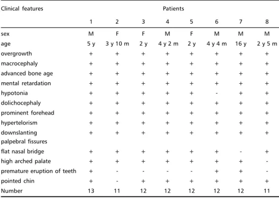
Table 1. Clinical features in our patientes with Soutos syndrome.
Clinical features Patients
1 2 3 4 5 6 7 8
sex M F F M F M M M
age 5 y 3 y 10 m 2 y 4 y 2 m 2 y 4 y 4 m 16 y 2 y 5 m
overgrowth + + + + + + + +
macrocephaly + + + + + + + +
advanced bone age + + + + + + + +
mental retardation + + + + + + + +
hypotonia + + + + + - + +
dolichocephaly + + + + + + + +
prominent forehead + + + + + + + +
hypertelorism + + + + + + + +
downslanting + + + + + + + +
palpebral fissures
flat nasal bridge + + + + + + - +
high arched palate + + + + + + +
-premature eruption of teeth + - - - - + +
-pointed chin + - + + + + + +
Number 13 11 12 12 12 12 12 11
Affected individuals are fertile and there is no evidence that life span is shortened6. The other
im-portant consideration is the risk of transmission. Sotos syndrome usually has been reported as spo-radic condition, but rarely genetic transmission as a dominant trait has been observed14, 15 including
male-to-male transmission16. Recessive inheritance has also
been suggested 17 and this syndrome has been
de-scribed in monosygotic twins2, first cousins18 and
sib-lings19. Therefore, in spite of the natural history of
Sotos syndrome has been elucidated over the years, its etiology and pathogenesis remains unknown4 .
Here, we report 8 patients with Sotos syndrome and relate their clinical features, neuroimaging and echocardiografic findings.
METHOD
All 8 patients (5 males and 3 females, age range be-tween 2 years and 16 years) had been seen in the Clinical genetics service of the University Hospital in Ribeirão Preto, University of São Paulo. The patients were seen by at least two clinical geneticists involved in this study (DGM, AXA, MAAS and JMPN). Only those patients with unambiguous classic Sotos syndrome 6 were included in this study. The
neuroimaging of the eight patients was performed by magnetic resonance imaging (MRI) scans, which were vi-sually inspected by two of the authors (ACS and JDVC), and the identified anomalies were tabulated. The
neu-roimaging findings were grouped into categories similar to those Schaefer et al.7. The criteria for ventriculomegaly
was a lateral ventricular ratio larger than 0.36 and was obtained by the width of the body of the lateral ventricle divided by half the greatest internal transverse diameter of the calvaria. All patients were also studied by echo-cardiographic method, including doppler, and the anoma-lies were registered.
Chromosomal analysis revealed normal in the 8 patients.
RESULTS
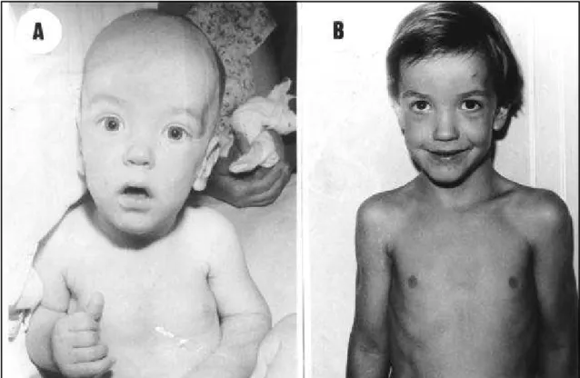
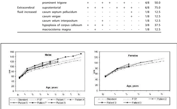
Clinical manifestations, including craniofacial cha-racteristics, in our patients with Sotos syndrome were reported in Table 1. The Figure 1 shows a patient with appearance considered typical of Sotos syn-drome, and the Figure 2 shows linear growth in all patients reported in this study.
Table 2 shows the neuroimaging findings in pa-tients, which were grouped into three categories: 1) ventricular abnormalities; 2) extracerebral CSF spa-ces; 3) midline variations.
The echocardiographic studies showed a small atrial septal defect (4.0 mm) without hemodynamic consequences in the sixth patient.
DISCUSSION
In a review, Cole and Hughes3 clinically assessed
Table 2. Neuroimaging findings in patients with Sotos syndrome
Neuroimaging findings Patients
1 2 3 4 5 6 7 8 N %
Ventricles large - + - - + - + + 4/8 50.0
prominent trigone + - + + - + - - 4/8 50.0
Extracerebral supratentorial + + + - + + + - 6/8 75.0
fluid increased cavum septum pellucidum - - - + - 1/8 12.5
cavum vergae - - - + - 1/8 12.5
cavum velum interpositum - - + - - - 1/8 12.5 hypoplasia of corpus callosum + + + - - - 3/8 37.5
macrocisterna magna - + - - - 1/8 12.5
Fig 2. A) Sotos syndrome height, males, birth to 8 years . B) Sotos syndrome height, females, birth to 5 years.
ages 1 and 6 years. Theses photographs, together with photographs of first-degree relatives, also at age 1 to 6 years, were reviewed by four clinical ge-neticists and in 41 probands, but no first-degree rela-tives the facial gestalt was thought to be character-istic of Sotos syndrome. Comparison of anthropo-metric measurements, bone age and developmen-tal delay in these 41 probands showed marker dif-ferences between them and the remaining 38 pro-bands. Length was identified as the most significantly increased prenatal parameter. In childhood, occipi-tofrontal head circumference (OFC), height and weight were all increased OFC remained above the 97th percentile in all but one case throughout
child-hood and adultchild-hood whereas height and weight had a tendency to return toward the mean. This normal-ization was more pronounced in females and was probably related to their early puberty. Early devel-opmental delay and an advanced bone age were seen in 100% and 84% of cases, respectively. Like this, the authors suggested that facial gestalt (Fig 1),
growth pattern (Fig 2), bone age and developmen-tal delay are the major diagnostic criteria7.
The neuroimaging abnormalities of Sotos syn-drome provide support for the hypothesis of delayed or disturbed development of the brain and particu-larly of midline structures20. For example, it has been
demonstrated that outside persistence of cavum sep-tum and cavum velum interposisep-tum are markers of disturb midline brain development and associated with increased risk of mental retardation21,22 .
The neuroimaging changes of Sotos syndrome appear to be at least compatible with the proposi-tion that the brain development, particularly in the midline, is delayed and/or disturbed and the enlarged CSF spaces and cerebral ventricles in these patients suggests that these children have normal size of brain inside a large head 7 .
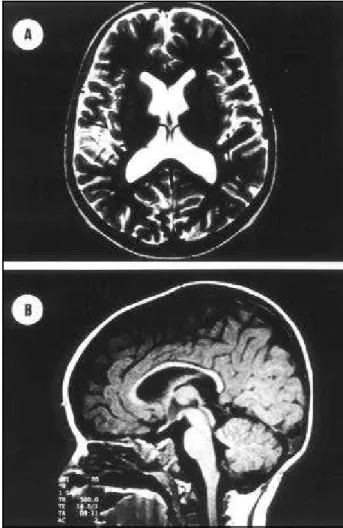
Our patients did not have migrational abnormali-ties as described by Schaefer et al.7 but we also
abnormal MRI scans (Fig 3) and we concluded, in agree-ment with Schaefer et al.7, that the neuroimaging
find-ings in Sotos syndrome are very distinctive and that MRI studies can aid in the confirmation of diagnosis7, 23.
Noreau et al. reviewed all Sotos syndrome pa-tients with congenital heart defects and related the incidence of it in approximately 8%, which is roughly a 10 fold increase over the population incidence of 0,6 to 1,0%2 . Atrial septal defect, like in our
pa-tient, is the most common defect described in Sotos syndrome23. We concluded that is just to research
CHD in Sotos syndrome patients.
Acknowledgments - We thank the Universitary Hos-pital, Faculty of Medicine of Ribeirão Preto, São Paulo Uni-versity for supporting this study.
REFERENCES
1. Sotos JF, Dodge PR, Muirhead D, Crawfort JD, Talbot NB. Cerebral gigantism in childhood: a syndrome of excessively rapid growth and acromegalic features and a non-progressive neurologic disorder. N Engl J Med 1964; 271:109-116.
2. Noreau DR, Al-Ata J, Jutias L, Teebi AS. Congenital heart defects in Sotos syndrome. Am J Med Genet 1998;79:327-328.
3. Hook EB, Reynolds JW. Cerebral gigantism: endocrinological and clini-cal observation of six patients including a congenital giant, concordant monozygotic twins, and a child who achieved adult gigantic size. J Pedriatr 1967;70:900-914.
4. Sotos JF. Overgrowth. Clin Pediatr 1997; 36:89-103.
5. Cole TRP, Hughes HE. Sotos syndrome. Am J Med Genet 1990; 27:571-576. 6. Cole TRP, Hughes HE. Sotos syndrome: a study of the diagnostic
crite-ria and the natural history. Am J Med Genet 1994;31:20-32. 7. Schaefer GB, Bodensteiner JB, Bueheer BA, Lin A, Cole TRP. The
neuroimaging findings in Sotos syndrome. Am J Med Genet 1997; 68:462-465.
8. Kaneko H, Tsukahara M, Tachibonar H, Kurashige H, Kiwano A, Kajii T. Congenital heart defects in Sotos sequence. Am J Med Genet 1987; 26:569-576.
9. Haeusler G, Guchev Z, Kohler I, et al. Constitutional chromosome anomalies in patients with cerebral gigantism (Sotos syndrome). Klin Padiatr 1993;205:351-353.
10. Opitz JM, Weaver DW, Reynolds Jr JF. The syndromes of Sotos and Weaver. Am J Med. Genet 1998;79:294-304.
11. Battaglia A, Ferrari AR. Cognitive and psychological profiles in dysmorphic syndromes. Pediatric Med Chir 1993;15:23-25.
12. Hersh JH, Cole TRP, Bloom AS, et al. Risk of malignancy in Sotos syn-drome. J Pediatr 1992;120:572-574.
13. Seyedabadi S, Bard DS, Zuna RE, et al. Epidermoid carcinoma of the vagina in a patient with cerebral gigantism. J Arkansas Med Soc 1981;78:123-127.
14. Zonana J, Sotos JF, Romshe CA, Fisher DA, Elders MJ, Rimoin DL. Dominant inheritance of cerebral gigantism. J Pediatr 1977; 91:251-256. 15. Mangano L, Palmeri S, Dotti MT, Moschini F, Faderico A. Macrossomia and mental retardation: evidence of autosomal dominant inheritance in four generations. Am J Med Genet 1989;32:67-71.
16. Halal F. Male to male transmission of cerebral gigantism. Am J Med Genet 1982;12:411-419.
17. Nevo S, Zeltzer M, Benderly A, Levy J. Evidence for autosomal reces-sive inheritance in cerebral gigantism. J Med Genet 1974;11:158-165. 18. Hooft C, Schotte H, Van Hooven G. Familial cerebral gigantism. Acta
Pediatr Belg 1968;22:173-183
19. Bejar RL, Smith GF, Park S, Spillacy WN, Wolfson SL, Nyhan WL. Ce-rebral gigantism: concentration of amino acids in plasma and muscles. J Pediatr 1970;76:105-111.
20. Garret WJ, Kossof G, Warren PS. Cerebral ventricular size in children: a two dimensional ultrasonic study. Radiology 1980; 136:711-713. 21. Breeding LM, Bodensteiner JB, Cowan L, Higgins WL. The cavum
sep-tum pellucidum: an MRI study of prevalence and clinical association in a pediatric population. J Neuroimag 1991;1:115-118.
22. Miller ME, Kido D, Horner F. Cavum vergae: association with neuro-logic abnormality and diagnosis by magnetic resonance imaging. Arch Neurol 1986; 43:821-823.
23. Melo DG, Pina-Neto JM, Acosta AX, Castro JDV, Santos AC. Neuroimaging and echocardiographic findings in Sotos syndrome. Am J Med Genet 1999;90:423-433.