CT SCAN FINDINGS IN MILD HEAD TRAUMA
A series of 2,000 patients
Kelly C. Bordignon
1, Walter Oleschko Arruda
2ABSTRACT - The present study describes the cranial computed tomography (CT) scan findings of 2,000 cases of mild head trauma (HT) in Curitiba, Southern Brazil. The mean age of the entire series was 30.8 ± 19 years. The overall male to female ratio was 2:1. The most common causes of head injury were interpersonal aggression (17.9%), falls (17.4%), automobile accidents (16.2%), falls to the ground (13.1%) and pedestrian injuries (13 %). Alcohol intoxication was associated with HT in 158 cases (7.9%). A normal CT scan was seen in 60.75% (1215) and an abnormal CT scan in 39.25% (785) of patients. Out of 785 abnormal CT scan, 518(65.9%) lesions were related to HT. The most common CT scan HT related findings were: soft tissue swelling (8.9 %), skull fractures (4.3 %), intracranial and subgaleal hematomas (3.4% and 2.4 %), brain swelling (2 %) and brain contusion (1.2%). Out of 785 abnormal CT scans, 267 (34.1%) lesions were not related to head trauma. Incidental CT scan findings included brain atrophy (5.9%), one calcification (5.2%) several calcifications (2.4%) (probably neurocysticercosis in most cases), ischemic infarct (1.9%) and leukoaraiosis (1.3%). These findings showed the importance of CT scan examination in mild head injuries. Further studies to identify mild HT patients at higher risk of significant brain injury are warranted in order to optimize its use.
KEY WORDS: head trauma, epidemiology, computed tomography, alcohol abuse.
Achados tomográficos no trauma cranioencefálico leve: análise de 2000 casos
RESUMO - São descritos os achados de tomografia computadorizada craniana (TC) de 2000 casos de trauma cranio-encefálico (TCE) leve em Curitiba, Paraná. A idade média de toda série de pacientes foi 30,8 ± 19 anos. A razão homem/mulher foi 2:1. A causas mais comuns de TCE foram agressão interpessoal (17,9%), quedas de nível (17,4%), acidentes automobilísticos (16,2%), queda ao solo (13,1%) e atropelamento (13%). Intoxicação por álcool foi um importante fator associado ao TCE e esteve presente em 158 casos (7,9% de 2000 pacientes). Uma TC normal ocorreu em 60,75% (1215) e uma TC anormal em 39,25% (785) dos pacientes. Das 785 TC anormais, os achados tomográficos mais comuns relacionados ao TCE foram: aumento de partes moles (8,9%), fraturas de crânio (4,3%), hematoma intracraniano e subgaleal (3,4% e 2,4%), “swelling” cerebral (2%) e contusão cerebral (1,2%). Os principais achados incidentais das TC anormais foram: atrofia cerebral (5,9%), uma calcificação (5,2%), múltiplas calcificações (2,4%), lesões isquêmicas vasculares (1,9%), leucoaraiose (1,3%). Achados mais incomuns foram calcificação de gânglios da base (0,8%), lesão ocupando espaço-neoplasia (0,4%) e cisto aracnóideo (0,5%). Estes achados mostram a importância da TC no TCE leve. Estudos para avaliar pacientes com TCE leve e com alto risco de lesão cerebral significativa são ainda necessários para otimizar o uso da TC.
PALAVRAS-CHAVE: trauma craniano, epidemiologia, tomografia computadorizada, alcoolismo.
Instituto de Neurologia de Curitiba, Curitiba PR, Brasil: 1Acadêmica de Medicina, Universidade Federal do Paraná (UFPR); 2Neurologista,
Professor Assistente de Neurologia, UFPR.
Received 10 May 2001, received in final form 12 November 2001. Accepted 13 November 2001.
Dr. Walter Oleschko Arruda - Rua Gonçalves Dias 713 - 80240-340 Curitiba PR – Brasil. E-mail: [email protected] Trauma is the leading cause of death in children
and young adults, and head trauma is the cause of death in more than 50% of trauma patients1. More
than 100,000 Brazilians, the majority between 5 and 40 years of age, lost their lives each year due to ex-ternal causes2, akin to what happens in other
West-ern Countries and USA, where injuries are also the leading cause of death under 45 years3. In Curitiba,
external causes were observed in 1251 deaths/year4.
Head trauma (HT) is any injury that cause lesion
or functional damage of cranium, meninges and brain. It is the most frequent lesion seen in trauma related-death5.
A CT scan is probably recommended for all pa-tients with mild head injury because one in five will have an acute lesion detectable by the scan6. The
METHOD METHOD
From September 1996 to June 1999, we prospectively registered all cases of mild head trauma (Glasgow Coma Scale score of 13,14,15), who were assisted at Emergency Services of two referal Hospitals for trauma in Curitiba: Hospital Evangelico and Hospital Cajuru. Curitiba is a 1,550,317 people city in Parana State, southern Brazil (Lati-tude 25° South, longi(Lati-tude 49° W-GR). Only the first CT-scan performed in each patient was analyzed. Most pa-tients reported loss of consciousness, though the time length could not be reliably retrieved in a few cases. Regar-ding cause of trauma, falls were considered in all patients who suffered fall from a higher level from ground. Direct trauma was defined as having the head struck by an ob-ject (e.g. stone, piece of wood, ball), and excludes cases of interpersonal aggression.
RESULTS
One thousand three hundred and six (67.3%) patients were male and 654 (32.7%) were female
(sex ratio M:F=2:1). Ages ranged from some days of life to 98 years, with a mean age of 30.5 ± 19 years. The mean age ± standard deviation (SD) of males and females patients were 30.2 ± 22.2 and 30.2 ± 17.2 years, respectively. The highest frequency of HT ocurred in the 21-30 years interval (25.1%), followed by the age groups 11-20 (21.6%) and 31-40 (17.5%) (Fig 1).
The most common causes of head injury were aggression (17.9%), falls (17.4%), automobile acci-dents (16.2%), falls to the ground (13.1%) and pe-destrians injuries (13%) (Fig 2). The medium age for each group of causes varied from 21 to 44 years. There was a pronounced difference in the distribu-tion of causes according to age and gender (Table 1). Fall to the ground was the most frequent cause of HT associated to alcohol abuse (17.6%), followed by aggression (12.3%) and motorcycle accidents (9.4%) (Fig 3).
Fig 1. Age distribution per decade in 2,000 HT patients.
Fig 2. Causes of trauma, distributed by gender.
0 50 100 150 200 250 300 350 400
0-1
0
11
-2
0
21
-30
31
-40
41
-50
51
-60
61
-70
71
-80
81
-90
91
-100
Female Male
0 50 100 150 200 250 300 350
Male 287 211 215 146 157 126 67 52 49 23 13 Female 70 136 109 116 102 27 33 19 15 26 1
Aggression Falls Automobile accident
Fall to the
ground Pedestrian Bicycle Direct
Table 1. Causes of HT according to age and gender.
Type of accident Number Age (mean±SD) Sex ratio
years (M:F)
Aggression 357 30.5 ± 13.0 4.1:1
Falls 347 27.9 ± 20.4 1.6:1
Automobile 324 29.2 ± 13.4 2.0:1
Fall to the ground 262 44.6 ± 25.9 1.3:1
Pedestrians 259 28.4 ± 19.1 1.5:1
Bicycle 153 21.5 ± 12.1 4.7:1
Direct impact 100 26.1 ± 17.7 2.0:1
Epilepsy 71 37.9 ± 16.1 2.7:1
Motorcycle 64 25.6 ± 9.4 3.3:1
Syncope 49 42.3 ± 21.7 1:1.1
Fig 3. Percentage of excessive alcohol intake and related forms of HT.
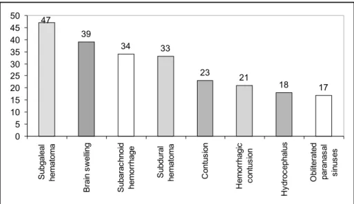
Fig 4. CT scan findings related to HT in 2,000 patients.
17,6
12,3
9,4
3,7 6,2
6,2 5,6
3,9
0 0
0 2 4 6 8 10 12 14 16 18 20
F
a
ll
to
the
gr
ound
A
g
g
re
ssio
n
M
o
to
rcycle
Pedes
tr
ian
A
ccid
e
n
t
E
p
ile
p
s
y
B
icycle Fa
lls
Dire
c
t
imp
a
c
t
Sy
nc
ope
% of alcool
39
34 33
23
21
18 17 47
0 5 10 15 20 25 30 35 40 45 50
S
ubgaleal
hem
at
om
a
B
rain
s
w
e
lling
S
u
bar
a
c
hnoid
hem
or
rhage
S
u
bdur
al
hem
at
om
a
Co
n
tu
s
io
n
H
e
m
o
rr
hagic
c
ont
us
ion
H
y
dr
oc
ephalus
O
b
lit
er
at
ed
par
anas
al
s
inus
Of 2,000 patients studied, 518 (25.9%) had 14 different lesions related to HT detected on CT scans. Soft tissue swelling was the most common finding (178), followed by fractures (86) (Fig 4). Fractures included facial fractures, skull base fractures, vault skull fractures, both lineal and depressed ones. Fig-ure 4 shows the different CT scan findings and their respective numbers. Ventricular dilatation and epi-dural hematoma were observed in 2 and 7 cases re-spectively. Obliterated paranasal sinusesand pneu-mocephalus were present in 17 and 8 victims, re-spectively.
Twelve different lesions not related to HT were present in 267 patients (13.3%) out of the total 2,000 patients. The most common incidental findings in-cluded brain atrophy, calcifications, vascular ischemic lesion, lacunes, leukoaraiosis, sinus disease and ara-chnoid cyst (Fig 5). Other findings were gliosis (7 cases), space-occupying lesion (7 cases), and active neurocysticercosis (3 cases).
DISCUSSION
Head injury is a major world health problem. Sev-eral reports point automobile accident as the most important cause of HT. Motor vehicle accident causes HT about 35% to 60% in diverse series, and is usu-ally a leading cause of serious injuries with head trauma in youth and middle age people and a com-mon cause of morbidity and mortality related to trau-ma7. They are also the most frequent cause of death
in individuals from 1 to 35 years of age8,9. Previous
studies of head trauma in the United States demon-strated traffic accidents accounting for about half the fatal cases10,11. Other series show automobile
acci-dents as a major cause of HT. In Massachussets, USA, 33.4% of causes were related to automobile acci-dents12. In our series, we only observed 16.2% of
automobile accidents in mild HT. Automobile acci-dents may be less common in low socioeconomic populations due to limitation of owning a car. In fact, a study of Taiwan did not find very significant figures for automobiles, because motorcycle is the most common means of transport in that country7.
Our findings are thus not in keeping with the abo-ve mentioned literature, for aggression, which in-cludes assaults and firearms injuries, were the com-monest cause of HT (17.9%). Assault, one form of aggression, is a common cause of head injury in some places, particularly in economically depressed, den-sely populated urban areas3. Alcohol abuse also
con-tributes with higher rates of aggression. In fact, it was present in 12.3% of all aggressions in our se-ries. A study from Scotland found 20% of HT causes related to assault3. In Chicago13 and Massachussets12,
40% and 24.9% (respectively) of mild HT were caused by assaults. In the Bronx14, assault was the leading
cause of head injury, but overall in the United States it accounted for only 10%3. Steven et al.32 found
in-terpersonal attacks (aggression) responsible for the largest proportion (40%) of head injuries in Blacks in an inner city community of Chicago. The authors again raised the possibility of socioeconomic status as an important factor related to HT in different com-munities15,16. Therefore, socioneconomic factors may
play a major role leading to higher prevalence of aggression observed in Curitiba. It is interesting to observe the correlation of excessive alcohol intake with aggression, as already commented.
Fig 5. Distribution of incidental findings.
117 104
47 37
26
19 15
12 10
0 20 40 60 80 100 120 140
Brain
a
tr
ophy
One
c
a
lc
if
ic
at
ion
Sev
e
ral
c
a
lc
if
ic
at
ions
Old
is
c
hem
ic
les
ion
Leuk
oaraios
is
Lac
unes
C
a
lc
if
ic
at
ion
bas
al
ganglia
Sinus
dis
eas
e
Arac
hnoid
cyst
num
ber
of
c
as
Falls are second only to motor vehicle accidents among unintentional traumatic causes of death in all ages17. A study from Scotland3 found 39% of HT
related to falls, in Chicago (29%)13 and in
Massachus-sets (30.5%)12. Our series observed falls as the
sec-ond cause of HT (17.4%). The higher percentage of falls in those series may be due to the separation between of falls and falls to the ground we made in our series. If we combine both conditions, we come with 30.45% of HT as caused by falls, a similar pro-portion observed in other countries.
Among elderly people, falls are particularly im-portant as causal factor of morbidity and mortality. In Massachusetts, a retrospective case series of 318 patients aged 60 years and over, falls causing HT were seen in 189 patients (59%)18. In our study, falls
reached an overall proportion of 17.35%, and the falls to the ground represented 13.1%. Taking in ac-count only patients aged 60 years and older, we also found a high proportion of falls (58.5%) as a major cause of HT in the elderly.
Although not very well documented, many falls in adults are related to alcohol. In our study, 17.6% of the falls to the ground were associated with alco-hol abuse. Nagurney et al.18 referred alcohol abuse
in 11% of elderly falls. Our study did not find alco-hol abuse in elderly falls. As a matter of fact, the rate of alcohol intake in elderly falls is quite varia-ble, ranging from 2% to 34%19.
Motorcycle or injuried pedestrians are less fre-quent causes of HT (5-10%)20,21. Nevertheless, in
Tai-wan the most common causes of head injury were motorcycle accident (53.6%) and pedestrian injuries (29.47%), since motorcycle is the most common means of transport in that country 7. Sallum and
Koizumi 22, in a study of 220 injuried patients in
traf-fic accidents (TA) in São Paulo, identified the follow-ing TA types: 50.46% pedestrians, 37.73% automo-bile, and 11.81% motorcycle. Our study showed simi-lar proportion of types of TA causing HT: pedestri-ans (50.1%), automobiles (40%), and motorcycle (9.9%). We observed alcohol intoxication in 9.4% of motorcyclists and in 6.2% of pedestrians.
There is increasing awareness of the importance of bycicle accidents, which are particularly common in children23,24. In this study there is a high frequency
of bicycle accidents (7.7%), probably related to the frequent use of bicycle by Brazilians, mainly during the second and third decade of the HT population (63.4% of all bicycle accidents). We observed alco-hol intoxication in 3.9% of the cyclists, although in
San Diego over half the brain injuried cyclists over 15 years-old were intoxicated24. These differences
may reflect biases of selection or of data retrieval.
Alcohol is an important contributory cause of in-jury. Its influence is best documented by automo-bile accidents, especially drivers3, since alcohol
in-toxication is more thoroughly investigated in such situation. In our series, alcohol abuse was present in 7.9% of the all HT cases. This relatively small frac-tion may be due to the smaller number (16.2%) of automobiles accidents causing HT in Curitiba. Not-withstanding, Schackford et al6 found that 28.2%
of all HT victims were drunk, but more than half of their 2,766 HT patients suffered accidents with ve-hicles motors. This could explain the larger propor-tion of alcohol intoxicapropor-tion in that series, since the screening for alcoholic intake is more systematic in these accidents.
The mean age in HT causes was relatively low in our series. For example, in Taiwan7, the mean age in
different HT groups were: motorcycle - 32.6 years, pedestrians - 32.5 years, automobile - 32.8 years, and fall - 34.5 years (Table 1). It can be explained for higher rate of smaller ages in Brazil. A statistics database observed that 35% of Brazilians were be-low 14 years old25.
In our series, men were more prevalent than wo-men in all groups of HT causes, except syncope, whe-re women pwhe-resented a slightly higher fwhe-requency (F:M ratio =1.1:1) (Table 1). In fact, another series26
pre-sented also a higher F:M ratio (1.51:1).
Cranial CT-scan is the most frequently performed radiological investigation in developed countries18.
Stein and Ross recommended routine and immedi-ate cranial CT scanning of all head injury patients who have lost conciousness and were amnesic, even if all other physical findings were normal27. They
re-ported a high risk of intracranial lesions (12.9%) in mild head injury28. In subsequent prospective
stud-ies of mild HT, the frequency of intracranial lesions on CT-scan was lower (6 to 9%)29,30. Haydel et al.
identified patients with mild HT who should undergo CT-scan based on clinical findings and, thus, did not recommend CT-scan in all patients with mild HT31 .
In our series, 518(25.9%) out of the 2,000 pa-tients presented CT-scan findings related to HT. Two studies observed 17.2%27 and 18%32 of abnormal
CT-scan in mild head trauma, numbers close to our observation. However, another study found a higher frequency (46%)6, but the authors included other
explain such discrepancy. On the other hand, Jeret et al.33 found 9.4% of abnormality on the CT-scan,
but they excluded lineal fractures and isolated soft tissue swelling. It’s conceivable that methodologi-cal differences among those studies and ours may account for such different proportions.
Regarding lesion types, soft tissue swelling was the most common (178 cases - 34% cases out of 518 abnormal CT scans). Skull fractures were also important, accounting for 74 (14.3%) cases. In other reports, the rates of skull fractures were much lower (8.5% and 4.1%)27,33. Only 2% of attenders at
Scot-tish accident departments for recent head injury have a skull fracture3. But, Shackford et al.6 observed
19.3% of skull fractures, a number closer to our study.
Intracranial hematoma after HT is a frequent cause of death and disability. Expeditious evaluation and adequate management of patients who initially seem at low risk are the most important factors to reduce their mortality34. Among our HT patients, 21
cases (4.1%) presented intracerebral hematoma. Jeret et al.33 found 10.4% of intracranial bleeding in
his study with mild head trauma and GCS score of 15. However, his work excluded patients younger than 18 years. Focal lesions are less common in chil-dren with HT when compared to adults35, what
prob-ably explains our lower number of intracranial he-matomas (23.7%) of our patients were younger than 18-year-old. Stein and Ross27 found 4.2% (11 cases)
of intracerebral hematomas, 7 of them with skull fracture. In our study, only one case of intracerebral hematoma was associated with skull fracture.
Stein and Ross27 found 6.8% of epidural
he-matoma (EH), 5.7% of subarachnoid hemorrhage (SAH), and 4.5% of subdural hematoma (SH). Our study showed similar numbers: 6.6% of SAH, 6.4% of SH, and 1.4% of EH.
Cerebral contusions were seen in 25%33 to 26.8%27.
The present study showed lower numbers (12.9%). The other series27,29 only included patients with loss
of consciousness or amnesia, therefore selecting pa-tients at higher risk of intracranial injuries. Stein and Ross27 obtained 30.9% for the brain swelling and
the literature varied from 5%6 to 11%37, and our
study found a number between these extremes (7.5%). We had a lower frequency of pneumoce-phalus (1.5%), when compared to Shackford6(5.4%)
and Feuerman et al. (13.2%)36.
As expected, a vast majority of 2,000 HT patients did not demonstrate incidental CT findings. As a
matter of fact, the relative young age of our all co-hort decreased the chance of revealing pathologic findings such as those related to age, in spite of having been the most common findings.
Katzman37 found 18% of incidental findings on
MRI of 1000 asymptomatic volunteers, a higher num-ber when compared to our 13.3% on CT scan. This difference may be due to the higher sensitivity of MR imaging when compared to CT scan.
Age related changes occurred in 173 (8.7%) pa-tients: brain atrophy (5.9%), ischemic vascular lesions (1.9%) and lacunes ( 1%). The small proportion of those findings are clue to the relative young age of our studied population (the vast majority between 10 and 50 year old.
One calcification (5.2%), several calcifications (2.3%) (probably mostly neurocysticercosis in our series ) and basal ganglia calcification (0.8%) were not reported by Katzman37. This is not surprising due
to the relative low MR sensitivity to calcifications and the different epidemiological profile regarding the neurocysticercosis/taeniasis between the series. Other findings were leukoaraiosis (1.3%) and space-occupying lesion (0.4%)(0.5% in Katzman’s work)37.
We found ten patients (0.5%) with arachnoid cyst (AC) comparing with 0.3% in the literature37. Other
series38 observed a higher prevalence of 1.4% of AC,
probably because only neurological patients were included. Another report found five in 3000 con-secutive fetal and neonatal autopsies, a much lower number39.
In conclusion, our study showed the epidemic profile of the head trauma in the city of Curitiba, Brazil. Aggression and falls played a major role as causes of HT. Automobile accident still has a great importance. The prevalence of abnormal CT-scan findings did not significantly change according to the HT cause. Alcohol intoxication played a major role as an associated factor related to HT. The high prevalence of HT related CT findings probably jus-tify the use of CT scan in mild HT. Further studies to identify which mild HT patients are at higher risk of significant cerebral injury are warranted in order to optimize its use.
REFERENCES
1. Castillo M, Harris JH. Skull and brain. In Harris JH, Harris WH, Novelline AR (eds).The radiology of emergency medicine. 3. Ed. Balti-more: Willians and Wilkins, 1993.
2. Birolini D. Trauma: uma epidemia esquecida ou o Brasil nos tempos do trauma. Rev AMB 1991;37:53-54.
4. Instituto Brasileiro de Geografia e Estatística (IBGE). Óbitos por grupo etário: principais causas. Brasil,1996.
5. Alberico AM, Ward JB, Choi SC, et al. Outcome after severe head in-jury: relationship to mass lesion, diffuse injury, and ICP course in pe-diatric and adult patients. J Neurosurg 1987;67:648-656.
6. Schackford SR, Wald SL, Ross SE, et al. The clinical utility of computed tomographic scanning and neurologic examination in the management of patients with minor head injuries. J Trauma 1992;33:385-394. 7. Lee ST, Lui TN, Chang CN, et al. Features of head injury in a
develop-ing country Taiwan (1977-1987). J Trauma 1990;30:194-199. 8. Champion HR, Copes WS, Sacco WJ, et al. The trauma outcome study:
establishing national norms for trauma care. J Trauma 1990; 30:1356-1365.
9. Thompson CT, Bickell WH, Siemens RA, Sacra JC . Community hospi-tal level II trauma center outcome. J Trauma 1992; 32:336-343. 10. Eisenberg HM. Outcome of head injury: general considerations and
neurobehavioral recovery: Part 1. General considerations. In Becker DP, Povlishock JT (eds). Central nervous system trauma status report. Rockville, MD: National Institutes of Health, 1985.
11. Jagger J, Levine JI, Jane JA, Rimel RW. Epidemiological features of head injury in a predominantly rural population. J Trauma 1984;24:40-44. 12. Borczuk P. Predictors of intracranial injury in patients with mild head
trauma. Ann Emerg Med 1995;25:731-736.
13. Whitman S, Cooley-Hoganson R, Desai BT. Comparative head trauma experience in two socioeconomically different Chicago-area commu-nities: a population study. Am J Epidemiol1984;119:570-580. 14. Cooper KD, Tabbador K, Hauser WA, et al. The epidemiology of head
injury in the Bronx. Neuroepidemiology 1983;2:70-88.
15. Jason J, Strauss LT, Tyler CW Jr. A comparison of primary and second-ary homicides in the United States. Am J Epidemiol 1983;117:309-319. 16. Centerwall BS. Race, socioeconomic status, and domestic homicide: Atlanta,1971-1972. Presented at Society for Epidemiologic Research meeting, Cincinnati, June 1982.
17. Accident Facts, 1988 edition. Chicago: National Safety Council, 1988. 18. Nagurney JT, Borczuk P, Thomas SH. Elderly patients with closed head
trauma after a fall: mechanisms and outcomes. J Emerg Med 1998;16:709-713.
19. Roy CW, Pentland B, Miller JD. The causes and consequences of minor head injury in the elderly. Injury 1986;17:220-223.
20. Annaegers JF, Grabow JD, Kurland LT, et al. The incidence, causes, and secular trends of head trauma in Olmsted county, Minnesota, (1935-1974). Neurology 1980;30:912-919.
21. Gale JL, Dikmen S, Wyler A, et al. Head injury in the Pacific North-west. Neurosurgery 1983;12:487-491.
22. Sallum AMC, Koizumi MS. Gravidade do trauma cranioencefálico em vítimas de acidente de trânsito. Rev Bras Neurol 1999;35:49-55.
23. Kraus J, Fife D, Conroy C. Incidence, severity and outcomes of brain injuries involving bicycles. Am J Public Health 1987;77:76-77. 24. Sacks JJ, Holmgreen P, Smith SM, Sosin DM. Bicycle-associated head
injuries and deaths in the United States from 1984 through 1988. JAMA 1991;266:3016-3018.
25. Marcílio ML. A lenta construção dos direitos da criança brasileira-século XX. São Paulo Biblioteca Virtual de direitos humanos-Universidade de São Paulo. [Disponível em http://www.direitos humanos.USP.br/ bibliografia/mluiza.html]
26. Kapoor WN. Evaluation and outcome of patients with syncope. Medi-cine 1990;69:160-175.
27. Stein SC, Ross SE. Mild head injury: a plea for routine early CT scan-ning. J Trauma 1992;33:11-13.
28. Stein SC, Ross SE. The value of computed tomographic scans in pa-tients with low-risk head injuries. Neurosurgery 1990;26:638-640. 29. Miller EC, Derlet RW, Kinser D. Minor head trauma: is computed
to-mography always necessary? Ann Emerg Med 1996;27:290-294. 30. Jeret JS, Mandell M, Anziska B, et al. Clinical predictors of abnormality
disclosed by computed tomography after mild head trauma. Neuro-surgery 1993;32:9-15.
31. Haydel MJ, Preston CA, Mills T, et al. Indications for computed to-mography in patients with minor head injury. N Engl J Med 2000;343:100-105.
32. Steven ER. Are CT scan for head injury patients always necessary? J Trauma 1991;31:801-805.
33. Jeret JS, Mandell M, Anziska B, et al. Clinical predictors of abnormality disclosed by computed tomography after mild head trauma. Neuro-surgery 1993;32:9-16.
34. Klauber MR, Marshall LF, Luerssen TG, et al. Determinants of head injury mortality: importance of the low risk patient. J Neurosurg 1989;24:31-36.
35. Committee on Trauma Research, Comission on Life Sciences, National Research Council, and institute of Medicine. Injury in America. Washigton, DC: National Academy Press, 1985.
36. Feuerman T, Wackym PA, Gade GF, Becker DP. Value of skull radiog-raphy, head computed tomographic scanning, and admission for ob-servation in cases on trauma of minor head injury. Neurosurgery 1988;22:449-453.
37. Katzman GL, Dagher AP, Patronas NJ. Incidental findings of brain mag-netic resonance imaging from 1000 asymptomatic volunteers. JAMA 1999;282:36-39.
38. Narata AP, Arruda WO, Ramina R. Cistos aracnóides: uma revisão de 36 casos. O Dendrito 1999;5:45-47.