IS LOW ANTIEPILEPTIC DRUG DOSE EFFECTIVE IN
LONG-TERM SEIZURE-FREE PATIENTS?
Tânia A.M.O. Cardoso
1, Fernando Cendes
2, Carlos A.M. Guerreiro
3ABSTRACT -Objective: To investigate the value of leaving seizure-free patients on low-dose medication. Method: This was an exploratory prospective randomized study conducted at our University Hospital. We evaluated the frequency of seizure recurrence and its risk factors following complete or partial antiepileptic drug (AED) withdrawal in seizure free patients for at least two years with focal, secondarily generalized and undetermined generalized epilepsies. For this reason, patients were divided into two groups: Group 1 (complete AED withdrawal), and Group 2 (partial AED withdrawal). Partial AED withdrawal was established as a reduction of 50% of the initial dose. Medication was tapered off slowly on both groups. Follow-up period was 24 months.
Results: Ninety-four patients were followed up: 45 were assigned to complete (Group 1) AED withdrawal and 49 to partial (Group 2) AED withdrawal. Seizure recurrence frequency after two years follow-up were 34.04% in group 1 and 32.69% in Group 2. Survival analysis showed that the probability of remaining seizure free at 6, 12, 18 and 24 months after randomization did not differ between the two groups (p = 0.8). Group 1: 0.89, 0.80, 0.71 and 0.69; group 2: 0.86, 0.82, 0.75 and 0.71. The analysis of risk factors for seizure recurrence showed that more than 10 seizures prior to seizure control was a significant predictive factor for recurrence after AED withdrawal (hazard ratio = 2.73). Conclusion:Leaving seizure free patients on low AED dose did not reduce the risk for seizure recurrence. That is, once the decision of AED withdrawal has been established, it should be complete.
KEY WORDS: epilepsy, antiepileptic drug withdrawal, seizure recurrence, prognosis.
Vale a pena manter baixas doses de droga antiepilética em pacientes com epilepsia controlada?
RESUMO - Objetivo: Investigar o valor da manutenção de baixas doses de medicação em pacientes com epilepsia controlada. Método: Trata-se de um estudo prospectivo aleatorizado exploratório, realizado em nosso Hospital Universitário. Nós avaliamos a freqüência de recorrência de crises e os fatores de risco associados após a retirada parcial ou completa da droga antiepiléptica (DAE), em pacientes com crises controladas por pelo menos dois anos. Os pacientes foram divididos em dois grupos: Grupo 1 (retirada completa da DAE) e Grupo 2 (retirada parcial da DAE). Retirada parcial da DAE foi estabelecida como uma redução de 50% da dose inicial. A medicação foi retirada lentamente nos dois grupos. O período de seguimento foi de 24 meses.
Resultados: Noventa e quatro pacientes foram seguidos: 45 foram selecionados para a retirada completa (Grupo 1) e 49 para a retirada parcial (Grupo 2). As taxas de recorrência de crises após dois anos de seguimento foram 34,04% para o Grupo 1 e 32,69% para o Grupo 2. A análise de sobrevivência mostrou que a probabilidade de permanecer livre de crises aos 6, 12, 18 e 24 meses após a aleatorização não diferiu entre os dois grupos (p = 0,8). Grupo 1: 0,89, 0,80, 0,71 e 0,69; Grupo 2: 0,86, 0,82, 0,75 e 0,71. A análise dos fatores de risco para a recorrência de crises demonstrou que mais que 10 crises antes do controle foi significante para a recorrência após a retirada da DAE (razão de risco = 2,73). Conclusões: A manutençãode baixas doses de medicação em pacientes com epilepsia controlada não reduz o risco de recorrência de crises. Portanto, caso a decisão de retirar a DAE tenha sido estabelecida, a retirada deve ser completa.
PALAVRAS-CHAVE: epilepsia, retirada de droga antiepiléptica, recorrência de crises, prognóstico.
Department of Neurology, State University of Campinas (UNICAMP), Campinas, SP Brazil: 1MD, 2Assistent Professor, 3Associated Professor. Received 11 December 2002. Accepted 3 March 2003.
Dr. Carlos A.M. Guerreiro Department of Neurology, University of Campinas, UNICAMP P. O. Box 6111 13083970 Campinas SP -Brasil. E-mail: [email protected]
It is widely known that a large majority of epileptic
patients benefit from the use of AED, which usually
bring about prompt seizure remission. Several studies
in newly diagnosed cases of epilepsies, treated with
AED, showed one-year remission rates between 65
and 80%
1. Retrospective studies based on general
populations also demonstrate elevated remission
rates in patients who were diagnosed with epilepsy
in the past and treated with AED
2,3. Epilepsy, as a
real effect of AED on the evolution of epilepsy. The
AED, apparently, do not modify the natural evolution
of the disease but seem to simply suppress seizures
during a vulnerable period
4. AED withdrawal is an
inevitable question when prolonged remission is
achieved. The toxicity of prolonged AED use is the
most powerful argument for drug withdrawal
5.
Te-ratogenic effect in pregnant women and AED
inte-raction with oral contraceptives should be considered
as well. Concern about weight gain, compromised
osseous metabolism, and reproductive-endocrine
disorders in women should also be addressed.
6.
Another important argument in favor of AED
inte-rruption is the implicit stigma associated with its use,
branding the patient as a chronic disease carrier.
Over the past decades, studies conducted on AED
withdrawal after prolonged seizure control in adults
and children demonstrated that a great proportion
of the patients remained seizure free
7. Various studies
report the evolution of patients with controlled
epilepsy after complete AED withdrawal, but no data
are available regarding the effectiveness of low AED
doses in these patients. Nevertheless, it is not
un-common to observe patients with controlled seizures
at “subtherapeutic” AED doses. Some authors state
that maybe the seizure threshold increases as the
duration of epilepsy increases, modifying minimal
therapeutic drug level. This is an important issue,
because maintenance of lower AED doses would
guarantee seizure control with reduced drug toxicity.
Nevertheless, patients would have other benefits
such as reduced treatment costs.
The objective of this pilot study is to investigate
the value of leaving seizure-free patients on low-dose
medication.
METHOD
This is an exploratory, randomized, prospective study that compares the risk of seizure recurrence after total and 50% partial AED withdrawal in patients with controlled epilepsy during a follow-up period of two years. This study was conducted at the Neurology Clinic of our University Hospital. The initial evaluation as well as the follow-up was exclusively performed by the first author, at a specific outpatient clinic. The period for patient inclusion extended from January 1991 to November 1995.
Patients included in this study were those who had partial or generalized tonic-clonic seizures (GTCS) and who have been seizure-free for two years or longer. They must have 14 years-old or more and be on monotherapy with AED conventional doses. Epilepsy was defined as the occur-rence of two or more unprovoked seizures with at least 24 hours between the seizures8. Patients with idiopathic partial and primary generalized epilepsies as well as acute
symptomatic and single seizure cases were not included in the study. Patients with confirmed psychiatric diseases and those with non-compliance were excluded.
The decision to withdraw AED was taken after the pa-tient was invited to participate in the study and after tho-roughly discussing the risks and benefits with the patient and the family. After the patients had agreed to participate in the study and before randomization, all the patients and/or their families (in the case of patients under 18 years of age) signed informed consents. The Ethic Committee of University of Campinas approved the study.
Patients were then randomly assigned to two groups: Group 1 – gradual and complete withdrawal within six months; Group 2 – partial withdrawal of 50% of the AED dose in two months. The technique of randomization used was alternating allocation of consecutive patients.
Before initiating withdrawal of the medication, a com-plete detailed clinical history was obtained in relation to epilepsy, personal and family antecedents, treatments and complementary investigations. All patients were subjected to clinical as well as neurological examinations.
1. Patients and Follow-up - From a total of 136 conse-cutive patients initially assessed, 117 patients fulfilled the inclusion criteria, agreed to participate in the study and were randomized. A group of 18 patients who were already randomly selected were later excluded because of inclusion errors: 10 patients due to either non–compliance or lost follow-up, two patients who later admitted a seizure control period of less than two years, one patient with primary generalized epilepsy (diagnosed after his inclusion in the study), one patient with a non-epileptic seizure (syn-cope), one patient had a single seizure with status epi-lepticus, one patient at withdrawal admitted that he was already on a sub-dose, one patient refused to sign the in-formed consent and one patient died due to gastro-intes-tinal cancer. The remaining 99 patients were followed up. From the 19 patients not included: six of them were already on a subdose and/or reducing AED dose, five pa-tients were concerned about the withdrawal and refuse to participate, four had less than two years of seizure con-trol, one was a chronic alcoholic with seizures due alcohol abstinence, one patient had psychiatric antecedents and probable non-epileptic seizures, one had unclear diagnosis and one had serious medical condition that required post-ponement of the procedure.
Five of the 99 patients that initiated the follow-up were excluded from the comparative analysis (two from Group 1 and three from Group 2) because they presented seizure recurrence during withdrawal while utilizing 75% of the AED dose. Than, the comparative analysis covered a total of 94 patients and none of the patients were lost during the follow- up period of two years.
Clinical characteristics of the patients - The clinical and demographic characteristics of 94 patients included in the comparative analysis were: Group 1 with 45 patients and Group 2 with 49 patients; 44 patients were females (46.81%); 74 patients were white (78.72%). The median age when withdrawal was initiated was 30.3 years (minimum = 15 and maximum = 76 years), the median age at epilepsy onset was 16.9 years (minimum = 0.1 and maximum = 62 years), the median active epileptic duration was 10.7 years (minimum = 0.1 anad maximum = 40 years). Seizure control period before withdrawal: 64 patients (68.09%) presented two to three years, 17 patients (18.09%) presented 3 to 4 years and 13 patients (13.83%) presented 4 years or more.
Recall visits took place every two months during the withdrawal phase and every four to six months during the follow-up phase, with telephone calls every six months to make sure that information regarding seizure recurrence was reported. All patients were also instructed to phone the physician in case of seizure recurrence. Precise infor-mation regarding seizure recurrence and type was obtai-ned through detailed interviews held with the patient and whenever possible with witnesses present.
2. Method and rate of withdrawal - Withdrawal began with a 25% of the total dose and then 25% every two months until withdrawal was complete for Group 1 (pre-dicted period of 6months) and up to 50% of the initial dose for Group 2 (predicted period of 2 months). According to the commercial preparations available, the real AED dose administered was as close as possible to the calculated dose.
3. Epileptic seizures and syndromes - The seizures were classified according to the revised ILAE Classification of Epileptic Seizures (1981)9. Classification of the type of epilepsy was based on the ILAE Classification of Epilepsies and Epileptic Syndromes (1989)10. A diagnosis of sympto-matic epilepsy was concluded when an etiological factor was clearly recognized or when clinical or imaging signs detected a cerebral lesion that was consistent with a type of epilepsy. The terms cryptogenic epilepsy were used when the etiology was unknown. The supposed localiza-tion of an epileptogenic zone was based on seizure semio-logy as well as electroencephalographic and imaging fin-dings. Patients without unequivocal features of focal or generalized seizures such as those with only GTCS that occurred exclusively while sleeping and without focal fin-dings by clinical, imaging or electroencephalographic tests were classified as undetermined epilepsy (10, 11).
Status epilepticus was defined as more than 30 minutes of continuous seizure activity or two or more sequential seizures without full recovery of consciousness between seizures9,12.
4. Electroencephalogram (EEG) - Electroencephalo-grams were obtained in accordance with the international norms related to the “ 10-20 System” electrode placement and montages recommended by the American EEG Society13. The exams were always performed utilizing activation methods (hyperventilation and intermittent photic stimulation) during wakefulness and whenever pos-sible, spontaneous sleep.
The EEG was performed in 88 of the 94 patients during AED withdrawal. Available EEGs performed before AED withdrawal (90/94 patients) and after withdrawal (92/94 patients) were also assessed.
EEGs were qualitatively analyzed by a team of electro-encephalographers at the Clinical Neurophysiology Depart-ment without previous knowledge about the randomiza-tion of AED withdrawal. In the analysis, the EEG results were classified as normal versus abnormal. Abnormal EEG was considered when epileptiform abnormalities and/or focal or diffuse slowing were present.
After AED discontinuation EEG results were assessed. It was considered worsening in EEG pattern when epilep-tiform activity was registered de novo or when it became abnormal.
5. Family history - Previous family history of epilepsy or an unprovoked single epileptic seizure that occurred in first-degree relatives and/or two or more second or third degree relatives were considered as positive.
6. Imaging exams - A computerized tomography (CT) was performed on all the patients and magnetic resonance imaging (MRI) was performed on 78 patients.
7. Epilepsy severity markers - We tabulated the total number of seizures and number of GTCS before seizure control; duration of active epilepsy (period between the first and last seizure before control); previous unsuccessful attempts at AED withdrawal (excluding abrupt withdra-wals); number of AED required in monotherapy to control seizures since the disease onset (to judge effectiveness, adequate doses of AED were utilized for adequate periods); previous history of status epilepticus before withdrawal; maximum seizure frequency before control (Fig. 8 -evidencing of the region of the body of an arachnoid granulation evidencing thick bundles of collagen fibers (arrows) associated to thinner bundles (arrows 1). 1100x
≥ 1/month and < 1/month).
The results were analyzed utilizing the parameters de-fined in the literature14 in the following therapeutic ran-ges: Carbamazepine – 4 to 12ug/ml, Phenytoin – 10 to 20 ug/ml and Phenobarbital - 10 to 40ug/ml.
Daily AED doses and plasma levels range as follows: carbamazepine 400 to 1400 mg/day (media = 692.7) and < 1.0 to 12.9ug/ml (media = 6.95), phenytoin 200 to 400 mg/day (media = 295.8) and < 1.0 to 37.4 ug/ml (media = 8.63), phenobarbital 100 to 150 mg/day (media = 101.8) and 1.0 to 34.6 ug/ml (media = 15.21) and valproate 500 to 1500 mg/day (media = 1000, only two cases).
9. Summary of the recurrence risk factors - Sex, age at AED withdrawal (up to 30 and above 30 years), age at onset of epilepsy (up to 12 and above 12 years), family history of epilepsy, etiology of epilepsy (localized sympto-matic, localized cryptogenic and undetermined), presence of deficits on neurological examination, seizure type (partial or generalized/ secondarily generalized), seizure control period (between two to three years and three years or more), EEG, CT, AED used at withdrawal, AED serum level at withdrawal and the epilepsy severity markers.
Statistical analysis - Statistical methods used took into consideration the duration of the follow up period, as it influences the probability of seizure recurrence. Therefore, the survival analysis was utilized to observe seizure recur-rence after AED withdrawal. The Kaplan-Meier curves pro-vided a cumulative probability of remaining seizure free (non-recurrence or remission) in relation to time. The uni-variate and multiuni-variate analysis were applied using the Cox proportional hazard regression model to assess the prognostic factors affecting seizure recurrence15.
RESULTS
Overall recurrence risk
During the first two years of follow-up, 16 out of
47 patients in Group 1 (34.04%) and 17 out of 52
patients in Group 2 (32.69%) presented seizure
re-currence. Two patients from Group 1 and three
pa-tients from Group 2 presented recurrence while using
75% of the initial AED dosage during the first six
months of follow-up.
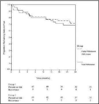
The survival analysis of 94 patients who were
subjected to two therapeutic strategies indicating
the time dependent cumulative probability of
pa-tients remaining seizure free is shown in Figure 1.
This cumulative probability of continuing in remission
at 6, 12, 18 and 24 months after initiating drug
with-drawal was 0.89, 0.80, 0.71, 0.69 for Group 1 and
0.86, 0.82, 0.75, 0.71 for Group 2 (Table 1).
Fig 1. Cumulative probability of remaining seizure free following discontinuation of AED in a 24 months follow-up, in patients with controlled epilepsy: overall recurrence risk - Kaplan-Meier survival curves comparing total with partial 50% dose.
Table 1. Cumulative probability of remaining seizure free among randomized groups after starting AED withdrawal (Survival analysis).
Survival Probability (Proportion of Remission) after starting AED withdrawal
6 months 12 months 18 months 24 months Group 1 0.8889 0.8000 0.7111 0.6889 Group 2 0.8571 0.8163 0.7551 0.7143
Total 0.8723 0.8085 0.7340 0.7021
(Group 1+ Group2) p = 0.8067
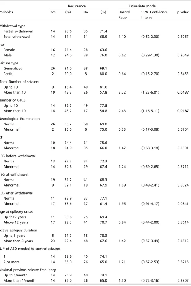
Table 2. Univariate analysis of risk factors for seizure recurrence after total or partial AED withdrawal – Cox proportional hazards regression model.
Recurrence Univariate Model
Variables Yes (%) No (%) Hazard 95% Confidence p-value
Ratio Interval
Withdrawal type
Partial withdrawal 14 28.6 35 71.4
Total withdrawal 14 31.1 31 68.9 1.10 (0.52-2.30) 0.8067
Sex
Female 16 36.4 28 63.6
Male 12 24.0 38 76.0 0.62 (0.29-1.30) 0.2049
Seizure type
Generalized 26 31.0 58 69.1
Partial 2 20.0 8 80.0 0.64 (0.15-2.70) 0.5453
Total Number of seizures
Up to 10 9 18.4 40 81.6
More than 10 19 42.2 26 57.8 2.72 (1.23-6.01) 0.0137
Number of GTCS
Up to 10 14 22.2 49 77.8
More than 10 14 45.2 17 54.8 2.43 (1.16-5.11) 0.0187
Neurological Examination
Normal 26 30.2 60 69.8
Abnormal 2 25.0 6 75.0 0.73 (0.17-3.08) 0.6704
CT
Normal 10 24.4 31 75.6
Abnormal 18 34.0 35 66.0 1.47 (0.68-3.18) 0.3301
EEG before withdrawal
Normal 13 27.7 34 72.3
Abnormal 14 32.6 29 67.4 1.24 (0.59-2.65) 0.5712
EEG at withdrawal
Normal 19 31.7 41 68.3
Abnormal 9 32.1 19 67.9 1.09 (0.49-2.41) 0.8324
EEG after withdrawal
Normal 11 22.9 37 77.1
Abnormal 17 38.6 27 61.4 1.95 (0.91-4.17) 0.0841
Age at epilepsy onset
Up to12 years 11 30.6 25 69.4
Above 12 years 17 29.3 41 70.7 0.94 (0.44-2.00) 0.8614
Active epilepsy duration
Up to 3 years 5 21.7 18 78.3
More than 3 years 23 32.4 48 67.6 1.42 (0.57-3.49) 0.4512
N. º of AED needed to control seizures
1 14 25.9 40 74.1
2 or more 14 35.0 26 65.0 1.21 (0.57-2.53) 0.6215
Maximal previous seizure frequency
Up to 1/month 14 25.9 40 74.1
More than 1/month 14 35.0 26 65.0 1.50 (0.72-3.16) 0.2807
Table 3. Multivariate analysis of risk factors for seizure recurrence after total or partial AED withdrawal – Cox proportional hazards regression model.
Multivariate Model
Variable Hazard 95% Confidence p-value Ratio Interval
Total number of seizures
Up to 10
More than 10 2.73 (1.22-6.07) 0.0142
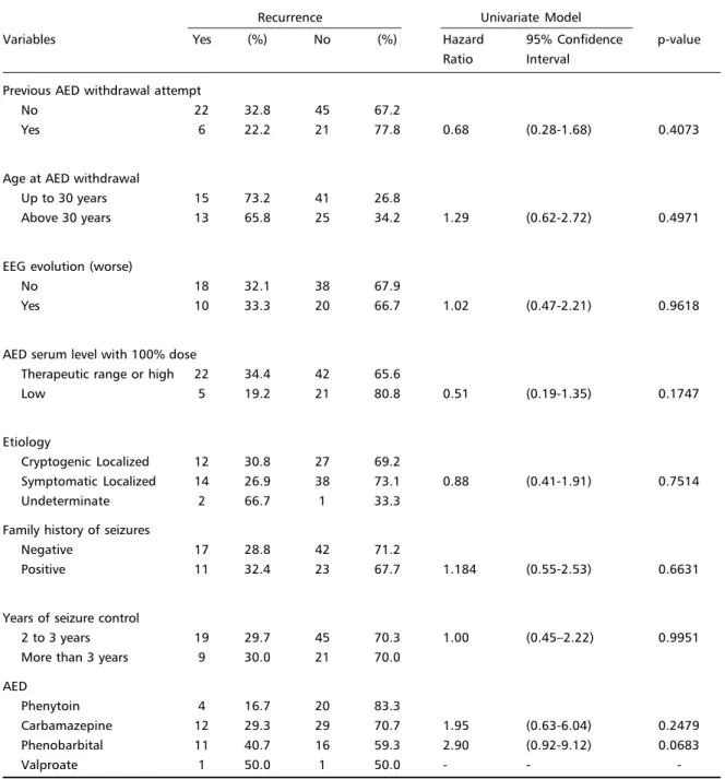
Table 2. Continued.
Recurrence Univariate Model
Variables Yes (%) No (%) Hazard 95% Confidence p-value
Ratio Interval
Previous AED withdrawal attempt
No 22 32.8 45 67.2
Yes 6 22.2 21 77.8 0.68 (0.28-1.68) 0.4073
Age at AED withdrawal
Up to 30 years 15 73.2 41 26.8
Above 30 years 13 65.8 25 34.2 1.29 (0.62-2.72) 0.4971
EEG evolution (worse)
No 18 32.1 38 67.9
Yes 10 33.3 20 66.7 1.02 (0.47-2.21) 0.9618
AED serum level with 100% dose
Therapeutic range or high 22 34.4 42 65.6
Low 5 19.2 21 80.8 0.51 (0.19-1.35) 0.1747
Etiology
Cryptogenic Localized 12 30.8 27 69.2
Symptomatic Localized 14 26.9 38 73.1 0.88 (0.41-1.91) 0.7514
Undeterminate 2 66.7 1 33.3
Family history of seizures
Negative 17 28.8 42 71.2
Positive 11 32.4 23 67.7 1.184 (0.55-2.53) 0.6631
Years of seizure control
2 to 3 years 19 29.7 45 70.3 1.00 (0.45–2.22) 0.9951
More than 3 years 9 30.0 21 70.0
AED
Phenytoin 4 16.7 20 83.3
Carbamazepine 12 29.3 29 70.7 1.95 (0.63-6.04) 0.2479
Phenobarbital 11 40.7 16 59.3 2.90 (0.92-9.12) 0.0683
Valproate 1 50.0 1 50.0 - -
-Risk factors for recurrence
more than 10 seizures before achieving control (19/
45 – 42.2%) presented a risk 2.72 times greater than
those who presented up to 10 seizures (9/49 – 18.4%)
(p = 0.014; Table 2). Figure 2 demonstrate the
survi-val curves for accumulated remission probability
related to time for patients with up to 10 seizures and
for those with more than 10 seizures before control.
Table 2 demonstrates that no significant change
occurred regarding the risk of seizure recurrence in
relation to the other variables analyzed.
Multivariate Analysis:
was performed using the
Cox proportional hazards model and those variables
with p < 0.20 in the univariate analysis were also
included. After AED withdrawal, only the number of
seizures before control once again presented
association with seizure recurrence risk (p = 0.014)
(Table 3).
DISCUSSION
In this study we found that the frequency of
sei-zure recurrence between patients who underwent
partial
versus
complete AED withdrawal was the
sa-me. The overall recurrence risk presented by both
groups was similar to that found in other studies
and those referred in Berg & Shinnar’s
meta-ana-lysis
16: 0.25 and 0.29, respectively, one and two years
after starting AED withdrawal.
It is interesting to note that two studies that
asses-sed seizure recurrence risk in patients with controlled
epilepsy for at least two years maintaining
medica-tion have similar results. The MRC Antiepileptic Drug
Withdrawal Study Group
17reported that the
fre-quency of seizure recurrence in these patients after
a two years period was 22%, and Specchio et al.
18reported a frequency of 18% for the same
observa-tion period.
Among the risk factors for seizure recurrence, only
more than 10 seizures prior to seizure control was
significant for recurrence after AED withdrawal at
the multivariate analysis. Patients who presented
mo-re than 10 seizumo-res befomo-re control demonstrated a
risk 2.73 fold greater than those who presented up
to 10 seizures, as verified by the multivariate analysis.
However, it is interesting to note that both the total
number of seizures and the number of GTCS affected
significantly the risk of seizure recurrence at the
uni-variate analysis. That is because there is a co-linearity
between them. This variable, which is an indicator
of seizure severity before control, has been shown
by other studies to be a predictive factor for seizure
recurrence after AED withdrawal
19-21. Other studies
have demonstrated that this factor has shown only
a tendency towards significance
22,23. Moreover,
others showed no relationship between number of
seizures prior to seizure control and increased
recurrence risk for seizure recurrence after
disconti-nuation of AED
24-26. However, the importance of these
studies might be limited to patients at tertiary
epi-leptic centers, since seizures are easily controlled in
a great majority of the patients with recently
diag-nosed epilepsy
7.
EEG result was not a risk factor in our study.
Ne-vertheless, studies often present conflicting results
and great variability regarding the EEG characteristics
as well as the manner in which these abnormalities
are reported and classified. Berg and Shinnar’s
16me-ta-analysis was used to assess 15 studies and
al-though they were substantially heterogeneous, the
relative recurrence risk for patients with abnormal
EEG before AED withdrawal was 1.45 fold that of
the patients with normal EEG, a small but significant
difference. In children, an abnormal EEG before AED
withdrawal is an important predictive factor for
seizure recurrence. In adults, however, the
relation-ship between abnormal EEG and risk of seizure
recur-rence has been established in a lesser degree
7.
We also found no difference in the risk for seizure
recurrence when AED withdrawal occurred after a
longer period of seizure control: 2-3 years versus >
3 years (p = 0.99). This observation was in
accordance with most reports in the literature
7and
suggests that a two-year period of seizure control is
sufficient to justify an attempt at AED withdrawal.
The major drawback of our study is the sample
size. Since there is no definite data about the risk of
seizure recurrence in adults with conventional or low
AED dose after a seizure free period of at least two
years, it is difficult to establish the exact sample size
needed to perform this task. Although a larger
sam-ple will be needed to investigate this issue, this would
be very difficult to be accomplished without a
mul-ticenter study.
REFERENCES
1. Sander JWAS. Some aspects of prognosis in the epilepsies: a review. Epilepsia 1993;34:1007-1016.
2. Annegers JF, Hauser WA, Elveback LR. Remission of seizures and relapse in patients with epilepsy. Epilepsia 1979;20:729-737. 3. Goodridge DMG, Shorvon SD. Epileptic seizures in a population of
6000, I: Demography, diagnosis and classification, and hole of the hos-pital services. BMJ 1983;287:641-644.
4. Pedley TA. Discontinuing antiepileptic drugs. N Engl J Med 1988;318:982-984.
5. Reynolds EH. Chronic antiepileptic toxicity: a rewiew. Epilepsia 1975;16:319-352.
6. Morrel M. Managing epilepsy in women across the reproductive cycle. Monograph. A CME Monograph for Neurologists. Secaucus-NJ: Projects in Knowledge, 2001.
7. Shinnar S, Gross-Tsur V. Discontinuing antiepileptic drug therapy. In Wyllie E. Ed. The treatment of epilepsy: principles and practice. 3.Ed. Philadelphia: Lippincott Williams & Wilkins, 2001:811-819. 8. Hauser WA, Kurland LT. The epidemiology of epilepsy in Rochester,
Minnesota, 1935 through 1967. Epilepsia 1975;16:1-66.
9. Commission on classification and terminology of the International League Against Epilepsy. Proposal for revised clinical and eletrencephalographic classification of epileptic seizures. Epilepsia 1981;22:489-501.
10. Commission on classification and terminology of the International League Against Epilepsy. Proposal for revised classification of epilepsies and epileptic syndromes. Epilepsia 1989;30:389-399. 11. Semah F, Picot M-C, Adam C, et al. Is the underlying cause of epilepsy
a major prognostic factor for recurrence? Neurology 1998;51:1256-1262. 12. Treatment of convulsive status epilepticus: recommendations of the Epilepsy Foundation of America’s Working Group on Status Epilepticus. JAMA 1993;270:854-859.
13. American Eletroencephalographic Society Guidelines in EEG and Evoked Potentials: Guideline seven: A proposal for standard montages to be used in clinical EEG. J Clin Neurophysiol 1986;3(Suppl 1):26-33.
14. Levy RH, Mattson RH, Meldrum BS (ed.). Antiepileptic drugs. 4.Ed. New York: Raven Press, 1995.
15. Collet D. Modeling survival data in medical research. London: Chapman & Hall, 1994.
16. Berg AT, Shinnar S. Relapse following discontinuaton of antiepileptic drugs: a meta-analysis. Neurology 1994;44:601-608.
17. Medical Research Council Antiepileptic Drug Withdrawal Study Group. Prognostic index for recurrence of seizures after remission of epilepsy. BMJ 1993;306:1374-1378.
18. Specchio LM, Tramacere L, Neve A La, Beghi E. Discontinuing antiepileptic drugs in patients who are seizure free on monotherapy. J Neurol Neurosurg Psychiatry 2002;72:22-25.
19. Emerson R, D’Souza BJ, Vining EP, et al. Stopping medication in children with epilepsy: predictors of outcome. N Engl J Med 1981;304:1125-1129.
20. Callaghan N, Garrett A, Goggin T. Withdrawal of anticonvulsant drugs in patients free of seizure for two years: a prospective study. N Engl J Med 1988;318:942-946.
21. Gherpelli JLD, Kok F, dal Forno S, Elkis LC, Lefevre BHW, Diament AJ. Discontinuing medication in epileptic children: a study of risk factors related to recurrence. Epilepsia 1992;33:681-686.
22. Juul-Jensen P. Frequency of recurrence after discontinuance of anticonvulsant therapy in patients with epileptic seizures. Epilepsia 1964;5:352-363.
23. Ehrhardt F, Forsythe WI. Prognosis after grand mal seizures: a study of 187 children with three-year remissions. Dev Med Child Neurol 1989;3:633-639.
24. Shinnar S, Vining EPG, Melits ED, et al. Discontinuing antiepileptic medication in children with epilepsy after two years without seizures: a prospective study. N Engl J Med 1985;313:976-980.
25. Shinnar S, Berg AT, Moshé S, et al. Discontinuing antiepileptic drugs in children with epilepsy: a prospective study. Ann Neurol 1994;35:534-545. 26. Tennison M, Greenwood R, Lewis D, Thorn M. Discontinuing