Arq Neuropsiquiatr 2003;61(3-A):639-641
SCHWANNOMA OF THE CRANIOCERVICAL JUNCTION
Surgical approach of two cases
Manoel Baldoino Leal Filho
1, Guilherme Borges
2, Arnaldo Ferreira
1,
Daniel França
3, Patricia Mello
4ABSTRACT- We report two cases of craniocervical junction schwannomas with a special focus on the surgical approach. The patients underwent a far-lateral approach in the sitting position that facilitated the lesion removal. This report is meant to improve the understanding of this surgical technique as well as improve awareness of its usefulness for similar cases.
KEY WORDS: craniocervical junction, far-lateral approach, schwannoma.
Schwanoma da transição craniocervical: nota técnica de dois casos
RESUMO - Relatamos dois casos de schwanoma da transição craniocervical e chamamos a atenção para alguns detalhes da técnica cirúrgica. Os pacientes foram submetidos ao acesso cirúrgico tipo extremo lateral, em posição sentada, o que facilitou a remoção da lesão. O presente relato é contribuição para melhor entendimento da técnica cirúrgica a ser empregada em casos semelhantes.
PALAVRAS-CHAVE: transição craniocervical, acesso cirúrgico extremo lateral, schwanoma.
1Neurosurgeon, Getúlio Vargas Hospital, Teresina PI, Brazil; 2Neurosurgeon, State University of Campinas (UNICAMP), Campinas SP,
Brasil; 3Medical Student of the Federal University of Piauí, Teresina PI, Brazil. 4Intensivist, Getulio Vargas Hospital, Teresina PI, Brazil.
Received 11 November 2002, received in final form 20 February 2003. Accepted 13 March 2003.
Dr. Manoel Baldoino Leal Filho - Rua Thomaz Tajra 1222, Ed. Excalibur/300 - 64048-380 Teresina PI - Brasil. E-mail: [email protected]
The craniocervical junction is the possible site of several pathological processes, such as degenerative disease, tumors, aneurysms, vascular malformations and traumatic lesions1,2. Salas et al., 19993 reported
51 patients with tumors of this region. There were 24 meningiomas, 10 chordomas, 7 paragangliomas, 3 schwannomas, 2 chondrosarcomas, 2 neurofibro-mas, and one each of hemangiopericytoma, osteo-chondroma, and metastatic carcinoma. The schwan-nomas are benign tumors of the spinal and cranial nerves4-6 and represent about 8% of all intracranial
tumors7. Schwannomas located at the craniocervical
region are rare3. The clinical presentation is variable,
depending on its location and can be associated with neurofibromatosis4. The structures that must be
con-sidered when planning the surgical approach to the craniocervical junction are the medulla and the spinal cord, the lower cranial and upper spinal nerves, the vertebral artery and its branches, the veins and dural sinuses, and the ligaments and muscles uniting the atlas, axis and occipital bone2,8.
A number of surgical access routes to this region
are currently in use, including the transoral, cervical, bifrontal transbasal, infratemporal, trans-petrosal, retromastoid, the far-lateral approach and its transcondylar, supracondylar, and paracondylar extensions and combined supra- and infratentorial approaches1,8,9. Lateral, far-lateral or extreme lateral
approaches are most widely used to remove tumors anterior or lateral to the brain stem. Bony resection is important to gain free exposure of the area and to limit manipulation of the vascular and neural struc-tures9,10. Samii et al., 199610 pointed out that the
far-lateral or extreme lateral approaches are a variant of the lateral suboccipital approach with opening of the foramen magnum. They explained that the angle of approach and the working space for the surgeon are sufficient when a standard lateral suboccipital craniotomy is combined with drilling the posterior third of the occipital condyle.
640 Arq Neuropsiquiatr 2003;61(3-A)
CASES
Case 1. A 43-year-old man developed a four-month history of progressive quadriparesis. There was a more recent history of headache. When he was admitted, he had been walking with difficulty and had weakness and dysesthesias of the upper extremities. He was submitted to a Gadolinium-enhanced, T1-weighted magnetic reso-nance imaging (MRI) which revealed a 2.2 X 3.8 cm lesion, located at the craniocervical junction, extending from the anterior portion of the foramen magnum to the right side up to the limit of the posterior arc and lateral mass of C1. There was no signal alteration in the brain stem, which would suggest anatomical lesion.
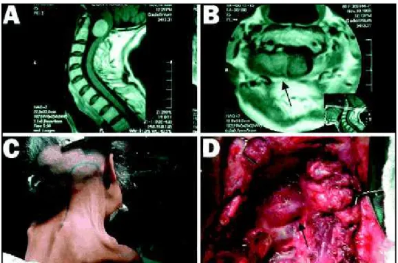
Case 2. A 59-year-old woman developed a six-month history of progressive quadriparesis and increasing diffi-culty in breathing and swallowing. When she was admitted she was no longer able to walk and had an episode of aspiration pneumonia thought to be secondary to a IX and X cranial nerves paresis. She was submitted to a Ga-dolinium-enhanced, T1-weighted MRI that showed a 3.5 X 1.7 cm lesion, located at the craniocervical junction, ex-tending from the anterior portion of the foramen magnum to the right side over to the vertebral artery (Fig 1-A and B). The lesion was leading to compression of the upper cervical cord and lower medulla.
Both patients were submitted to the same procedure described here. The illustrations shown below were recor-ded from Case 2. Under general anesthesia the head was fixed with a Mayfield device. In the sitting position, using
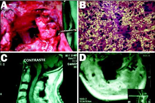
a hockey stick incision extending from the mastoid process and through the superior nuchal line as far as the midline, continuing inferiorly down to C4 vertebral level, a far-late-ral approach was performed (Fig 1-C). The myocutaneous flap was elevated to expose the posterior craniovertebral junction, deviating laterally on the subocciput toward the right side. Ipsilateral posterior arc of the C1 vertebrae was removed (Fig 1-D). The lesion originating from the right C1 root was totally removed (Fig 2-A). The C1 root ipsila-teral to the tumor was cut in both cases because of the great extension of the lesion originated from that root.
The patients had a prompt recovery upon total removal of the tumor with marked improvement of their neurolo-gical status. Post-operatively they were submitted to reha-bilitation and at a six-month follow up visit both patients were able to walk, talk, swallow and breathe without any difficulties. Pathology analysis reported the tumors to be a schwannoma (Fig 2-B) in both cases. Postoperative Gado-linium-enhanced, T1-weighted MRI showed no residual tumor (Figs 2-C and 2-D).
DISCUSSION
This report describes a surgical approach to an anterolateral placed schwannoma of the craniocer-vical junction. In our two cases the patients were operated on from the sitting position.
Some authors prefer to place the patient in the supine position1,3,9,11 with the body rotated 45° to
the opposite side11. Others prefer the sitting position,
Arq Neuropsiquiatr 2003;61(3-A) 641
with the head flexed ventrally and rotated approxi-mately 30° ipsilaterally and tilted slightly to the con-tralateral site of the surgical approach12. The
extreme-lateral craniocervical technique developed progressi-vely as a more lateral approach to lesions in the mid-line on the anterior aspects of the foramen magnum3,9.
The surgical anatomy determines the access gai-ned through lateral approaches and the intradural structures are visualized through windows between 9th and 12th cranial nerves and C28. Lateral,
far-late-ral or extreme latefar-late-ral approaches are most widely used to remove tumors anterior or lateral to the brain stem. Bony resection is important to gain free expo-sure of the area and to limit manipulation of the vascular and neural structures9,10.
Samii et al., 199610, reported that the far-lateral
or extreme lateral approach is a variant of the late-ral suboccipital approach with opening of the fora-men magnum. They demonstrated that the angle of approach and the working space for the surgeon are sufficient once a standard lateral suboccipital cra-niotomy is combined with drilling the posterior third of the occipital condyle.
Regarding the neural structures, the C1 root can be cut without any apparent adverse effects3, as in
these case reports.
Fig 2. (A) Operation site after total removal of the lesion. (B) Photomicrograph of the lesion (hematoxylin and eosin; original magnification, X 40) showing a schwannoma. (C and D) Postoperative gadolinium-enhanced T1-weighted MRI showed no residual tumor (sagital and axial view respectively).
The far-lateral approach with the patients in the sitting position was markedly important and facilita ted the total removal of the schwannoma. This report is meant to increase awareness of the usefulness of this surgical technique when approaching these cases.
REFERENCES
1. Baldwin H, Christopher G. The far lateral/combined supra- and infratentorial approach. J Neurosurg 1994;81:60-68.
2. Oliveira E, Rhoton AL Jr, Peace D. Microsurgical anatomy of the region of the foramen magnum. Surg Neurol 1985;24:293-352.
3. Salas E, Sekhar LN, Ziyal IM, Caputy AJ, Wright DC. Variations of the extreme-lateral craniocervical approach: anatomical study and clinical analysis of 69 patients. J Neurosurg 1999;90(Spine 2):206-219. 4. Martins RS, Suzuki SH, Sanematsu P Jr, Plese JPP. Acoustic neuroma
in children without association with neurofibromatosis: report of two cases. Arq Neuropsiquiatr 1999;57:96-100.
5. Paiva MA Neto, Stamm AC, Braga FM. Mandibular trigeminal schwannoma: case report. Arq Neuropsiquiatr 2001; 59:959-963. 6. Siqueira MG, Jennings E, Moraes OJS, et al. Naso-ethmoid schwannoma
with intracranial extension: case report. Arq Neuropsiquitr 2001;59:421-423. 7. Andrade GC, Paiva MA Neto, Braga FM. Thalamic intracerebral
schwannoma: case report. Arq Neuropsiquiatr 2002;60:308-313. 8. Rhoton AL Jr. The far-lateral approach and its transcondylar, supracondylar,
and paracondylar extensions. Neurosurgery 2000;47:195-210.
9. Spetzler RF, Grahm TW. The far-lateral approach to the inferior clivus and the upper cervical region technical note. BNI Quarterly 1990;6:35-38. 10. Samii M, Klekamp J, Carvalho G. Surgical results for meningiomas of
the craniocervical junction. Neurosurgery 1996;39:1086-1095. 11. Al-Mefty O, Borba LAB, Aoki N, Angtuaco E, Pait TG. The
transcon-dylar approach to extradural nonneoplastic lesions of the craniover-tebral junction. J Neurosurg 1996;84:1-6.