Review Article
THE ROLE OF OSTEOMEATAL COMPLEX ANATOMICAL
VARIANTS IN CHRONIC RHINOSINUSITIS*
Severino Aires de Araújo Neto1, Paulo de Sá Leite Martins2, Antônio Soares de Souza3, Emílio Carlos Elias Baracat4, Lívio Nanni5
* Study developed at Faculty of Medical Sciences, Universidade Estadual de Campinas,
Campinas, SP, Brazil.
1. MD, Radiologist at Centro de Atenção Integral à Saúde da Mulher, Universidade
Estadual de Campinas.
2. Coordinator for the Tomovale’s Residence Program in Radiology, São José dos
Campos, SP.
3. Head of the Radiology Service at Hospital de Base de São José do Rio Preto, SP.
4. Professor at the Department of Pediatrics, Faculty of Medical Sciences, Universidade
Estadual de Campinas.
5. Professor at the Department of Radiology – Faculty of Medical Sciences
-Universidade Estadual de Campinas, SP.
Mailing address: Dr. Severino Aires de Araújo Neto. Rua Maria Helena Rocha, 113, ap.
1102/A. João Pessoa, PB, Brazil, 58036-670. E-mail: [email protected]
Received May 18, 2004. Accepted after revision December 14, 2004.
Abstract
Currently, computed tomography is the method of choice for assessment of paranasal sinuses,
nasal fossae and their anatomical variants. Presumably, these variations might induce osteal
obstruction, preventing mucus drainage and predisposing to chronic rhinosinusitis. However,
this concept is still controversial and the presence of any anatomical variant does not necessarily
establish an etiology for rhinosinusitis. Among three subtypes of concha bullosa, only the
bulbous type seems to be strongly associated with symptoms. Size and obliteration of
the mucus drainage process. Computed tomography offers detailed study of anatomical
variations and is an invaluable tool for managing clinical decisions and planning surgical
strategies. Imaging assessment must be based on identification of variants, definition of their
dimensions, as well as on their association with obstruction of drainage ostia and tomographic
signs of sinus disease.
Keywords: Anatomical variations; Paranasal sinuses; Sinusitis; Computed tomography.
INTRODUCTION
The approach to patients with chronic rhinosinusitis has been changed with the arrival of
the endoscopic functional surgery of paranasal sinus and nasal cavity. As far as it is concerned,
the computed tomography (CT) has become indispensable to the surgical planning, since it
allows a detailed study of the whole structure of this region, which would not be possible with
plain X-rays. In most cases, the endoscopic surgery aims at removing the obstruction of the
main drainage pathway – the osteomeatal complex –, based essentially on the concept that such
obstruction perpetuates the sinus disease(1–3).
Paranasal sinus and nasal cavity anatomical variants are usual findings with an estimated
prevalence of 65%(4). Some authors propose the hypothesis that anatomical variants may be obstructive factors, predisposing to sinusitis(5). Notwithstanding, a consensus on this matter has
not been reached yet but many recent studies have contributed with new information. This
article provides an updated review, showing points that seem to be of agreement about what
remains undefined on this theme.
DEVELOPMENT AND ANATOMY OF PARANASAL SINUS AT CT
The development of paranasal sinus starts early in the fetal period as nasal cavity
invaginations. Only the maxillary and ethmoidal sinuses are present and can pneumatize at
birth. The sphenoidal and frontal sinuses develop from the first years of life(6), expanding
progressively and maturing up to the age of 12–14 years(7,8). The ethmoidal cells pneumatization process may originate some variant cells like agger nasi, concha bullosa and Haller’s(7,8).
The anatomical relations of relevant bone structures and soft tissues with sinus drainage
ostia can be more easily understood on coronal tomographic images(9) (Figure 1). The nasal
septum is an osseocartilagenous wall dividing the nasal cavity into right and left sides. The
lateral nasal wall consists of inferior and middle turbinates and, occasionally, a superior or
supreme turbinate bone with their respective meatus. The middle meatus is the most important
from the paranasal sinuses. The drainage to this fissure is made by the frontal sinus, through the
frontal recessus, and the maxillary sinus, through the infundibula (medially limited by the
unciform process and laterally limited by the ethmoidal bulla), middle and anterior ethmoidal
cells. The semilunar hiatus and surrounding structures together compose the osteomeatal
complex(9). One believes that the obstruction of this narrow region is a key factor in the
development of chronic sinusitis(1–3). The drainage of the sphenoidal sinus and posterior ethmoidal cells is performed through the sphenoethmoidal recess and through the superior
meatus. Only occasionally this posterior group of sinuses is affected by inflammatory
processes(3,4,6,9).
ANATOMICAL VARIANTS
The role of anatomical variants in the sinusitis genesis is controversial. Theoretically,
these variants could shift and compress osteomeatal complex components, determining an
obstruction to the paranasal sinuses mucus drainage(9,10). Researches on this theme consider that,
if anatomical variants really predispose to sinusitis, one should expect that these variants were
more frequently found at CT in patients with sinus disease (symptomatic) than in the general
population (asymptomatic). Findings of several studies on the theme are summarized in Tables
1 to 6. Tonai and Baba(1) have analyzed tomographic studies of 75 adult patients. Comparison of anatomical variants prevalences in the symptomatic and asymptomatic groups has showed no
significant difference (Table 1). In another study(4), of all the evaluated anatomical variants
(Table 2), only one specific type of middle concha bullosa has presented association with the
clinical disease (Table 3). Four studies have described the anatomical variants prevalence on CT
examinations in children with chronic or recurrent sinusitis.(Table 4)(6,11–13). However, in these studies there was not a control group for statistical correlation. Liu et al.(14) have demonstrated that the greater the size of the anatomical variant, the higher the frequency of association with
paranasal sinus mucosal alterations at CT (Table 5). Scribano et al.(10) have studied patients with anatomical variants by means of CT, aiming at identifying patients in which an anatomical
variant determined a contact between the mucosal surfaces of the osteomeatal complex (aerial
space obliteration). One has observed that the maxillary sinus opacification was significantly
more frequent in cases where the concha bullosa determined osteomeatal complex obliteration
(Table 6), when compared with cases of concha bullosa without osteomeatal complex
obliteration. For these authors, the contact of mucosal surfaces would be more significant for the
Different types of anatomical variants present distinct relations with either clinical or
tomographic sinus disease. Main anatomical variants are middle concha bullosa, Haller and
agger nasi cells, nasal septum deviation and enlarged ethmoidal bulla.
The middle concha bullosa is a result of pneumatization of the osseous plate due to
ethmoidal extention (Figures 2 and 3). A prevalence up to 80% is observed(4) and, besides Haller
and agger nasi cells and deviated nasal septum, the middle concha bullosa is one of the most
frequent anatomical variants(1,4,6,11–13) (Tables 1 and 2). In children diagnosed with sinusopathy,
the frequency of this anatomical variant ranged between 4.2% and 24% of cases(6,11–13) (Table 4). Milczuk et al. have found an association with ipsilateral ethmoid-maxillary sinusopathy in the childhood in 63% of cases studied(13) (Figure 2). Contradictorily, Lusk et al.(12) have not observed any association of concha bullosa with sinus disease in children (Figure 3). Tonai and
Baba(1) (Table 1) and Zinreich et al.(15) also have not detected any relation of concha bullosa with sinusopathy. It is important to note that the degree of pneumatization found in the
population studied by Tonai and Baba(1) was low, which they attribute possibly to a racial factor
(the study has been developed in Japan). The concha pneumatization may occur at several
degrees, from that affecting only the bulbous portion (distal) (Figure 2) or lamellar portion
(proximal), or the called true variant where there is pneumatization of both portions(4) (Figure 4). In one of these studies(4), the bulbous-type middle concha bullosa was the only anatomical variant presenting a relation with sinusopathy (Table 3). The association of anterior ethmoidal
cells and maxillary sinuses with tomographic alterations may depend not only on the subtype,
but also on dimensions of the concha bullosa(14). Scribano et al.(10) have demonstrated that when the concha bullosa does not interfere with the amplitude of paranasal sinuses drainage paths at
CT, it possibly cannot cause a sinus disease, and probably the rhinosinusitis genesis involves an
osteomeatal complex aerial column obliteration by an anatomical variant (Table 6).
Haller cells are found in up to 45% of the general population(1,4,16,17)and in 1.4% to 18%
of children(6,11–13) (Tables 1, 2 and 4). Haller cells are anterior ethmoidal cells that project beyond the limits of the bulla ethmoidal under the orbital floor, forming the infundibulum
lateral wall between the papyracea lamina and the unciform process(1,13,18) (Figure 4). There was no difference in its prevalence between patients with and without sinusopathy (Tables 1 e 2)(1,4).
However, larger Haller cells are more likely related to tomographic alterations of maxillary
sinuses(14,18) (Table 5).
Middle concha may present a curvature contrary to the one normally seen; this is another
anatomical variant named paradoxical concha (Figure 5) and present at CT in 30% of patients
4). Depending on the curvature degree, the infundibulum may be compressed, causing sinusal
obstruction(1,9). There is no consistent data about its relation with sinusopathy(1,4,11–13).
The agger nasi cell is the most anterior ethmoidal cell, situated below the frontal sinus,
next to the frontal recess, representing the lacrimal bone pneumatization due to ethmoidal
extension (Figure 6). This may be an important factor in the genesis of symptoms like
lacrimation and frontal sinus disease. Reported prevalence is quite variable (10% to 98.5%)(1,4,9) (Tables 1 and 2), possibly due to two factors: 1) different definitions are assigned to this
anatomical variant; 2) the small size of the agger nasi cell might make its detection difficult in
former researches involving anatomic pieces. Therefore, higher frequencies are described in CT
studies in which agger nasi cell can be easily identified(1,4). A positive relation between the cell size and the presence of frontal sinus disease has been described by Liu et al. (Table 5)(14). Nassar Filho et al.(19) also have observed that this cell presented hyperpneumatized more frequently in the group with sinusopathy than in the control group. However, this anatomical
variant has not been considered as an obstructive factor in the study of Voegels et al.(20).
In the junction of the nasal cartilage with the vomer, an acute angulation occurs in 20% to
30% of the population(7,16), constituting the nasal septum deviation (Figure 7), one of the most
frequent anatomical variants(19,11,12). Its prevalence in children ranges from 10.4% to 14%(6,11,12). In older children (on average 9 years of age) septum deviation tends to be more pronounced(12). This last observation might indicate an acquired nature of this condition(11). It may determine
compression of the middle concha with consequential infundibulum obstruction(9). One has
demonstrated the association of higher grades of septum deviation with ipsilateral sinusopathy
in adults(9,21,22).Some authors(1,4) do not mention this anatomical variant in their studies.
An enlarged ethmoidal bulla may obstruct the infundibulum or the middle meatus. The
exact prevalence of enlarged ethmoidal bulla is not known(9,23). Its size is an important factor when associated with opacification of anterior ethmoidal cells at CT in patients diagnosed with
sinusopathy(14). However, one has not found in the literature an objective description of what could be considered an enlarged ethmoidal bulla(9,10,19,23). Late in its development, the ethmoidal
sinus measures on average 36 x 18 x 14 mm (length, height and width) in measurements
performed in MRI studies(24). These measures were similar in cadaver skulls(25). The ethmoidal
bulla, however, has not been separately evaluated in theses studies. In measurements at CT in
adults, the average area of each ethmoidal cell is 0.73 ± 0.42 cm², the larger ones, situated in the
Other anatomical variants appear less frequently. The superior extremity of the unciform
process may deviate laterally, medially or anteriorly and may interfere in the middle meatus
drainage(14). Its prevalence was of 6.9% in a study with children(11). The unciform process
pneumatization also is hardly frequent (prevalence up to 2.5%)(1,4,11,12,16). Hypoplastic maxillary sinus appears in about 6.9% to 17.5% of the pediatric population(11–13). According to Lusk et al.(12),the hypoplasia may be a result of the loss of pneumatization by the infundibulum. The correlation of these anatomical variants with sinusopathy has been not determined(4,9,12,13).
DISCUSSION
Most probably, the rhinosinusitis genesis is multifactorial, and the physiological factor
(mucociliary clearance disorders) possibly is as much significant as the mechanical obstructive
factor(19). Indications for surgical correction of the sinusal drainage deal with the possibility of
an anatomical variant constituting an obstructive factor, principally at the level of the
osteomeatal complex, but there is no reference to objective parameters like anatomical variant
dimensions or drainage pathways amplitude as specific indicators(27,28).
The role of anatomical variants in the chronic or recurrent sinusitis pathogenesis can be
evaluated by comparison between anatomical variants prevalence in populations with
sinusopathy and prevalence in populations free from sinusal problems. If anatomical variants
determine any effect on the chronic rhinosinusitis genesis, it is assumed that they are found
more frequently in groups of patients with sinusopathy. Some studies on the prevalence of
anatomical variants have failed in identifying a significant relation with rhinosinusitis symptoms
or with mucosal alterations of paranasal sinus at CT(1,4,19,20). However, Bolger et al.(4) have found out that the pneumatization of the bulbous portion of the middle concha presented a
prevalence significantly increased in patients with sinusopathy (Table 3). Likewise, larger
anatomical variants present higher probability of association with tomographic alterations of
paranasal sinus (Table 5)(14). Finally, even disregarding factors like subtype or size, Scribano et al.(10) have observed that, if the anatomical variant determines obliteration of the aerial space of the osteomeatal complex drainage paths, the sinusal disease is more frequently detected at CT
(Table 6) than when the anatomical variant does not obstruct these pathways.
The prevalence of anatomical variants seems to increase with the age (Table 4). Lower
prevalences are found in the study including lower age ranges (1 to 7 years of age)(6). These data
suggest the hypothesis of some anatomical variants being of acquired nature. Besides the fact
that in children the sinus disease is usually bilateral and symmetrical(12), one may infer that anatomical variants have less influence on the sinusitis etiopathogenesis in this age range than in
Some disparities between frequencies in different studies(1,4,6,10–14,19,20) can be explained
by some controversial factors, definitions and ratings of the anatomical variant(4,9), the utilization of evaluation methods with different sensitivities (anatomical pieces versus CT) and also racial or population factors(1). Also, it is necessary establish the difference between clinical sinusopathy and tomographic sinusopathy, since sinusal alteration at CT does not mean
necessarily clinical disease(4,29–33). Finally, since each variant seems to have a different influence on the development of the sinus disease, it would be convenient to determine the risk of each
variant independently. A few studies involve a sufficient number of cases for a statistically
satisfactory data analysis, as some anatomical variants present a very low incidence. Lusk et al.(12), for example, have examined 115 children and observed that the frequency of anatomical variants was not sufficiently high to allow a statistical correlation with sinusopathy.
CONCLUSION
There is no consensus in the literature on the role of anatomical variants in the chronic
rhinosinusitis physiopathogenesis. The single detection of an anatomical variant itself does not
establish the genesis of the disease. Before the suggestion of a causal relation between the
anatomical variant and the sinusopathy in the tomographic analysis of a patient with
sinusopathy and one anatomical variant, these conditions should be considered in conjunction
with the clinical picture, its type and size, its association with obliteration of osteomeatal
complex drainage paths and the presence of ipsilateral sinusal mucosa alterations.
REFERENCES
1. Tonai A, Baba S. Anatomic variations of the bone in sinonasal CT. Acta Otolaryngol Suppl
1996;525:9–13.
2. Isaacson G. Sinusitis in childhood. Pediatr Clin North Am 1996;43:1297–1318.
3. Zinreich J. Imaging of inflammatory sinus disease. Otolaryngol Clin North Am 1993;26:535–
547.
4. Bolger WE, Butzin CA, Parsons DS. Paranasal sinus bony anatomic variations and mucosal
abnormalities: CT analysis for endoscopic sinus surgery. Laryngoscope 1991;101:56–64.
5. Stammberger H, Wolf G. Headaches and sinus disease: the endoscopic approach. Ann Otol
Rhinol Laryngol Suppl 1988;134:3–23.
6. Dutra LD, Marchiori E. Tomografia computadorizada helicoidal dos seios paranasais na
criança: avaliação das sinusopatias inflamatórias. Radiol Bras 2002;35:161–169.
7. Kronemer KA, McAlister WH. Sinusitis and its imaging in the pediatric population. Pediatr
8. Scuderi AJ, Harnsberger HR, Boyer RS. Pneumatization of the paranasal sinuses: normal
features of importance to the accurate interpretation of CT scans and MRI images. AJR Am J
Roentgenol 1993;160:1101–1104.
9. Laine FJ, Smoker WR. The ostiomeatal unit and endoscopic surgery: anatomy, variations,
and imaging findings in inflammatory diseases. AJR Am J Roentgenol 1992;159:849–857.
10. Scribano E, Ascenti G, Cascio F, Racchiusa S, Salamone I. Computerized tomography in
the evaluation of anatomic variations of the ostiomeatal complex. Radiol Med (Torino)
1993;86:195–199.
11. April MM, Zinreich SJ, Baroody FM, Naclerio RM. Coronal CT scan abnormalities in
children with chronic sinusitis. Laryngoscope 1993;103:985–990.
12. Lusk RP, McAlister B, el Fouley A. Anatomic variation in pediatric chronic sinusitis: a CT
study. Otolaryngol Clin North Am 1996;29:75–91.
13. Milczuk HA, Dalley RW, Wessbacher F, Richardson M. Nasal and paranasal sinus
anomalies in children with chronic sinusitis. Laryngoscope 1993;103:247–252.
14. Liu X, Han D, Zhou B. Relationship between anatomic variations of nasal sinus and chronic
sinusitis. Zhonghua Er Bi Yan Hou Ke Za Zhi 1998;33:149–152.
15. Zinreich SJ, Mattox DE, Kennedy DW, Chisholm HL, Diffley DM, Rosenbaum AE.
Concha bullosa: CT evaluation. J Comput Assist Tomogr 1988;12:778–784.
16. Arslan H, Aydinlioglu A, Bozkurt M, Egeli E. Anatomic variations of the paranasal sinuses:
CT examination for endoscopic sinus surgery. Auris Nasus Larynx 1999;26:39–48.
17. Kennedy DW, Zinreich, SJ. Functional endoscopic approach to inflammatory sinus disease:
current perspectives and technique modifications. Am J Rhinol 1988;2:89–96.
18. Stackpole SA, Edelstein DR. The anatomic relevance of the Haller cell in sinusitis. Am J
Rhinol 1997;11:219–223.
19. Nassar Filho J, Anselmo-Lima WT, Santos AC. Participação das variações anatômicas do
complexo ostiomeatal na gênese da rinossinusite crônica, analisadas por tomografia
computadorizada. Rev Bras Otorrinolaringol 2001;67:489–495.
20. Voegels RL, Goto EY, Chung D, Nita LM, Lessa MM, Butugan O. Correlação etiológica
entre variações anatômicas na tomografia computadorizada e a rinossinusite crônica. Rev Bras
Otorrinolaringol 2001;67:507–510.
21. Kainz J, Stammberger H. The roof of the anterior ethmoid: a place of least resistant in the
skull base. Am J Rhinol 1989;3:191–199.
22. Elahi MM, Frenkiel S, Fageeh N. Paraseptal structural changes and chronic sinus disease in
23. Castro Jr NP, Takara CK. Anatomia cirúrgica da parede lateral do nariz. Rev Bras
Otorrinolaringol 1998;64(Supl 2):5–10.
24. Dos Santos RM. Desenvolvimento dos seios paranasais: estudo por ressonância magnética
do crânio. (Tese de Mestrado). São Paulo: Universidade Federal de São Paulo, 2002.
25. Wolf G, Anderhuber W, Kuhn F. Development of the paranasal sinuses in children:
implications for paranasal sinus surgery. Ann Otol Rhinol Laryngol 1993;102:705–711.
26. Saheki T. The investigation of the structures in the ethmoidal air cells on CT. Nippon
Jibiinkoka Gakkai Kaiho 1989;92:827–836.
27. Kennedy DW, Zinreich SJ, Rosenbaum AE, Johns ME. Functional endoscopic sinus
surgery: theory and diagnostic evaluation. Arch Otolaryngol 1985;111:576–582.
28. Zinreich SJ, Kennedy DW, Rosenbaum AE, Gayler BW, Kumar AJ, Stammberger H.
Paranasal sinuses: CT imaging requirements for endoscopic surgery. Radiology 1987;163:769–
775.
29. Havas TE, Motbey JA, Gullane PJ. Prevalence of incidental abnormalities on computed
tomography scans of the paranasal sinuses. Arch Otolaryngol Head Neck Surg 1988;114:856–
859.
30. Manning SC, Biavati MJ, Phillips DL. Correlation of clinical sinusitis signs and symptoms
to imaging findings in pediatric patients. Int J Pediatr Otorhinolaryngol 1996;37:65–74.
31. Diament MJ, Senac MO, Gilsanz V, Baker S, Gillespie T, Larsson S. Prevalence of
incidental paranasal sinus opacification in pediatric patients: a CT study. J Comput Assist
Tomogr 1987;11:426–431.
32. Lesserson JA, Kieserman SP, Finn DG. The radiographic incidence of chronic sinus disease
in the pediatric population. Laryngoscope 1994;104:159–166.
Table 1 Prevalence of anatomical variants at CT (%). (Tonai & Baba(1)).
Agger nasi cell Haller cell
Middle concha bullosa Middle paradoxical concha Pneumatized uncinate
Total (n = 75)
86.7 36.0 28.0 25.3 0 Symptomatic (n = 57)
86.0 33.3 28.1 29.8 0 Asymptomatic (n = 18)
88.9 38.9 27.8 11.1 0 p > 0.05 > 0.05 > 0.05 > 0.05 > 0.05
Table 2 Prevalence of anatomical variants at CT (%). (Bolger et al.(4)).
Agger nasi cell Haller cell
Middle concha bullosa Middle paradoxical concha Pneumatized uncinate
Total (n = 202)
98.5 45.1 53.0 26.1 2.5 Symptomatic (n = 166)
(a) 45.9 53.6 27.1 3.0 Asymptomatic (n = 36)
(a) 41.6 50.0 22.3 5.6 p (a) > 0.05 > 0.05 > 0.05 > 0.05
(a)Not reported by the authors.
Table 3 Prevalence of middle concha bullosa subtypes in symptomatic and asymptomatic patients at CT (%). (Bolger et al.(4)).
Lamellar True Bulbous*
Total (n = 202)
46.2 15.7 31.2
Symptomatic (n = 166)
45.9 17.4 35.3
Asymptomatic (n = 36)
47.2 8.4 13.9 p 0.99 0.38 0.04*
* Statistically significant difference.
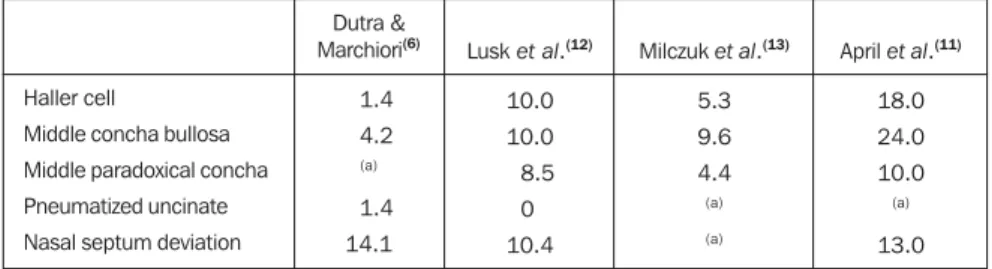
Table 4 Prevalence of anatomical variants in symptomatic children at CT (%).
Haller cell
Middle concha bullosa Middle paradoxical concha Pneumatized uncinate Nasal septum deviation
Dutra & Marchiori(6) 1.4 4.2 (a) 1.4 14.1
Lusk et al.(12)
10.0 10.0 8.5 0 10.4
Milczuk et al.(13)
5.3 9.6 4.4
(a)
(a)
April et al.(11)
18.0 24.0 10.0
(a)
13.0
(a)Not reported by the authors.
Table 5 Size of the anatomical variant in relation to the tomographic alteration of respective paranasal sinus (between brackets in the first column). (Liu et al.(14)).
Agger nasi cell (frontal) Haller cell (maxillary)
With contact (n = 44) Without contact (n = 69)
35 17
9 52
< 0.05* < 0.05*
(a)n = 73 (113 anatomical variants). * Statistically significant difference.
Figure 1. Coronal CT slice at the level of the osteo-meatal complex. Maxillary sinus (1), ethmoidal cells (2), middle concha (3), inferior concha (4), middle meatus (5), hard palate (6), nasal septum (7), unci-form process (+), ethmoidal bulla (*), infundibulum (arrow). Note the slight nasal septum deviation to the left.
Figure 3. Coronal CT at the level of the osteomeatal complex. Child with a true middle concha bullosa to the right (*), with dimensions similar to that presented in Figure 2, without associated sinus disease.
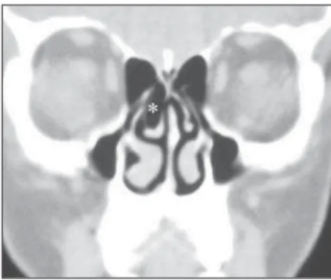
Figure 2. Coronal CT at the level of the osteomeatal complex. Right ethmoid-maxillary sinusopathy (*), probably determined by obstruction of the osteomeatal complex by bulbous-type middle concha bullosa (+).
Figure 5. Coronal CT. Paradoxical middle concha to the right (arrow), posteriorly to the osteomeatal com-plex. There is a discrete mucous thickening on the floor of the maxillary sinus.
Figure 4. Coronal CT. Haller cell to the left (arrow), associated with bilateral conchas bullosas (*), a true concha bullosa at the left and a lamellar concha bullosa to the right, both small-sized. The osteomeatal