Magnetic resonance imaging of the prostate:
an overview for radiologists*
Ressonância magnética da próstata: uma visão geral para o radiologista
Ronaldo Hueb Baroni1, Maria Ines Novis2, Ângela Hissae Motoyama Caiado3, Luciana Mendes de Oliveira Cerri1, Claudia da Costa Leite4, Giovanni Guido Cerri5
Prostate adenocarcinoma is the second tumor in incidence and mortality among malignant neoplasms in men. The differentiation between tumors confined to the organ and those with extraprostatic extension is critical for an appropriate therapeutic planning. Different studies have demonstrated that magnetic resonance imaging of the prostate with endorectal coil is useful in the local staging of these tumors. The present article presents information on the prostate gland anatomy, the tumor aspect at magnetic resonance imaging, specific signs of extracapsular extension and seminal vesicles invasion, protocol suggestions, general principles and relevance of proton spectroscopy, perfusion and diffusion imaging, role of magnetic resonance imaging in the postoperative and post-radiotherapy detection of local tumor recurrence, and also in the detection of lesions in patients with clinical/laboratory suspicion of prostate adenocarcinoma. Additionally, the present article describes differential diagnoses and limitations of the method.
Keywords: Prostate cancer; Adenocarcinoma; Magnetic resonance imaging; Cancer staging.
O adenocarcinoma prostático é o segundo tumor em incidência e mortalidade dentre as neoplasias malignas masculinas. Para adequada programação terapêutica é importante a distinção entre tumores confinados à próstata e aqueles com extensão extraprostática. Diferentes estudos têm demonstrado que a ressonância magnética da próstata com bobina endorretal auxilia no estadiamento local destes pacientes. Este artigo apresenta informações sobre a anatomia prostática, o aspecto tumoral à ressonância magnética, sinais de extensão tumoral extraprostática e invasão de vesículas seminais, sugestões de protocolo, princípios gerais e importância da espectroscopia de prótons, do estudo perfusional e da difusão, indicações da ressonância magnética na investigação de recidiva pós-operatória e pós-radioterapia, seu papel na detecção de lesões suspeitas em pacientes com suspeita clínico-laboratorial de adenocarcinoma prostático, além de apresentar os diagnósticos diferenciais e limitações do método.
Unitermos: Neoplasias da próstata; Adenocarcinoma; Imagem por ressonância magnética; Estadiamento de neoplasias.
Abstract
Resumo
* Study developed at Instituto de Radiologia do Hospital das Clínicas da Faculdade de Medicina da Universidade de São Paulo (InRad/HC-FMUSP), São Paulo, SP, Brazil.
1. PhDs, Physician Assistants at Instituto de Radiologia do Hospital das Clínicas da Faculdade de Medicina da Universidade de São Paulo (InRad/HC-FMUSP), São Paulo, SP, Brazil.
2. MD, Fellow PhD degree, Instituto de Radiologia do Hospi-tal das Clínicas da Faculdade de Medicina da Universidade de São Paulo (InRad/HC-FMUSP), São Paulo, SP, Brazil.
3. Physician Assistant at Instituto de Radiologia do Hospital das Clínicas da Faculdade de Medicina da Universidade de São Paulo (InRad/HC-FMUSP), São Paulo, SP, Brazil.
4. Private Docent, Professor at Instituto de Radiologia do Hospital das Clínicas da Faculdade de Medicina da Universidade de São Paulo (InRad/HC-FMUSP), São Paulo, SP, Brazil.
5. Titular Professor, Division of Radiology, Chief of the Radiol-ogy Service at Instituto de Radiologia do Hospital das Clínicas da Faculdade de Medicina da Universidade de São Paulo (InRad/ HC-FMUSP), São Paulo, SP, Brazil.
PSA levels and the Gleason score deter-mined by biopsy (the highest the Gleason score, the highest is the tumor aggressive-ness), with a relatively high accuracy in local staging of PCa(3).
Several studies have demonstrated that magnetic resonance imaging (MRI), with its high anatomical resolution, adds rel-evant information to the clinical nomo-grams in the local staging of PCa. Endorectal coil MRI presents an accuracy of 85% in the prediction of extracapsular extension and up to 97% for seminal vesicles invasion(4,5).
New prospects include the 3 tesla MRI, that may be utilized with endorectal coil (possibly increasing even further the sen-sitivity and specificity in the local staging of PCa), or without the endorectal coil with
Baroni RH, Novis MI, Caiado AHM, Cerri LMO, Leite CC, Cerri GG. Magnetic resonance imaging of the prostate: an over-view for radiologists. Radiol Bras. 2009;42(3):185–192.
surpassed only by non-melanoma skin can-cer in incidence, and by lung cancan-cer in number of deaths(1,2).
The methods employed in the screening for PCa in the population are prostate-spe-cific antigen (PSA) test and digital rectal examination. Transrectal ultrasonography-guided biopsy is the method of choice for histological diagnosis of the disease.
An accurate staging of the disease is of paramount importance for an appropriate therapeutic planning. The nomograms in-troduced by Partin combine clinical stag-ing based on digital rectal examination,
Mailing address: Dr. Ronaldo Hueb Baroni. Avenida Albert Eins-tein, 627/701, 4º andar, Bloco D, Morumbi. São Paulo, SP, Brazil, 05651-901. E-mail: [email protected]
Received May 7, 2008. Accepted after revision January 12, 2009.
INTRODUCTION
consequential reduction in discomfort for the patient(6,7).
The present study is aimed at present-ing the most relevant aspects of the evalu-ation of the prostate by means of MRI, par-ticularly those related to the detection and staging of PCa.
ANATOMY
Zonal anatomy of the prostate
The prostate is an inverted cone-shaped gland with the base adjacent to the vesical floor and the apex postero-inferior to the pubic symphysis. It is divided into four glandular zones: peripheral, transitional, central and periurethral(8). The transitional,
central and periurethral zones, hardly dif-ferentiated by imaging methods, are jointly denominated central gland. The limit be-tween the peripheral zone and the central gland is called “surgical capsule”, and the
discontinuous fibromuscular layer covering the gland is the “prostatic capsule” (Figure 1). For a better localization of lesions and biopsy guidance, the prostate is convention-ally divided into three regions by imaginary cross sectional lines (base, middle and apex), and in two sides by an imaginary longitudinal median line (left and right), thus configuring the prostatic sextants.
Seminal vesicles
Normal seminal vesicles present a typi-cal ductal pattern and hyperintense signal on MRI T2-weighted sequences.
DESIGN AND OPTIMIZATION OF THE PROTOCOL
General recommendations
A four-hour fasting and rectal prepara-tion is recommended for greater comfort of the patient. Shortly before the examination
1 mL of N-butylescopolamine (20 mg/mL) shall be administered to reduce intestinal peristalsis.
Study of the pelvis
The images shall be obtained from the aortic bifurcation to the lower limit of the pubic symphysis, utilizing the torso or pel-vic phased-array coil.
Suggested protocol – Axial T2-weighted
fast spin echo (FSE) with fat saturation, and axial T1-weighted gradient echo (GRE), 7 mm slice thickness, 1–2 mm interslice gap, 256 × 192 matrix, number of excitations (NEX) = 3 for T2-weighted sequences, NEX = 1 for T1-weighted sequences, field-of-view (FOV) of approximately 30 cm.
Study of the prostate
Initially, the patients shall be given an explanation about the procedure. After that, a digital rectal examination is performed to evaluate the prostate size to guide an appro-priate positioning of the coil. The coil (which is disposable) is covered by a con-dom and lubricated with local anesthetic gel (xylocaine). After the coil is in place, it is filled with 40 to 80 ml of air or perfluoro-carbon, with the objective of keeping the coil in place and reducing sphincter con-traction artifacts. Perfluorocarbon is pre-ferred in examinations with spectroscopy, and its advantages will be further detailed. The protocol for specific study of the prostate and seminal vesicles after endo-rectal coil insertion must comprise high-resolution FSE T2-weighted sequences in the axial, coronal and sagittal planes, from the bottom of the seminal vesicles to the prostate apex (Figure 2).
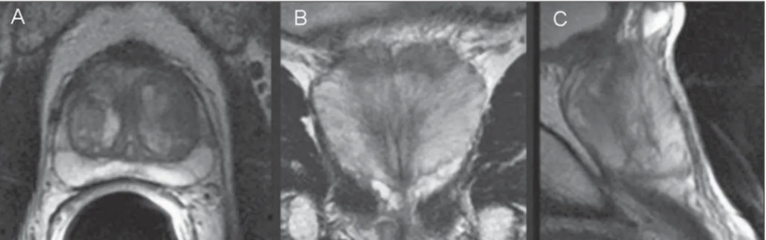
Figure 1. Correlation between MR T2-weighted image (A) and macroscopic aspect of a surgical speci-men (B).
➡
Fibromuscular stroma➡
Central gland➡
Prostatic capsule➡
Peripheral zone➡
Vascular-nervous bundleSuggested protocol – The sequences
may be performed with 3–4 mm thick slices, 0–1 mm interslice gap, FOV from 12 to 16 cm, 256 × 256 matrix, NEX = 3 or 4 and TE = 120–160.
TUMOR STAGING
Pathological staging
The systems accepted for PCa staging are TNM and Jewett-Whitmore, TNM be-ing the most widely utilized. The TNM classification describes the primary tumor extent (T), presence or absence of locore-gional lymph node involvement (N) and presence or absence of metastases (M)(9).
The appropriate preoperative PCa stag-ing presents therapeutic and prognostic im-plications. Specifically, the detection of ex-tracapsular extension and seminal vesicles invasion are of great importance, as they allow the differentiation between T2 and T3 stages(10).
Local staging – MRI
Tumor identification – PCa is most
fre-quently found in the peripheral zone (65%
to 74%). Usually the lesion presents like a poorly defined nodule with hypointense signal intensity on T2-weighted sequences, in contrast with signal hyperintensity in the normal peripheral zone (Figure 3).
Detection of extracapsular extension – The detection of extracapsular extension
can be inferred from both specific and non-specific signs at MRI(11) .
Specific signs of extracapsular exten-sion (Figure 4): solid tissue in the
peripro-static fat; irregular bulging of the properipro-static
capsule; obliteration of the rectoprostatic angle.
Non-specific signs of extracapsular ex-tension: regular capsular thickening;
cap-sular discontinuity; regular bulging of the prostatic capsule.
Invasion of seminal vesicles – Invasion
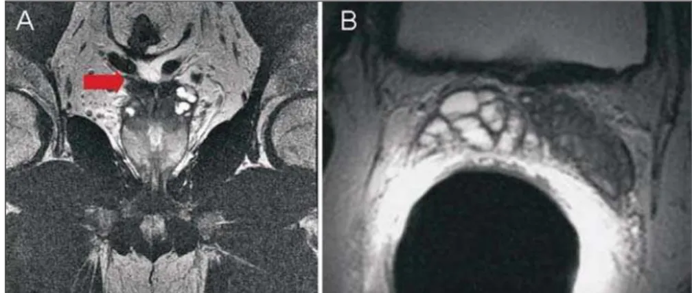
of seminal vesicles may occur by direct extension of the tumor localized in the base of the prostate gland, through the ejacula-tory duct, or by hematogeneous spreading (Figure 5).
Figure 5.A: MR coronal T2-weighted image dem-onstrating PCa invading the seminal vesicles by contiguity. B: MR axial T2-weighted image dem-onstrating extensive tumor canalicular invasion of the left seminal vesicle through ejaculatory ducts.
Figure 4. Endorectal coil MR – axial T2-weighted images demonstrating examples of PCa with extracapsular extension appearing as solid extraprostatic tissue (A), irregular capsular contour bulge (B) and rectoprostatic angle obliteration (C).
Diagnostic findings of seminal vesicle invasion are: hypointense signal on T2-weighted sequences and loss of the typical ductal pattern(12).
Regional staging
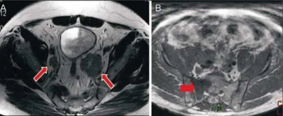
It is also important to evaluate the se-quences of the pelvis and of the prostate in the search for regional lymphadenopathies (> 1 cm in the smallest diameter), invasion of periprostatic structures and bone lesions (Figure 6).
SPECTROSCOPY
General principles
Proton spectroscopy allows a nonin-vasive evaluation of anatomic and biologi-cal characteristics of the tumor, emphasiz-ing the detection, localization and stagemphasiz-ing of PCa. Spectroscopy utilizes a powerful magnetic field and radiofrequency waves to obtain metabolic data, based on the rela-tive concentration of endogenous prostatic metabolites. It is always performed in as-sociation with endorectal coil MRI, in-creasing total examination time by 10 to 17 minutes.
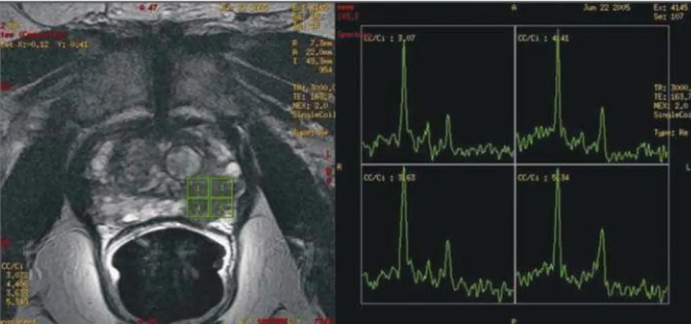
Voxels of 0.3 cm3 are programmed in
the whole prostate extent, utilizing T2-weighted images as a reference, which al-lows the joint analysis of anatomical and metabolic alterations. The metabolites ana-lyzed are: choline, creatine, citrate and, more recently, polyamine. The analysis is
performed by means of amplitude ×
fre-quency graphs, the frequencies being spe-cific for each metabolite. The areas under the peaks correlate with the concentration of each metabolite in the prostatic tis-sue(13,14) (Figure 7).
Citrate – peak: 2.6 ppm. Metabolite
synthesized by a healthy prostate, most re-markably in the peripheral zone. Hyper-plastic nodules may present citrate levels as high as those observed in the peripheral zone. In the presence of cancer, citrate lev-els are reduced or undetectable.
Choline – peak: 3.22 ppm. Cell
metabo-lism marker. Increased choline levels are observed in malignant tissues, because of high phospholipid metabolism of the cell membrane.
Creatine – peak: 3.03 ppm. Energy re-serve with constant concentration. Because
of the proximity between creatine and cho-line peaks, they may be inseparable on the spectral line; therefore, the (choline + cre-atine)/citrate ratio is utilized in the spectral analysis. (Choline + creatine)/citrate ratios > 0.5 are suggestive of malignancy (the higher the ratio, the higher is the possibil-ity of cancer).
Polyamine – peak: 3.1 ppm. This peak
resolution is better observed when the endorectal coil is filled with perfluoro-carbon. It is reduced in suspicious areas,
being identified as an acute decreasing curve between the choline and creatine peaks.
Perfluorocarbon, a fluorine and carbon compound without hydrogen atoms, can be utilized to inflate the endorectal coil, reduc-ing the magnetic susceptibility difference between the air and the prostatic tissue. It improves the characterization of peaks of low spectral signals in the peripheral zone, as well as the peak resolution of each me-tabolite(15).
Figure 6.A: MR axial T2-weighted image demonstrating bilateral obturatory lymphadenopathy. B: MR axial T2-weighted image demonstrating a metastatic bone lesion in the sacrum.
Jung et al.(16) described a score system
in an attempt to standardize and system-atize the prostatic metabolic evaluation. This five-score system utilizes progres-sively more suspicious spectral patterns, with good accuracy (74.2% to 85.0%) and excellent interobserver agreement, using a score ≥ 4 for tumor detection. Additional criteria, such as polyamine peak inversion, should be considered.
Role of proton spectroscopy
To increase the MRI specificity –
Le-sions with hypointense signal on T2-weighted sequences may be observed in other non-tumor conditions such as hem-orrhage and chronic prostatitis, among oth-ers. The addition of spectroscopy to endorectal coil MRI increases specificity and reduces interobserver variability(17).
To improve local staging (number of tumor voxels × risk for extracapsular extension) – Studies demonstrate that the
higher the number of tumor voxels at spec-troscopy, the higher is the risk for extra-capsular extension. Patients with small tu-mors at MRI spectroscopy (0–1 tumor voxel/slice) presented a risk of 6% for ex-tracapsular extension, while those with extensive tumors (> 4 tumor voxels/slice) presented a risk of 80%(18) (Figure 8).
To predict tumor aggressiveness –
MRI spectroscopy has the potential to noninvasively infer tumor aggressiveness. Tumor changes vary from a subtle increase in choline levels and moderate citrate lev-els in low grade tumors, to a sharp rise in choline levels and reduction/absence of citrate in high grade tumors(19).
To predict tumor volume –
Three-di-mensional spectroscopy in combination with MRI increases global accuracy of tu-mor volume estimates. The prediction of tumor volume by MRI spectroscopy in le-sions > 0.5 cm3 obtained a significant
cor-relation with histopathologic tumor vol-ume(20) .
PERFUSION STUDY (DYNAMIC CONTRAST-ENHANCED MRI)
Dynamic contrast-enhanced MRI is use-ful in the detection of areas under suspicion for neoplastic involvement. It is performed with 3D T1-weighted sequences, before
and after intravenous gadolinium injection (0.1 mmol of gadopentate dimeglumine/kg of weight), administered by injection pump at least 2.5 mL/s, followed by 15 mL saline solution. Initially, a non-contrast enhanced series is obtained (“mask”), followed by repeated high temporal resolution, con-trast-enhanced series (< 20 seconds/series) during 5 to 10 minutes, in order to evalu-ate the hemodynamic behavior of the whole prostatic tissue.
The contrast-enhancement peak corre-sponds to the concentration at which the exponential curve is at its highest. The clearance is defined as the decreasing ex-ponential delay curve. Tumors, particularly in the peripheral zone, demonstrate faster, more intense and short-lasting contrast-enhancement than healthy tissues, mainly due to newly formed vessels with greater
permeability. The combination of early enhancement peak (wash-in) and the pres-ence of clearance (wash-out) are strong indicator of PCa(21–25) (Figure 9).
DIFFUSION-WEIGHTED MRI
Diffusion-weighted MRI may be uti-lized to increase MRI sensitivity and speci-ficity in the evaluation of prostate tumors, considering that neoplasms usually restrict the diffusion of water molecules. Se-quences acquisition should be performed with high b values (500 to 1000) and al-ways with analysis of images at the appar-ent diffusion coefficiappar-ent map for tumor detection. Recent studies have shown that diffusion adds accuracy to MRI in tumors detection, particularly when combined with spectroscopy(26,27).
Figure 9. Paramagnetic contrast-enhanced MR axial T1-weighted image demonstrating a lesion with early enhancement in the right peripheral zone. Perfusion graph: early enhancement peak, followed by wash-out in the tumor area (purple line) and progressive and persistent enhancement of the normal peripheral zone at left (green line).
CENTRAL GLAND
Approximately 65% to 74% of tumor nodules originate in the prostatic peripheral zone. Although frequently tumors origi-nated in the central gland present extension to the peripheral zone at the time of the diagnosis, this region may hide PCa in more than 25% of the cases, as demon-strated in specimens from radical prostate-ctomy(27). Therefore a significant
percent-age of all PCa’s may not be diagnosed in case the radiologist focuses on the periph-eral zone only. Prostate cancer detection in the central gland is difficult, as this region presents a high incidence of benign hyper-plastic nodules, whose signal intensity is heterogeneous, and sometimes similar to the one in cases of cancer.
One may suspect of tumors in the cen-tral gland by the presence of a homoge-neously hypointense area on T2-weighted images, with poorly defined or spiculate margins, lenticular shape conditioning the poor definition of the surgical capsule, in-vasion of the urethra or anterior
fibromus-cular stroma, (choline + creatine)/citrate ratio > 0.7 at spectroscopy, and/or early en-hancement peak and prominent clearance at the perfusion study (Figure 10). In con-trast, benign prostatic hyperplasic nodules generally present well defined contours, hypointense halo and signal intensity het-erogeneity on T2-weighted images(28,29).
TUMOR DETECTION
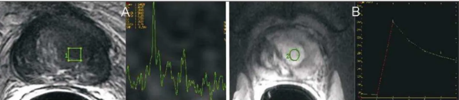
Patients with high PSA levels and negative biopsy
Approximately 66% of patients with PSA levels > 4 ng/ml present negative pros-tate biopsies, many of them being submit-ted to rebiopsy with a higher number of fragments. MRI spectroscopy and/or per-fusion studies have demonstrated good re-sults in the identification of suspicious foci in patients with clinical-laboratory suspi-cion of PCa and at least one previous nega-tive biopsy, targeting ultrasound-guided rebiopsy with additional samples of those areas (Figure 11). Accuracy between 80% and 90% has been demonstrated for
spec-troscopy and perfusion in the detection of tumors ≥ 0.5 cm3(30,31).
Postoperative tumor recurrence
MRI may demonstrate post prostatec-tomy local recurrence and/or residual tu-mor, which are seen as soft tissue masses with subtle hyperintense signal on T2-weighted images as compared with the ad-jacent musculature (Figure 12). The main differentiation to be considered is with post-operative fibrosis, which shows marked hypointensity on T2-weighted images. The main locations for tumor recurrence include: retrovesical (40%), perianastomotic (29%) and in retained seminal vesicles (22%)(32).
Post-radiotherapy tumor recurrence
Approximately 25% of patients with prostate cancer are treated with radiother-apy, with the rate of PSA recurrence after 5 years ranging from 15% to 67%, depend-ing on each patient’s risk.
After radiotherapy, a diffuse reduction in the signal intensity on T2-weighted se-quences is observed, as a consequence of
Figure 10. MR axial T2-weighted image demonstrating an ill-defined lesion markedly hypointense signal in the central gland at left, with elevated choline peak spectroscopy (A). Perfusion study: neoplastic pattern with early enhancement followed by washout (B). Biopsy revealed PCa.
fibrosis and glandular atrophy. The follow-ing imagfollow-ing criteria are adopted in case tumor recurrence is suspected(33):
– MRI criterion: nodular area with marked hypointense signal on T2-weighted sequences.
– Spectroscopy criterion: voxels with a (choline + creatine)/citrate ratio ≥ 0.5; or any voxel with increased choline levels, when there is too much noise limiting the appropriate individualization of the other metabolites.
DIFFERENTIAL DIAGNOSIS
Other conditions with hyposignal inten-sity in the peripheral zone on T2-weighted images may mimic prostate cancer, such as hemorrhage, prostatitis, benign prostatic hyperplasia, trauma, other tumors such as lymphoma and sarcoma, among other causes.
Post-biopsy hemorrhage is character-ized by hypointense signal on T1-weighted images and variable signal intensity on T2-weighted images. Hemorrhagic foci present a significant decrease 21 days after the pro-cedure, so that this minimum interval should be utilized between biopsy and MRI.
METHOD LIMITATIONS
MRI is limited as far as small tumors (<
0.5 cm3) and low grade tumors (Gleason
sum < 6) are concerned, and such limita-tions persist even with the utilization of spectroscopy and perfusion. MRI study is
also impaired in the presence of extensive hemorrhage, hence the already mentioned recommendation of a minimum interval of 21 days between biopsy and MRI.
Artifacts caused by sphincter contrac-tion or body mocontrac-tion may also affect study quality. For this reason, the patient must be carefully instructed on how to avoid these factors prior to the examination. Intestinal peristalsis artifacts can also be minimized with the use of antiperistaltic medication (N-butylescopolamine bromide).
Artifacts may also be found on spectros-copy studies (for example: lipid contami-nation, partial water peak suppression, low signal-to-noise ratio, susceptibility artifacts affecting the metabolite signals), particu-larly in patients with prostheses or other metal devices. A correct planning of the spectroscopy sequence, eventually comple-mented with manual calibration of the se-quence, can minimize the effect of such artifacts.
CONCLUSION
MRI plays a relevant role in the study of the prostate, particularly in the detection and staging of tumors, with the potential to provide valuable information in the thera-peutic planning of prostate cancer. There-fore, radiologists must have the knowledge on the indications, potential and limitations of the method.
REFERENCES
1. Instituto Nacional de Câncer. Estimativas da
in-cidência e mortalidade por câncer no Brasil, 1996-2002. Rio de Janeiro: INCA; 2002. 2. American Cancer Society. Cancer facts and
fig-ures. Atlanta: ACS; 2004.
3. Partin AW, Kattan MW, Subong EN, et al. Com-bination of prostate-specific antigen, clinical stage, and Gleason score to predict pathological stage of localized prostate cancer. A multi-insti-tutional update. JAMA. 1997;277:1445–51. 4. Ogura K, Maekawa S, Okubo K, et al. Dynamic
endorectal magnetic resonance imaging for local staging and detection of neurovascular bundle in-volvement of prostate cancer: correlation with histopathologic results. Urology. 2001;57:721–6. 5. Wang L, Hricak H, Kattan MW, et al. Prediction of organ-confined prostate cancer: incremental value of MR imaging and MR spectroscopic im-aging to stim-aging nomograms. Radiology. 2006; 238:597–603.
6. Fütterer JJ, Heijmink SW, Scheenen TW, et al. Prostate cancer: local staging at 3-T endorectal MR imaging – early experience. Radiology. 2006; 238:184–91.
7. Heijmink SW, Fütterer JJ, Hambrock T, et al. Prostate cancer: body-array versus endorectal coil MR imaging at 3T – comparison of image qual-ity, localization, and staging performance. Radi-ology. 2007;244:184–95.
8. McNeal JE. The zonal anatomy of the prostate. Prostate. 1981;2:35–49.
9. Sobin L. TNM classification of malignant tumors. New York: Willey-Liss; 2002.
10. Carroll PR, Benaron DA, Blackledge G, et al. Third international conference on innovations and challenges in prostate cancer: prevention, detection and treatment. J Urol. 2003;170(6 Pt 2):S3–5. 11. Yu KK, Hricak H, Alagappan R, et al. Detection
of extracapsular extension of prostate carcinoma with endorectal and phased-array coil MR imag-ing: multivariate feature analysis. Radiology. 1997;202:697–702.
12. Sala E, Akin O, Moskowitz CS, et al. Endorectal MR imaging in the evaluation of seminal vesicle invasion: diagnostic accuracy and multivariate feature analysis. Radiology. 2006;238:929–37. 13. Claus FG, Hricak H, Hattery RR. Pretreatment
evaluation of prostate cancer: role of MR imag-ing and 1H MR spectroscopy. Radiographics. 2004;24 Suppl 1:S167–80.
14. Kurhanewicz J, Vigneron DB, Hricak H, et al. Three-dimensional H-1 MR spectroscopic imag-ing of the in situ human prostate with high (0.24-0.7-cm3) spatial resolution. Radiology. 1996;198:
795–805.
15. Choi H, Ma J. Use of perfluorocarbon compound in the endorectal coil to improve MR spectros-copy of the prostate. AJR Am J Roentgenol. 2008; 190:1055–9.
16. Jung JA, Coakley FV, Vigneron DB, et al. Pros-tate depiction at endorectal MR spectroscopic im-aging: investigation of a standardized evaluation system. Radiology. 2004;233:701–8. 17. Scheidler J, Hricak H, Vigneron DB, et al.
Pros-tate cancer: localization with three-dimensional proton MR spectroscopic imaging – clinicopatho-logic study. Radiology. 1999;213:473–80. 18. Yu KK, Scheidler J, Hricak H, et al. Prostate
can-cer: prediction of extracapsular extension with endorectal MR imaging and three-dimensional
proton MR spectroscopic imaging. Radiology. 1999;213:481–8.
19. Zakian KL, Sircar K, Hricak H, et al. Correlation of proton MR spectroscopic imaging with Gleason score on step-section pathologic analy-sis after radical prostatectomy. Radiology. 2005; 234:804–14.
20. Coakley FV, Kurhanewicz J, Lu Y, et al. Prostate cancer tumor volume: measurement with endo-rectal MR and MR spectroscopic imaging. Radi-ology. 2002;223:91–7.
21. Fütterer JJ, Engelbrecht MR, Huisman HJ, et al. Staging prostate cancer with dynamic contrast-enhanced endorectal MR imaging prior to radi-cal prostatectomy: experienced versus less expe-rienced readers. Radiology. 2005;237:541–9. 22. d’Arcy JA, Collins DJ, Padhani AR, et al.
Infor-matics in Radiology (infoRAD). Magnetic Reso-nance Imaging Workbench: analysis and visual-ization of dynamic contrast-enhanced MR imag-ing data. Radiographics. 2006;26:621–32. 23. Buckley DL, Roberts C, Parker GJ, et al. Prostate
cancer: evaluation of vascular characteristics with dynamic contrast-enhanced T1-weighted MR
im-aging – initial experience. Radiology. 2004;233: 709–15.
24. Engelbrecht MR, Huisman HJ, Laheij RJF, et al. Discrimination of prostate cancer from normal pe-ripheral zone and central gland tissue by using dynamic contrast-enhanced MR imaging. Radi-ology. 2003;229:248–54.
25. Bloch BN, Furman-Haran E, Helbich TH, et al. Prostate cancer: accurate determination of extra-capsular extension with high-spatial-resolution dynamic contrast-enhanced and T2-weighted MR imaging – initial results. Radiology. 2007;245: 176–85.
26. Reinsberg SA, Payne GS, Riches SF, et al. Com-bined use of diffusion-weighted MRI and 1H MR
spectroscopy to increase accuracy in prostate can-cer detection. AJR Am J Roentgenol. 2007;188: 91–8.
27. Haider MA, van der Kwast TH, Tanguay J, et al. Combined T2-weighted and diffusion-weighted MRI for localization of prostate cancer. AJR Am J Roentgenol. 2007;189:323–8.
28. Fütterer JJ, Heijmink SW, Scheenen TW, et al. Prostate cancer localization with dynamic
con-trast-enhanced MR imaging and proton MR spec-troscopic imaging. Radiology. 2006;241:449–58. 29. Akin O, Sala E, Moskowitz CS, et al. Transition zone prostate cancers: features, detection, local-ization, and staging at endorectal MR imaging. Radiology. 2006;239:784–92.
30. Prando A, Kurhanewicz J, Borges AP, et al. Pro-static biopsy directed with endorectal MR spec-troscopic imaging findings in patients with el-evated prostate specific antigen levels and prior negative biopsy findings: early experience. Radi-ology. 2005;236:903–10.
31. Beyersdorff D, Winkel A, Hamm B, et al. MR im-aging-guided prostate biopsy with a closed MR unit at 1.5 T: initial results. Radiology. 2005;234: 576–81.
32. Sella T, Schwartz LH, Swindle PW, et al. Sus-pected local recurrence after radical prostatec-tomy: endorectal coil MR imaging. Radiology. 2004;231:379–85.