J. Evid. Based Med. Healthc., pISSN- 2349-2562, eISSN- 2349-2570/ Vol. 3/Issue 75/Sept. 19, 2016 Page 4053
A CLINICAL STUDY OF OPERATIVE TREATMENT FOR LUMBAR INTERVERTEBRAL DISC
PROLAPSE
Anilkumar S. D1
1Assistant Professor, Department of Orthopaedics, Mount Zion Medical College, Adoor.
ABSTRACT
BACKGROUND
Amongst painful diseases, sciatica occupies a foremost place by reason of its prevalence, its production by a great variety of conditions, the great disablement it may produce and its tending to relapse all of which have led to its recognition as one of the great scourges of humanity. Intervertebral disc prolapse is the important and common cause of low back pain and sciatica. Here, the subject of laminectomy and discectomy in the treatment of proven intervertebral disc prolapse in the lower lumbar region is reviewed and its results examined.
AIM OF STUDY
This study was undertaken in order to evaluate the following objectives.
PRIMARY
Analysis of clinical parameters and per operative findings of lumbar intervertebral disc prolapse.
SECONDARY
Analysis of clinical parameters and surgical outcome in lumbar intervertebral disc prolapse with respect to improvement in pain and neurological status.
MATERIALS AND METHODS
The study was undertaken in 22 patients who attended the Orthopaedic Department of Mount Zion Medical College, Adoor, between August 2014 to July 2015. All of them were suffering from a prolapsed lumbar vertebral disc as shown by clinical examination and investigations. Lumbar laminectomy and discectomy constituted the operative procedure for all of them.
RESULTS
In acute onset cases and cases with short duration, results were good. By six months, 80% of patients recorded of good pain relief, 80% of patients returned to work within six 6 months, 60% of patients showed good neurological recovery by 6 months. Laminectomy and discectomy in proven cases of lumbar intervertebral disc prolapse is a rewarding procedure. LT definitely relieved pain in all cases and improved morbidity and neurological deficits in most of the cases.
CONCLUSION
1. Laminectomy and discectomy is an effective method of treatment in herniation of lumbar intervertebral disc. 2. The procedure is ideally done in those with the disc prolapse proved with the help of investigations.
3. A course of conservative treatment should be tried initially.
4. The subjective relief of pain is taken as the most important factor in deciding the result of treatment. Good result is seen in those with shorter duration of symptoms. In those with acute onset of symptoms, the results were good.
5. With passage of time, there was increase in percentage of good results of pain relief. Neurological recovery starts by 4 weeks to 12 weeks.
KEYWORDS
Laminectomy and Discectomy, Herniation of Lumbar Intervertebral Disc, Magnetic Resonance Imaging, Straight Leg Raising Test.
HOW TO CITE THIS ARTICLE: Anilkumar SD.A clinical study of operative treatment for lumbar intervertebral disc prolapse.
J. Evid. Based Med. Healthc. 2016; 3(75), 4053-4058. DOI: 10.18410/jebmh/2016/866
INTRODUCTION: Amongst painful diseases, sciatica
occupies a foremost place by reason of its prevalence, its production by a great variety of conditions, the great disablement it may produce and its tending to relapse all of which have long age led to its recognition as one of great scourges of humanity. It has been known as long as medicine has been studied, but it has only been recognised as a clinical entity since the Italian physician Dominic
Financial or Other, Competing Interest: None. Submission 12-08-2016, Peer Review 21-08-2016, Acceptance 11-09-2016, Published 16-09-2016. Corresponding Author:
Dr. Anilkumar S. D,
Aiswarya, TC 8/2072(1), Mahila Samajam Lane, Bapuji Nagar, Pongumoodu, Trivandrum-695011. E-mail: [email protected]
J. Evid. Based Med. Healthc., pISSN- 2349-2562, eISSN- 2349-2570/ Vol. 3/Issue 75/Sept. 19, 2016 Page 4054
Cotungo gave a description of it in 1764, which came in his "De Ischiade Nervosa Commentarius."
Intervertebral disc prolapse is the important and common cause of low back pain and sciatica. Though anon fatal condition, it causes much morbidity and runs a very prolonged course. One of the major causes of loss of manpower and man hour in industry is the back and leg pain. Hence, it demands much attention from the authorities and is a suitable field of research.
More often, the patient with back and leg pain comes to the orthopaedic surgeon only after many consultations with the general practitioner. The neurosurgeon takes a definite role in the surgical treatment of disc prolapse. But, still the main stay of treatment is with the orthopaedic surgeon since the disc prolapse causes problem not only with the root, but with the integrity of the vertebral column. So, every orthopaedic surgeon has to be well-versed with various aspects of lumbar intervertebral disc prolapse.
Various causes of back and leg pain other than lumbar intervertebral disc prolapse has to be thought of. Here, one has to think of problems of gynaecology, sacroiliac subluxation, postural fault, congenital anomalies, fasciitis, contractures, spastic piriformis, thickened ligamentum flavum, narrowed intervertebral spaces and spinal canal stenosis due is a variety of causes. The orthopaedic surgeon recognises the aetiological features of the symptom complex and chooses from a wide variety therapeutic measures in prescribing for his patient. The subject of laminectomy and discectomy in the treatment of proven intervertebral disc prolapse in the lower lumbar region is reviewed and its results examined.1,2,3
AIMS OF STUDY: This study was undertaken in order to
evaluate the following objectives.
Primary:
1. Analysis of clinical parameters and per operative findings of lumbar intervertebral disc prolapse. 2. Determination of efficacy and role of diagnostic
studies like Myelography, computerized Tomography and Magnetic resonance imaging. In terms of accuracy indices like sensitively specificity and predictive value of positive test in lumbar intervertebral disc prolapse by correlating their results with surgical findings.
Secondary: Analysis of clinical parameters and surgical outcome in intervertebral disc prolapsed with respect to improvement in pain, morbidityand neurological status.
MATERIALS AND METHODS: The study was undertaken
in 22 patients who attended the Orthopaedic Department Mount Zion Medical College Hospital, Adoor, between August 2014 and July 2015. All of them were suffering from prolapsed lumbar intervertebral disc as shown by clinical examination and investigations.
Inclusion Criteria:
1. All patients suffering from prolapsed lumbar intervertebral disc.
2. All Patients had pain after six weeks of conservative treatment.
3. Laminectomy and discectomy done in all patients.
Exclusion Criteria:
1. Age below 20 excluded. 2. Age above 60 excluded.
3. Failed back with multiple reasons excluded.
4. Patients with systemic rheumatic diseases excluded.
All of these patients had severe pain and were forced to stay in bed. They were unable to move about without pain even to the bathroom. Initially, all of them underwent various modes of treatment, which included indigenous methods like massage, oil application or manipulation or drugs, skin traction, spinal support and local infiltration from a doctor of modern medicine. Fifteen of them had received epidural injection with normal saline, local anaesthetic and hydrocortisone acetate. Almost, all patients had sciatica. This was bilateral in 5 patients and two were shown to have a central disc prolapse during operation. The patients under this study were unable to pursue their occupation.
In the history, the mode of onset of symptoms was noted as to whether acute or insidious. Any related trauma was enquired. The aggravating and relieving factors for pain were noted. The nature of occupation was recorded according to whether of a physically demanding type or sedentary. All the female patients were housewives.
General examination was made with special note in the build of the patient and the weight was recorded. Almost, all patients had very good physique. The spine was examined systematically. All the patients had a guarded gait with the hip and knee kept in mild degrees of flexion. Any tilt of lumbar spine was noted as to its presence and side. Paraspinal muscle spasm, tenderness in the lumbosacral junction and restriction of movements of the lumbar spine was noted in all cases and were comparable.
The straight leg raising test was performed and the degree recorded. This was used to group patients and assess the improvement.
The neurological status of the patient was assessed. The absence or blunting of sensation was recorded according to the dermatome. The presence of wasting and motor weakness was noted. The extensor hallucis longus weakness indicated the level of lesion, the deep tender reflexes were elicited and any differences noted. Any weakness of the ankle or knee jerk was of importance. No patients of this series had any bladder or bowel dysfunction. No difficulty in sexual function complained of.4
J. Evid. Based Med. Healthc., pISSN- 2349-2562, eISSN- 2349-2570/ Vol. 3/Issue 75/Sept. 19, 2016 Page 4055
Anteroposterior and lateral plain roentgenograms of the lumbar spine were taken. Any other pathology as source of pain or concomitant lesion was ruled out. They were evaluated for the presence of features of degenerative disc disorders (disc space narrowing, osteophytosis, traction spurs, facetal hypertrophy and retrolisthesis and vacuum sign) and anomalies of bony segmentation. The main diagnostic studies include myelography, computerized tomography (with or without contrast) and Magnetic Resonance Imaging. Because these investigation were mainly done outside the institution, the selection of particular investigation was at random and dependent mainly on the financial status and affordability of the patient. These results were analysed and help from the Radiology Department was sought in cases of difficult interpretation.7
Myelogram was evaluated for signs of filling defect and root compression or cut off. Computerized tomography and MRI were studied in detail including level of disc protrusion (single or multiple). Location, containment and extent of thecal and nerve root compression. The states of disc hydration was observed in MRI. The levels of disc prolapse as revealed by diagnostic studies were recorded.8,9,10
Patients were given bed rest. All of them had already received enough of various types of conservative treatment with no sustained relief. Operation as a method of treatment was suggested. Various aspects of this form of treatment were discussed with the patient. Patient was assessed for the fitness of operations, presence of any focus of infection and the local condition of skin was also noted. At least two units of blood were made ready.
Lumbar laminectomy and discectomy constituted the operative procedure. General anaesthesia was used in all patients. Knee chest position was used with pillows under chest and pelvis to leave the abdomen free.
Postoperatively, patient was placed under close observation. All patients had some difficulty in passing urine in the immediate postoperative period. Some had features of intestinal ileus also. They responded to usual measures of treatment and were not a problem later.11
The patient was allowed to turn in bed by himself on second day. From 4 or 5 days postoperatively, he was allowed out of bed. Sutures were removed on the tenth day. Then on, exercises of the spine were started as tolerated. Usually, they were discharged on the tenth day.
Assessment was made on 10th day as to subjective relief of pain and any neurological deficits getting improved. Patients were asked for review at 4 weeks, 8 weeks, 3 months and 6 months.
Based on these data, proforma were made and data were compiled to come to conclusion.12,13,14
OBSERVATION AND ANALYSIS: In this series, 22
patients with lumbar intervertebral disc prolapse, which proved either by Myelography, CT, CT with contrast or MRI studied prospectively with laminectomy and discectomy as the treatment in all cases. Assessment was done initially at 10th day and followup at 4 weeks, 8 weeks, 3 months and
after 6 months. All the patients regularly attended the followup. The percentage of investigation used in Table 1.
Method of Investigation Number Percentage
Myelogram 4 19
CT 4 19
CT Myelogram 1 5
MRI 13 57
Table 1: Investigations
The investigation findings compared with intraoperative findings in Table 2.
Investigation False Positive False Negative
Number Percentage Number Percentage
MRI 4 31 Nil Nil
CT Nil Nil 1 25
CT
Myelogram Nil Nil Nil Nil
Myelogram Nil Nil Nil Nil
Table 2: Comparison between Investigative and Intraoperative Findings
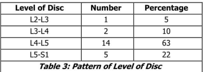
Level of prolapse was compared in Table 3.
Level of Disc Number Percentage
L2-L3 1 5
L3-L4 2 10
L4-L5 14 63
L5-S1 5 22
Table 3: Pattern of Level of Disc
Presentation of disc usually shoulder (Lateral to root) are shown in Table 4.
Type of Prolapse Number Percentage
Lateral to Root 15 67
Medial to Root 5 23
Central 2 10
Table 4: Type of Prolapse
The nature of root involvement was noted. Majority were found to be pressing on the root as shown below.
The common intraoperative complication was dural tear. The CSF leak stopped by repairing the dura. Later, it caused no problem. The list of intraoperative complication shown in Table 5.
Complication Number Percentage
Dural tear 3 15
Neural injury 1 5
Missed level 1 5
J. Evid. Based Med. Healthc., pISSN- 2349-2562, eISSN- 2349-2570/ Vol. 3/Issue 75/Sept. 19, 2016 Page 4056
The majority of patients postoperatively complains of urinary retention. Twenty percent of patients developed mild intestinal illness. Wound infection and wound gaping was below 20%. Table 6 show the frequency of postoperative complications.
Complications Number Percentage
Urinary retention 7 32
Wound infection 4 18
Wound gaping 3 14
Intestinal ileus 6 27
Cauda equina syndrome 1 5
Thromboembolism 2 10
Discitis 1 5
Table 6: Postoperative Complications
Duration after Laminectomy
and Discectomy
Good Moderate Poor
No. Per. No. Per. No. Per
Date of discharge
(10th day) 1 5 17 76 4 19
4 weeks 1 5 20 90 1 5
8 weeks 7 32 14 64 1 5
3 months 12 54 9 41 1 5
6 months 18 81 3 14 1 5
Table 7: Relief of Pain
Neurological improvement is shown below in table 8.
Duration after Laminectomy
and Discectomy
Good Moderate Poor
No. Per. No. Per. No. Per.
Date of
discharge 0 0 0 0 22 100
4 weeks 0 0 2 10 20 90
8 weeks 0 0 11 50 11 50
3 months 1 5 16 72 5 23
6 months 10 45 10 45 2 10
Table 8: Neurological Improvement
The percentage of pain relief with each followup is shown below in Table 9.
Duration Good (%) Moderate (%) Poor (%)
10 days 5 76 19
4 weeks 5 90 5
8 weeks 32 63 5
3 months 54 41 5
6 months 81 14 5
Table 9: Pain Relief and Duration of Followup
The percentage of neurological improvement in each followup is shown below in Table 10.
Duration of
Followup Good (%) Moderate (%) Poor
10 days 0 0 100
4 weeks 0 10 90
8 weeks 0 50 50
3 months 5 72 23
6 months 45 45 10
Table 10: Duration and Neurological Improvement
DISCUSSION: The prevalence of leg and back pain in the
general population is very high. It is said that above 50% of the population has back pain during their lifetime. It is the cause of considerable disability and loss of work resulting in economic hardship to the patients.
The results of conservative treatment were not satisfactory and sustained. It is prolonged and costly in a proved case of intervertebral disc prolapse in the lumbar region. Removal of the disc brings about considerable relief of symptoms. Disc surgery began to be popular after the report of Mixter and Barn. In this study, L4-L5 disc was maximally involved (63%) followed by L5-SI (22%), L3-L4 and L2-L3 disc were minor contributions. Out of these, 38% of patient had neurogenic claudications.
Regarding clinical findings and surgical corroboration, weakness of extensor hallucis longus was found to be 90% correlating with L5 root involvement. L5 sensory deficit was found to be strong evidence of L4-L5 level prolapse. Sensory involvement in SI dermatome was found in both L5-SI and L4-L5 prolapse. So, SI sensory changes require full interpretation.
Unilateral absence of ankle jerk was fully consistent with SI nerve root involvement and L5-SI disc prolapse. Knee reflexes was diminished only in high lumbar disc prolapse. The most common plain x-ray findings were reduction in disc space and obliteration of lumbar lordosis. The list of spine helped to know the type of disc prolapsed whether auxiliary or shoulder. This was 80% consistent with diagnostic study and surgery.
Another part of study was to know accuracy of diagnostic studies in relation to per operative findings. But, most of the patients were done MRI investigations, so a comparison between MRI CT scan and Myelogram may not be accurate. MRI is found to be 100% sensitive in demonstrating disc prolapses. This may be due to depiction of mild and early disc herniation because of its superior soft tissue resolution. CT has least sensitivity of the three studies with high false negativity. Because of high sensitivity, MRI had the highest incidence of false positivity whereas Myelogram has less false positivity. So, the predictive value of positive test was found to be highest with Myelogram and lowest with MRI.
J. Evid. Based Med. Healthc., pISSN- 2349-2562, eISSN- 2349-2570/ Vol. 3/Issue 75/Sept. 19, 2016 Page 4057
In this study, there were more patients with duration of symptom less than 2 years. In the final assessment of pain relief and neurological deficit improvement, this was found to pose significance. With duration of less than 6 months, there was 60% good results in the follow up. This may be due to the fact that there was not secondary changes in the facet joints, but in those with more than 2 years, there was 20% of poor results. This indicates that the duration of disease should be taken into consideration in planning treatment.
The quality of relief of pain was noted to get increased with passage of time. The 5% good result within 10 days, 54% good results within 3 months and 81% good results within 6 months. 5% of patients show no pain relief after 3 months or fresh pains after 3 months. This is due to the fact that with removal of disc, the body of vertebrae comes close to each other and causes increased strain in the facet joint. To prevent this, the regular practice of spinal exercises maybe of value.
The improvement of neurological deficit was found to be increases with time. The percentage of good result at 10 days was nil at 4 weeks, 10% at 8 weeks, 50% moderate improvement. This get increased at 3 months to 72% moderate improvement. This make marked improvement by 6 months showing good improvement of 45%, moderate improvement of 45% and only 10% of poor status.
The mode of onset was insidious in majority of cases. The acute cases amount to be about 46%. Of these, 60% had good improvement of pain at final follow up. This may be due to the fact that in acute attack, patient had taken rest and seeks medical advice early with insidious onset cases. The percentage of good result was 40, but the acute onset patient had 30% of good neurological recovery. In insidious onset, 40% of good neurological recovery percent. This may be due to the fact that with sudden prolapse there is more damage to the nerve roots.
The SLR test has a value in the improvement of pain. Those with less than 30° had 60% of good results in contrast to 50% in other groups. The nerve root involvement as seen during operation has some role in the neurological improvement. In those with nerve root adherent to the disc, 60% remained without neurological improvement. While in those with only pressure on the nerve root, there was 60% good improvement.
In this series, patients with sedentary occupation were able to pursue the same job. Those with jobs, which were physically demanding had to change to less strenuous occupation in the same surroundings.
In this study, 80% of patients had good improvement in morbidity and can return to work within 6 months. They can do works without pain. The quality of life of 80% of patient was same as before the very onset of disease. Poor improvement was seen in 10% of cases. Moderate improvement in 30% of patients.
In conclusion, proper selection of cases and proper procedure in expert hands brings rewarding benefits for patients by laminectomy and discectomy in proved cases of lumbar intervertebral disc prolapses.17,18,19,
CONCLUSIONS:
MRI is 100% sensitive in diagnosing disc disorders. It depicts early mild disc prolapse. It has high false positivity when correlated with surgical findings.
Laminectomy and discectomy is an effective method of treatment in herniation of the lumbar intervertebral disc.
The procedure is ideally done in those with the disc prolapse proved with the help of investigations. The level and side of lesion should be identified.
The indications of surgery should be present.
A course of conservative treatment should be tried initially.
Laminectomy and discectomy has to be carried out in a well-equipped theatre by trained personnel.
The subjective relief of pain is taken as the most important factor in deciding the result of treatment.
Good result is seen in those with shorter duration of symptoms.
In those with acute onset of symptom, the result were good.
With passage of time, there was increase in the percentage of good results of pain relief.
The neurological recovery starts by four weeks to 12 weeks.
By 6 months, 60% of good neurological recovery was obtained.
80% of good improvement in morbidity by 6 months after operation.
By 6 months, 80% of patients returned to their jobs without pain.
REFERENCES
1. Arnoldi C. Brodsky AE, Cauchoix J, et al. Lumbar spine stenosis and nerve root entrapment syndromes. Definition and classification. Clin Orthop Relat Res 1976;115:4-5.
2. Boden SD, Davis DO, Dina TS, et al. Abnormal MRI of lumbar spine in symptomatic subjects. A prospective investigation. J of Bone and Joint Surgery 1990;72(3):403-408.
3. Crenshaw AH. Campbell's operative orthopaedics. 8th
edn. Mosby 1992.
4. Bavadekar AV. Treatment of lumbar disc prolapse. Clinical Orthopaedics India 1988;2:21.
5. Getty CJ. Management of the problem back. In: Catterall A, ed. Recent advances in orthopaedics. Vol. 5. Edinburgh, London: Churchill. Livingstone1987. 6. Kampmann H, Schroed lP, Spranger Metal. Diagnosis
of lumbar disc prolapse by CT and myelogram-a comparative study with surgical results. Rontgenblatter 1985;38(12):387.
7. Call MIW. Recent advances in imaging in orthopaedics. In: Catterall A, ed. Recent advances in orthopaedics. 6th edn. Vol. 6. Churchill Livingstone
J. Evid. Based Med. Healthc., pISSN- 2349-2562, eISSN- 2349-2570/ Vol. 3/Issue 75/Sept. 19, 2016 Page 4058
8. Post MJD, Brown MD, Gargagno FP. The technique and interpretation of lumbar myelogram. Spine 1977;2(3):214-230.
9. Rai J. Correlation between clinical, CT and surgical findings in lumbar disc prolapse. Paper presented in Indian Orthopaedic Association National Conference 1994.
10. Djukicetal S, Lang P, Morris J, et al. MRI of postoperative lumbar spine. Radiology Clinics of North America 1990;28:341-360.
11. Verbiest H. Results of surgical treatment of developmental stenosis of lumbar vertebral canal. A review of twenty seven years’ experience. J Bone Joint Surgery Br 1977;59(2):181-188.
12. Kirkaldy-Willis WH, Hill RJ. A more precise diagnosis of low back pain. Spine 1979;4(2):102-109.
13. Kratzer M, Hipp E. Value of myelogram, CT in problem cases of lumbar intervertebral disk diagnosis. Z Orthop Ihre Grenzgeb 1986;124(1):107-111.
14. Mixter WJ, Barr JS. Rupture of intervertebral disc with involvement of spinal canal. N Engl J Med 1934;211:210-215.
15. Modic T. MRI in evaluation of low back pain. Orthopaedics Clinics of North America 1991;22(2):283-301.
16. Postacchini F, Pezzeri G, Montanaro A, et al. CT in lumbar stenosis-a preliminary report. J of Bone and Joint Surgery 1980;62-B(1):78-82.
17. Cailliet R. Low back pain syndrome. 4th edn.
Philadelphia, PA: FA Davis Company 1988.
18. Rothman RH. The clinical syndrome of lumbar disc disease. Orthopaedic Clinics of North America 1971;2(2):463-475.