Anagen, Catagen, and Telogen Phases in Tibetan Sheep
Guangbin Liu
1, Ruize Liu
1, Qinqun Li
1, Xiaohui Tang
2, Mei Yu
1, Xinyun Li
1, Jianhua Cao
1*, Shuhong Zhao
1*1 Key Laboratory of Agricultural Animal Genetics, Breeding and Reproduction, Ministry of Education, Huazhong Agricultural University, Wuhan, Hubei, China, 2 Agriculture and Animal Husbandry College of Tibet, Linzhi, Tibet, China
Abstract
Background: Wool quality is one of the most important economic traits in sheep. The wool fiber is derived from specialized skin cells that are referred to as wool follicles. To understand the roles of microRNAs (miRNAs) in wool fiber growth, we detected the expression patterns of miRNAs in wool follicles at the anagen, catagen, and telogen stages from Tibetan sheep through Solexa sequencing.
Results: A total of 244 mature miRNAs were identified. Of these, only five miRNAs are listed in the database of sheep miRNAs (miRBase Database V19), and the other 239 miRNAs have not been previously described in this species. Further analyses indicated that 204 miRNAs are evolutionarily conserved among mammal species, whereas 35 of the identified miRNAs were first found specifically in sheep. The expression pattern analyses showed that the expression levels of 39, 34, and 20 of the miRNAs significantly change between anagen and catagen, between anagen and telogen, and between catagen and telogen, respectively. The results of the bioinformatics analysis show that these differentially expressed miRNAs might regulate wool follicle development by targeting genes in many different pathways, such as the MAPK and Wnt pathways, as well as the pathways that regulate the actin cytoskeleton, focal adhesion, and tight junctions. Furthermore, we identified six differentially expressed miRNAs (oar-miR-103-3P, oar-miR-148b-3P, oar-miR-320-3P, oar-miR-31-5P, oar-novel-1-5P, and oar-novel-2-3P) that might target the key genes of the Wnt pathway. It has been reported that the Wnt pathway is critical for wool follicle development. Therefore, these miRNAs may regulate wool development through the Wnt pathway.
Conclusions: Our results provide new information on the identification and expression pattern of miRNAs in wool follicles. Our data might therefore aid in the understanding of the mechanisms of wool follicle development in sheep.
Citation: Liu G, Liu R, Li Q, Tang X, Yu M, et al. (2013) Identification of microRNAs in Wool Follicles during Anagen, Catagen, and Telogen Phases in Tibetan Sheep. PLoS ONE 8(10): e77801. doi:10.1371/journal.pone.0077801
Editor: Huaijun Zhou, University of California, Davis, United States of America Received April 11, 2013; Accepted September 4, 2013; Published October 17, 2013
Copyright: © 2013 Guangbin Liu. This is an open-access article distributed under the terms of the Creative Commons Attribution License, which permits unrestricted use, distribution, and reproduction in any medium, provided the original author and source are credited.
Funding: This work was supported by a grant (2011ZX08009-004) from the National Program of Transgenic Variety Development of China and Regional project of the National Natural Science Foundation (30960163) of China. The funders had no role in study design, data collection and analysis, decision to publish, or preparation of the manuscript.
Competing interests: The authors have declared that no competing interests exist. * E-mail: shzhao@mail.hzau.edu.cn (SZ); jhcao@mail.hzau.edu.cn (JC)
Introduction
MicroRNAs (miRNAs) are a class of noncoding small RNAs. A mature miRNA is usually single-stranded and 21-24 nt (nucleotides) in length. It can bind the 3’UTR of mRNA through pairing with the miRNA seed region and can block gene expression by inhibiting the translation or degradation of the mRNA [1]. Researchers have revealed that a miRNA can target many different sites on the same or different genes and that approximately 30% of genes are regulated by miRNAs [2]. Since the first miRNA was discovered in 1993 [3], thousands of miRNAs have been identified in different species. Increasing evidence shows that miRNAs participate in many biological processes, particularly cell proliferation, differentiation, apoptosis, and immune responses [4].
Wool, as one of the most valuable products from sheep, is an important material in the textile industry. An improvement in the wool quality will result in marked economic value in the field of animal husbandry. As the direct tissue from which wool is derived from, the wool follicle plays a vital role in the production of better-quality wool [5]. In general, the development of the wool follicle could be divided into three stages: anagen, catagen, and telogen. During these three stages, the wool follicle undergoes growth, regression, and rest phases [6]. The hair follicle is also a regenerating system, and each mature hair follicle develops under a growth cycle [7-10].
that miRNAs could be important regulatory factors in hair follicle development. However, the molecular mechanism of miRNAs in hair follicle development has not been illustrated.
To understand the functions of miRNAs in wool follicle development, wool follicles at the anagen, catagen, and telogen stages were collected in this study. The miRNAs of wool follicles and the expression patterns of these miRNAs during the anagen, catagen, and telogen stages were investigated through Solexa sequencing. A number of miRNAs were found to be differentially expressed between the three hair follicle developmental stages. Our study could provide new knowledge regarding the development of wool follicles in the sheep.
Results
Overview of the Solexa sequencing data
To understand the expression pattern of miRNAs during wool follicle development, three small RNA libraries were constructed from the total RNA of wool follicles at the anagen, catagen, and telogen stages. Each library pooled the RNA of the wool follicles at the same phase from three Tibetan sheep. We detected the expressions of two marker genes, LEF1 and TGFB1, to confirm that our samples were collected from the right phases. Previous studies have reported that the expression of LEF1 in hair follicles is higher in anagen compared with the catagen and telogen phases, whereas the expression level of TGFB1 is upregulated in catagen and downregulated in telogen [12,13]. The QPCR (Real-time Quantitative PCR) results (Figure 1) show that the expression patterns of these two genes are consistent with the expected phases. Subsequently, Solexa sequencings were performed for each library. A total of 12,740,200 (anagen), 16,768,947 (catagen), and 16,564,009 (telogen) raw reads were obtained
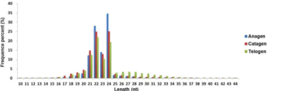
from the three small RNA libraries. After removing the low-quality reads and adapter fragments, 12,197,976 (anagen), 15,623,402 (catagen), and 15,079,495 (telogen) clean reads were obtained, which corresponded to 97.37%, 95.05%, and 92.81% of the raw reads from the three small RNA libraries, respectively. The length distribution of these clean reads is shown in Figure 2. The majority of the reads are 21-24 nt in length, and the number of 22-nt and 24-nt reads are significantly greater than the number of reads of other sizes. This result coincides with the length range for Dicer-derived products.
Identification of miRNAs from sequencing data using the miRDeep software and annotation
The sequencing data from the three small RNA libraries were analyzed using the miRDeep software (Version 2.0.0.3) (http:// mdc.helmholtz.de/8551903/en/research/research_teams/ systems_biology_of_gene_regulatory_elements/projects/ miRDeep). The reference genome sequence of sheep used in this study was OAR2.0 Released by CSIRO Australia (http:// www.livestockgenomics.csiro.au/), and the miRNA database used is the miRBase database V19 (http://www.mirbase.org/). In total, 244 mature miRNAs were identified, and these miRNAs belong to 208 unique miRNA precursors. In addition, 218, 229, and 221 miRNAs are expressed in the anagen, catagen, and telogen phases, respectively. Of these, 198 miRNAs overlapped among the three libraries, whereas seven, eight, and three miRNAs are specifically expressed in anagen, catagen, and telogen libraries, respectively.
The annotation work was performed using the miRBase database V19 (Figure 3). Only five of the sheep miRNAs in our dataset have been deposited into the miRBase database V19: oar-miR-379, oar-miR-127, oar-miR-411a, oar-miR-493, and oar-miR-382. The other 239 miRNAs have not been reported in
Figure 1. Confirmation of the samples from the three phases through detection of the expression of marker genes. The two maker genes LEF1 and TGFB1 were detected by QPCR. The LEF1 gene was more highly expressed in anagen than in catagen and telogen (P < 0.01), and the TGFB1 gene was more highly expressed in catagen than in anagen and telogen (P < 0.01).
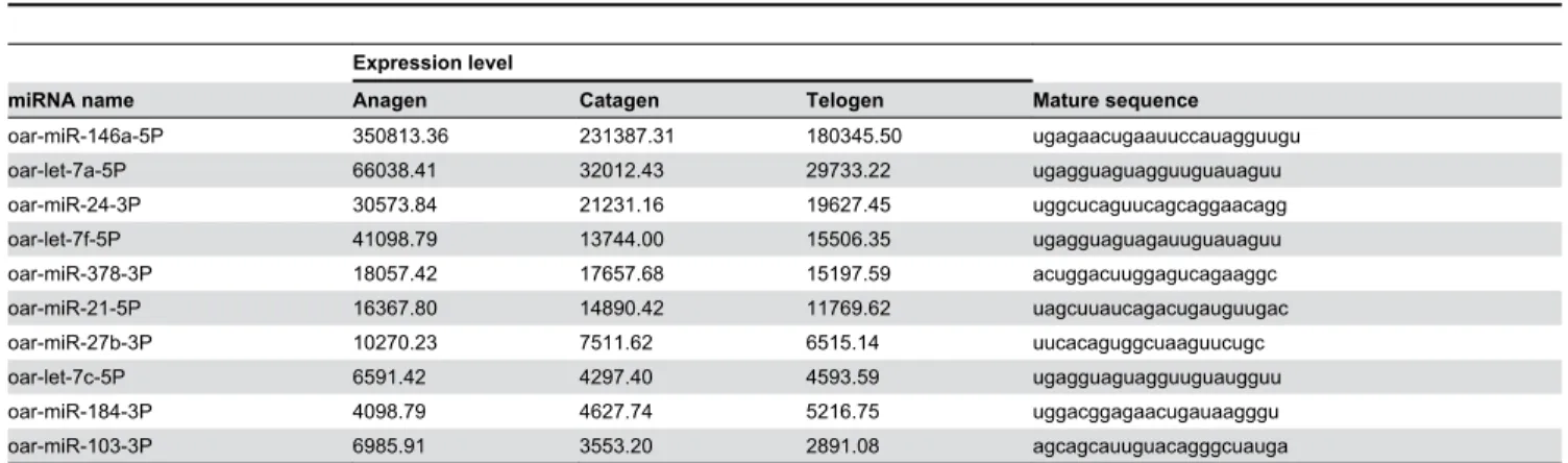
sheep (miRBase database V19). Among these 239 candidate miRNAs, 204 exhibit evolutionary conservation with the mature miRNAs from other species, and 35 miRNAs are novel candidates. All of the information on the 244 mature miRNAs is shown in Table S1, and we list the 10 most abundant miRNAs in Table 1. We found that the expression levels of these 10 miRNAs constitute approximately 90% of the total expression level of all of the miRNAs. Oar-miR-146a-5P is the most abundantly expressed miRNAs in wool follicles. Its expression level is nearly six-fold higher than that of the second most abundantly expressed miRNA (oar-let-7a-5P) in our data, and constitutes more than a half of the total expression level of all of the miRNAs.
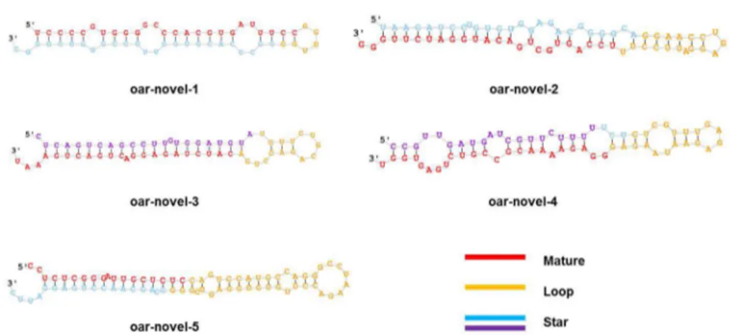
The precursor sequences and secondary structures of the 35 novel miRNAs identified from our sequencing data using miRDeep software (Table S2) were predicted (Figure S1). The five most abundantly expressed novel miRNAs are shown in Tables 2 and 3 and Figure 4. The results show that most of the novel miRNAs in our dataset exhibit relatively low expression levels. In addition, many of the novel miRNAs present a phase-specific expression pattern. For example, oar-novel-1 is specifically expressed in catagen, whereas oar-novel-2 is not
detected in anagen. In fact, 23 of the novel miRNAs are either expressed or not expressed in a phase-specific manner. This number accounts for 65.7% of the total number of novel miRNAs in our dataset.
Validation of the sequencing data by QPCR
To validate the sequencing data, we randomly selected eight different miRNAs with expression levels of more than 500 per million for QPCR analyses (Figure 5). As anticipated, the QPCR results are consistent with the sequencing data. For example, both the sequencing data and the QPCR results show that the expression level of oar-miR-184-3P is upregulated gradually from anagen to telogen. In addition, the expression level of oar-miR-183-5P is upregulated from anagen to catagen and downregulated from catagen to telogen.
Identification of differentially expressed miRNAs between different wool follicle development phases and prediction of miRNA target genes and pathways
To understand the expression patterns of miRNAs during wool follicle development, we performed a cluster analysis
Figure 2. The distribution of the read length from the Solexa sequencing data. The majority of the reads in the three libraries are 21-24 nt in length.
doi: 10.1371/journal.pone.0077801.g002
Figure 3. Annotation of miRNAs. The number of miRNAs is indicated.
(Figure 6A). The results demonstrate that the expression levels of most of the miRNAs change during the development of the wool follicle from anagen to telogen. Approximately 50% of the miRNAs are downregulated from anagen to catagen and telogen, although a few miRNAs are upregulated in telogen.
We then compared the expression level of each miRNA in the three phases to identify miRNAs that are differentially expressed between two different phases (anagen compared with catagen, anagen compared with telogen, and catagen compared with telogen). The miRNAs that satisfied the criteria log2FoldChange ≥ 1 or ≤ -1 and P ≤ 0.01 were denoted as differentially expressed miRNAs. As a result, we found that the expression level of 39, 34, and 20 miRNAs significantly change between anagen and catagen, between anagen and telogen, and between catagen and telogen, respectively (Figure 6B). We list all of the differentially expressed miRNAs in Table S3.
The results show that the number of differentially expressed miRNAs between anagen and catagen are similar to that between anagen and telogen, but higher than the number of miRNAs that are differentially expressed between catagen and telogen. Additionally, more miRNAs are downregulated as the wool follicles develop and transition from the anagen to the telogen phase.
To understand the function of these differentially expressed miRNAs in wool follicle development, we predicted the target genes of the miRNAs using the RNAhybrid software. A dataset of the sequenced sheep wool follicle transcriptome from a previous work (unpublished) was used as the candidate target gene reference. As a result, 3,223 genes were found to be targeted by 48 of the differentially expressed miRNAs (Table
S4). We subsequently analyzed these 3,223 genes through the DAVID website (http://david.abcc.ncifcrf.gov/), and the results
Table 1. The 10 most abundantly expressed miRNAs in sheep wool follicles.
Expression level
miRNA name Anagen Catagen Telogen Mature sequence
oar-miR-146a-5P 350813.36 231387.31 180345.50 ugagaacugaauuccauagguugu oar-let-7a-5P 66038.41 32012.43 29733.22 ugagguaguagguuguauaguu oar-miR-24-3P 30573.84 21231.16 19627.45 uggcucaguucagcaggaacagg oar-let-7f-5P 41098.79 13744.00 15506.35 ugagguaguagauuguauaguu oar-miR-378-3P 18057.42 17657.68 15197.59 acuggacuuggagucagaaggc oar-miR-21-5P 16367.80 14890.42 11769.62 uagcuuaucagacugauguugac oar-miR-27b-3P 10270.23 7511.62 6515.14 uucacaguggcuaaguucugc oar-let-7c-5P 6591.42 4297.40 4593.59 ugagguaguagguuguaugguu oar-miR-184-3P 4098.79 4627.74 5216.75 uggacggagaacugauaagggu oar-miR-103-3P 6985.91 3553.20 2891.08 agcagcauuguacagggcuauga doi: 10.1371/journal.pone.0077801.t001
Table 2. The five most abundantly expressed novel miRNAs in sheep wool follicles.
Expression level
miRNA name Anagen Catagen Telogen Mature sequence Rank
oar-novel-1-5P 0.00 543.61 0.00 uccccguggggcccacgugauuucc 52
oar-novel-2-3P 0.00 38.21 414.54 uccagugcugacauggaucuuggg 55
oar-novel-3-3P 117.07 98.76 92.05 caucuagaggacugacugaaau 68
oar-novel-4-3P 65.01 43.14 34.22 ggagaaaacgccgucugaguggu 91
oar-novel-5-5P 2.05 5.89 79.38 ccucucgggauugcucuc 101
doi: 10.1371/journal.pone.0077801.t002
Table 3. Predicted precursor sequences and genome locations of novel miRNAs.
miRNA name miRNA precursor sequence Chromosome Strand
show that 176 pathways could be involved in the corresponding miRNA regulation (Table S5). We list 20 pathways associated with the most number of target genes in Figure 7. Some pathways, such as the MAPK and Wnt signaling pathways, regulate cell proliferation and differentiation. We also identified pathways that are involved in the maintenance of cell and tissue structures, e.g., the regulation of the actin cytoskeleton pathway, the focal adhesion pathway, and the tight junction pathway. Of these, we focused on the Wnt pathway because many reports have revealed that the Wnt pathway can regulate hair follicle development. In our results, we found that 22 differentially expressed miRNAs can target 29 genes in the Wnt pathway (Table 4); these miRNAs include mir-16a-5P, oar-mir-31-5P, and oar-mir-103-3P. In addition, some novel miRNAs, such as novel-1-5P, novel-2-3P, oar-novel-5-5P, oar-novel-8-5P and oar-novel-10-5P, might be involved in the Wnt pathway. We also found the some miRNAs,
such as oar-miR-103-3P, oar-miR-148b-3P, oar-miR-320-3P, oar-miR-31-5P, oar-novel-1-5P, and oar-novel-2-3P, can directly or indirectly affect the expression of the TCF7 or LEF1 genes to regulate the cell cycle (Figure 8).
Discussion
Many studies have proven that miRNAs play important roles in cell proliferation and differentiation [4,14,15]. There is growing evidence that miRNAs take part in the regulation of hair follicle development [16]. To understand the roles of miRNAs in wool follicle development, we identified 244 mature miRNAs in wool follicles from three different hair follicle growth phases (anagen, catagen, and telogen). Compared with other species, few studies have focused on sheep miRNAs. The most recent miRBase database version 19 contains 2,042 mature miRNAs from human, 1,281 miRNAs from mouse, 755
Figure 4. Predicted secondary structures of novel miRNAs. The red color indicates the mature sequence, the yellow color indicates the loop sequence, the blue color indicates the predicted star sequence, and the purple indicates the miRNA star sequences.
doi: 10.1371/journal.pone.0077801.g004
Figure 5. Validation of sequencing data by QPCR. The blue lines indicate the expression patterns of miRNAs in the sequencing data, and the red lines indicate the QPCR results.
miRNAs from cow, and only 103 miRNAs from sheep. Therefore, our study not only demonstrates the expression pattern of miRNAs during wool follicle development but also enriches the information in the miRNA database.
Our study found that miR-146a is the most abundantly expressed miRNA (in all three phases). This miRNA occupies
more than 50% of the total reads (the sum of the three phases), and its expression level is nearly six-fold higher than that of oar-let-7a, which is the second most abundantly expressed miRNA in our dataset. Studies have shown that miR-146a can regulate the resolution of T cell responses [17,18] and that this miRNA also participates in inflammatory
Figure 6. Identification of differentially expressed miRNAs among different wool follicle development phases. A: Cluster analysis of the expression levels of miRNAs in the three phases. B: The number of miRNAs that exhibited a significant change in expression level between different phases. The red color indicates upregulation, and the blue color indicates downregulation.
doi: 10.1371/journal.pone.0077801.g006
Figure 7. Top 20 pathways predicted to be targeted by differentially expressed miRNAs. The column indicates the unique gene number.
responses [19]. In addition, some studies have found that miR-146a might induce cell proliferation and differentiation [20,21]. These evidences suggested that the function of
oar-miR-146a might be related to the immune response or cell growth. We predicted the target genes of miR-146a in the present study. As a result, we found that miR-146 may regulate
Table 4. Target genes of differentially expressed miRNAs in the Wnt pathway.
Expression level
miRNA name Anagen Catagen Telogen Target genes in Wnt pathway
oar-miR-103-3P 6985.91 3553.2 2891.08 TCF7, NLK, MAPK8, CTBP2, AXIN1, BTRC, RHOA oar-miR-16a-5P 4407.53 2168.61 1896.28 AXIN1, NFATC3
oar-miR-148b-3P 1483.61 470.06 623.1 AXIN1, MAP3K7 oar-miR-320-3P 1010.17 195.54 192.12 TP53, RAC1, LRP5, LEF1
oar-miR-31-5P 605.43 217.05 231.84 CREBBP, TCF7, MAPK9, AXIN1, CACYBP, MAP3K7, DAAM1
oar-novel-1-5P 0 543.61 0 TP53, TCF7, NLK, FZD1, NFATC1, PPP2R5B, CTBP2, FZD7, AXIN1, DAAM1 oar-novel-2-3P 0 38.21 414.54 TP53, NLK, LEF1, AXIN1
oar-miR-6529-5P 207 52.23 45.36 TP53, FZD1, NFATC1 oar-miR-125b-3P 62.8 5.76 65.32 TP53, SENP2, FZD1, AXIN1 oar-novel-5-5P 2.05 5.89 79.38 FZD1, PPP2R5B
oar-miR-503-5P 46.73 15.04 13.4 SENP2, TCF7, MAPK8, AXIN1, BTRC, NFATC3 oar-miR-17-3P 0.82 22.53 38.86 FZD1, JUN, NFATC3, DAAM1
oar-novel-8-5P 11.23 17.6 26.86 SMAD4, TCF7, FZD1, NFATC1, CTBP2, AXIN1, NFATC3 oar-miR-193b-3P 26.81 11.78 1.33 TP53, BTRC, DAAM1
oar-miR-122-5P 10.17 0.7 20.43 SMAD4, DAAM1
oar-novel-10-5P 0 31.24 0 DVL3, SENP2, TCF7, FZD1, LEF1, CTBP2, NFATC3, PPP2CA, CTNNBIP1, MAP3K7, DAAM1 oar-miR-330-3P 20.58 4.8 3.78 RAC1, JUN
oar-miR-331-5P 15.25 0 11.27 TP53, SENP2, FZD1, PPP2R5B, AXIN1
oar-miR-877-5P 10.82 4.03 8.36 SMAD4, MAPK8, FZD1, LEF1, CAMK2G, CTBP2, AXIN1, DAAM1, TP53, DVL3, SENP2, TCF7, NLK, PPP2R5B, JUN, BTRC, PPP2CA, NFATC3
oar-miR-3604-3P 13.53 6.27 2.72 CREBBP, CHD8, AXIN1
oar-miR-194-3P 6.15 10.5 0.66 TP53, SENP2, NLK, CHD8, LEF1, AXIN1, CACYBP, BTRC, PPP2CA, CTNNBIP1 oar-miR-331-3P 7.62 0 8.22 TCF7, NLK, MAPK8, FZD1, PPP2R5B, CAMK2G, AXIN1, JUN, CTNNBIP1, MAP3K7 doi: 10.1371/journal.pone.0077801.t004
Figure 8. Differentially expressed miRNAs that potentially target genes in the Wnt pathway.
the Notch signaling pathway by inhibiting the target gene NOTCH2. Many studies have reported that the Notch signaling pathway plays an important role in regulating the differentiation of the epidermis and hair follicles, particularly the control of cell fate in hair follicles. Members of the Notch pathway are expressed in the epidermis and hair follicles during embryonic development and the adult stage. The loss of Notch function in fair follicles leads to alopecia [22,23]. Because of the high expression level of miR-146a in all three phases, we suspect that miR-146a may take participate in the basal regulation of wool follicle development and maintenance.
In this study, we found that the expression levels of some miRNAs significantly change during wool follicle development from anagen to telogen. Further analyses show that the target genes of these miRNAs may be involved in pathways that regulate cell proliferation and differentiation and in pathways that maintain tissue and/or cell structure. These findings suggest that these miRNAs may take part in the regulation of wool follicle formation and development. Many reports have revealed that the Wnt pathway is one of the important pathways associated with wool follicle development [24]. It has been proven that the Wnt pathway plays multiple roles in cell migration, proliferation, and differentiation. When Wnt pathway was inhibited, the structure of hair follicle cannot maintain [25-28]. The LEF1 and β-catenin genes are two key factors in the Wnt pathway that could affect the cell cycle. The absence of LEF1 and β-catenin could lead to the hairless phenotype as a result of abnormal follicle formation [25,29,30]. Conversely, the overexpression of these genes in the ectoderm could induce hair follicle morphogenesis [31]. In the present study, we found 22 differentially expressed miRNAs that could be involved in the regulation of the Wnt pathway. In addition, 16 of these miRNAs may directly or indirectly regulate the expression of the LEF1 gene to control the wool follicle growth cycle. Based on the miRNA expression levels, we hypothesize that miR-103-3P, miR-148b-3P, miR-320-3P, oar-miR-31-5P, oar-novel-1-5P, and oar-novel-2-3P, which exhibited normalized expression levels greater than 100 per million (sum of the three phases), play particular roles in the regulation of the wool follicle growth cycle. Some studies have revealed that these miRNAs participate in cell proliferation and differentiation and in cell cycle control. For example, it has been reported that miR-103 is associated with erythroid differentiation control [32] and participates in cellular migration through the regulation of CDK5R1 expression [33]. MiR-148b is related to osteogenesis [34]. In addition, miR-148a, which shares the same seed sequence with miR-148b, has been proven to promote cell proliferation by targeting p27 in gastric cancer cells [35]. These studies have indicated the miR-148b may have the same function. Some studies have shown that miR-320 is involved in cardiac injury and protection [36] and affects the cell cycles of primary murine bronchial epithelial cells exposed to benzo[a]pyrene [37]. It has been revealed that miR-31 plays an important role in the control of anagen-associated gene expression programs in the hair follicle. The absence of miR-31 results in the acceleration of the anagen phase and alterations in the hair shaft formation and outer root sheath morphology [11]. Our study revealed that
miR-103-3P, miR-148b-3P, miR-320-3P, and oar-miR-31-5P may directly or indirectly target the TCF7 and LEF1 genes, which are important transcription factors that regulate the cell cycle and could regulate the lineage differentiation of multipotent stem cells in skin [26]. Thus, we hypothesize that these four miRNAs may play multiple roles in the regulation of the wool follicle cycle. Because novel-1-5P and oar-novel-2-3P are novel miRNAs found in the present study, their functions have not been previously studied. However, our study found that these two novel miRNAs might be able to regulate the cell cycle by targeting LEF1 and NLK, which is an inhibitor of LEF1. In addition, oar-novel-1-5P is specifically expressed in catagen, and oar-novel-2-3P is more highly expressed in telogen than in catagen and is not expressed in anagen. We thus hypothesize that these two novel miRNAs may take part in the regulation of phase changes in wool follicle development.
Conclusions
We identified 244 mature miRNAs from wool follicles. Of these miRNAs, 204 are conserved with other species, and 35 are novel. In addition, we found that the expression levels of some of the miRNAs significantly change during wool follicle development, and we hypothesize that miR-103-3P, oar-miR-148b-3P, oar-miR-320-3P, oar-miR-31-5P, oar-novel-1-5P, and oar-novel-2-3P may be involved in the regulation of the follicle growth cycle through the Wnt pathway. Our study could expand the database on sheep miRNAs and provide reference information on sheep and wool follicle development.
Materials and Methods
Ethics statement
All research involving animals was conducted according to Regulation No. 5 of the Standing Committee of Hubei People's Congress and was approved by the Standing Committee of Hubei People's Congress and the ethics committee of Huazhong Agricultural University, P. R. China. The ethics committee of Huazhong Agricultural University, P. R. China approved this study, and the approved permit number for this study is "HBAC20091138”.
Animal and sample collection
provided by the manufacturer of the TRIzol Reagent (Invitrogen). The RNA samples from the three different sheep and the same developmental phases were pooled together based on an equal RNA quantity. In the end, three RNA libraries from nine total RNA samples were built, and these represented the samples from the three different phases. Subsequently, the small RNA sequencing libraries were prepared using a “Small RNA Sample Prep Kit (Illumina)”, and all of the procedures and standards were performed according to the kit protocol. The Solexa sequencing work was performed by Beijing Genomics Institute (BGI).
Sequencing data analysis by miRdeep2 software
The miRNA sequencing data were analyzed using the miRDeep software (version 2.0.0.3) [38]. The miRNA reference was obtained from the miRBase database (version 19; release August 2012) [39-43]. The genome reference used was the OAR V2.0 release by CSIRO Australia (http:// www.livestockgenomics.csiro.au/) [44]. The expression level of each miRNA was normalized by the following formula: Normalized expression (NE) = Actual miRNA count/Total count of clean reads * 1,000,000. We removed the miRNAs with a normalized expression level of less than 1 in each of the three libraries and the miRNAs with an estimated probability value of less than 0.95. The fold-change in the expression level and the P-value between two libraries were calculated using the following formulas, respectively:
Fold change = log2 (normalized expression 1/ normalized
References
1. Ambros V (2004) The functions of animal microRNAs. Nature 431: 350-355. doi:10.1038/nature02871. PubMed: 15372042.
2. Lewis BP, Shih IH, Jones-Rhoades MW, Bartel DP, Burge CB (2003) Prediction of mammalian microRNA targets. Cell 115: 787-798. doi: 10.1016/S0092-8674(03)01018-3. PubMed: 14697198.
3. Lee RC, Feinbaum RL, Ambros V (1993) The C. elegans heterochronic gene lin-4 encodes small RNAs with antisense complementarity to lin-14. Cell 75: 843-854. doi:10.1016/0092-8674(93)90529-Y. PubMed: 8252621.
4. Bartel DP (2004) MicroRNAs: genomics, biogenesis, mechanism, and function. Cell 116: 281-297. doi:10.1016/S0092-8674(04)00045-5. PubMed: 14744438.
5. Rogers GE (2006) Biology of the wool follicle: an excursion into a unique tissue interaction system waiting to be re-discovered. Exp Dermatol 15: 931-949. doi:10.1111/j.1600-0625.2006.00512.x. PubMed: 17083360.
6. Alonso L, Fuchs E (2006) The hair cycle. J Cell Sci 119: 391-393. doi: 10.1242/jcs02793. PubMed: 16443746.
7. Stenn KS, Paus R (2001) Controls of hair follicle cycling. Physiol Rev 81: 449-494. PubMed: 11152763.
8. Greco V, Chen T, Rendl M, Schober M, Pasolli HA et al. (2009) A two-step mechanism for stem cell activation during hair regeneration. Cell Stem Cell 4: 155-169. doi:10.1016/j.stem.2008.12.009. PubMed: 19200804.
9. Van Mater D, Kolligs FT, Dlugosz AA, Fearon ER (2003) Transient activation of β-catenin signaling in cutaneous keratinocytes is sufficient to trigger the active growth phase of the hair cycle in mice. Genes Dev 17: 1219-1224. doi:10.1101/gad.1076103. PubMed: 12756226. 10. Sick S, Reinker S, Timmer J, Schlake T (2006) WNT and DKK
determine hair follicle spacing through a reaction-diffusion mechanism. Sci Signal 314: 1447–1450. PubMed: 17082421.
11. Mardaryev AN, Ahmed MI, Vlahov NV, Fessing MY, Gill JH et al. (2010) Micro-RNA-31 controls hair cycle-associated changes in gene expression programs of the skin and hair follicle. FASEB J 24: 3869-3881. doi:10.1096/fj.10-160663. PubMed: 20522784.
12. Foitzik K, Lindner G, Mueller-Roever S, Maurer M, BOTCHKAREVA N et al. (2000) Control of murine hair follicle regression (catagen) by TGF-β1 in vivo. FASEB J 14: 752-760. PubMed: 10744631.
13. Zhou P, Byrne C, Jacobs J, Fuchs E (1995) Lymphoid enhancer factor 1 directs hair follicle patterning and epithelial cell fate. Genes Dev 9: 700-713. doi:10.1101/gad.9.6.700. PubMed: 7537238.
14. Wu Z, Sun H, Zeng W, He J, Mao X (2012) Upregulation of MircoRNA-370 Induces Proliferation in Human Prostate Cancer Cells by Downregulating the Transcription Factor FOXO1. PLOS ONE 7: e45825. doi:10.1371/journal.pone.0045825. PubMed: 23029264. 15. Wu N, Sulpice E, Obeid P, Benzina S, Kermarrec F et al. (2012) The
miR-17 Family Links p63 Protein to MAPK Signaling to Promote the Onset of Human Keratinocyte Differentiation. PLOS ONE 7: e45761. doi:10.1371/journal.pone.0045761. PubMed: 23029228.
16. Ning MS, Andl T (2012) Control by a hair’s breadth: the role of microRNAs in the skin. Cell Mol Life Sci: 1-21.
17. Yang L, Boldin MP, Yu Y, Liu CS, Ea CK et al. (2012) miR-146a controls the resolution of T cell responses in mice. J Exp Med 209: 1655-1670. doi:10.1084/jem.20112218. PubMed: 22891274.
18. Rusca N, Monticelli S (2011) MiR-146a in immunity and disease. Molecular. Biol Int: 2011.
19. Iyer A, Zurolo E, Prabowo A, Fluiter K, Spliet WGM et al. (2012) MicroRNA-146a: A Key Regulator of Astrocyte-Mediated Inflammatory Response. PLOS ONE 7: e44789. doi:10.1371/journal.pone.0044789. PubMed: 23028621.
20. He Y, Huang C, Sun X, Long X, Lv X et al. (2012) MicroRNA-146a modulates TGF-beta1-induced hepatic stellate cells proliferation by targeting SMAD4. Cell Signal.
21. Hung PS, Chen FC, Kuang SH, Kao SY, Lin SC et al. (2010) miR-146a induces differentiation of periodontal ligament cells. J Dent Res 89: 252-257. doi:10.1177/0022034509357411. PubMed: 20110513. 22. Aubin-Houzelstein G (2012) Notch Signaling and the Developing Hair
Follicle. Notch Signal Embryol Cancer, 727: 142-160. PubMed: 22399345.
23. Lin HY, Kao CH, Lin KMC, Kaartinen V, Yang LT (2011) Notch signaling regulates late-stage epidermal differentiation and maintains postnatal hair cycle homeostasis. PLOS ONE 6: e15842. doi:10.1371/ journal.pone.0015842. PubMed: 21267458.
24. Park PJ, Moon BS, Lee SH, Kim SN, Kim AR et al. (2012) Hair growth-promoting effect of< i> Aconiti Ciliare Tuber</i> extract mediated by the activation of Wnt/β-catenin signaling. Life Sci.
25. Huelsken J, Vogel R, Erdmann B, Cotsarelis G, Birchmeier W (2001) beta-Catenin controls hair follicle morphogenesis and stem cell differentiation in the skin. Cell 105: 533–545. doi:10.1016/ S0092-8674(01)00336-1. PubMed: 11371349.
26. Merrill BJ, Gat U, DasGupta R, Fuchs E (2001) Tcf3 and Lef1 regulate lineage differentiation of multipotent stem cells in skin. Genes Dev 15: 1688-1705. doi:10.1101/gad.891401. PubMed: 11445543.
27. Andl T, Reddy ST, Gaddapara T, Millar SE (2002) WNT signals are required for the initiation of hair follicle development. Dev Cell 2: 643– 653. doi:10.1016/S1534-5807(02)00167-3. PubMed: 12015971. 28. Niemann C, Owens DM, Hülsken J, Birchmeier W, Watt FM (2002)
Expression of ΔNLef1 in mouse epidermis results in differentiation of hair follicles into squamous epidermal cysts and formation of skin tumours. Development 129: 95-109. PubMed: 11782404.
29. Zhang Y, Andl T, Yang SH, Teta M, Liu F et al. (2008) Activation of β-catenin signaling programs embryonic epidermis to hair follicle fate. Development 135: 2161-2172. doi:10.1242/dev.017459. PubMed: 18480165.
30. Petersson M, Brylka H, Kraus A, John S, Rappl G et al. (2011) TCF/ Lef1 activity controls establishment of diverse stem and progenitor cell compartments in mouse epidermis. EMBO J 30: 3004-3018. doi: 10.1038/emboj.2011.199. PubMed: 21694721.
31. Celso CL, Prowse DM, Watt FM (2004) Transient activation of β-catenin signalling in adult mouse epidermis is sufficient to induce new hair follicles but continuous activation is required to maintain hair follicle tumours. Development 131: 1787-1799. doi:10.1242/dev.01052. PubMed: 15084463.
32. Yang GH, Wang F, Yu J, Wang XS, Yuan JY et al. (2009) MicroRNAs are involved in erythroid differentiation control. J Cell Biochem 107: 548-556. doi:10.1002/jcb.22156. PubMed: 19350553.
33. Moncini S, Salvi A, Zuccotti P, Viero G, Quattrone A et al. (2011) The role of miR-103 and miR-107 in regulation of CDK5R1 expression and in cellular migration. PLOS ONE 6: e20038. doi:10.1371/journal.pone. 0020038. PubMed: 21625387.
34. Schoolmeesters A, Eklund T, Leake D, Vermeulen A, Smith Q et al. (2009) Functional profiling reveals critical role for miRNA in differentiation of human mesenchymal stem cells. PLOS ONE 4: e5605. doi:10.1371/journal.pone.0005605. PubMed: 19440384. 35. Guo SL, Peng Z, Yang X, Fan KJ, Ye H et al. (2011) miR-148a
promoted cell proliferation by targeting p27 in gastric cancer cells. Int J Biol Sciences 7: 567–574. PubMed: 21552422.
36. Kukreja RC, Yin C, Salloum FN (2011) MicroRNAs: new players in cardiac injury and protection. Mol Pharmacol 80: 558-564. doi:10.1124/ mol.111.073528. PubMed: 21737570.
37. Duan H, Jiang Y, Zhang H, Wu Y (2010) MiR-320 and miR-494 affect cell cycles of primary murine bronchial epithelial cells exposed to benzo [a] pyrene. Toxicol Vitro 24: 928-935. doi:10.1016/j.tiv.2009.11.013. 38. Friedländer MR, Chen W, Adamidi C, Maaskola J, Einspanier R et al.
(2008) Discovering microRNAs from deep sequencing data using miRDeep. Nat Biotechnol 26: 407-415. doi:10.1038/nbt1394. PubMed: 18392026.
39. Kozomara A, Griffiths-Jones S (2011) miRBase: integrating microRNA annotation and deep-sequencing data. Nucleic Acids Res 39: D152-D157. doi:10.1093/nar/gkr817. PubMed: 21037258.
40. Griffiths-Jones S, Saini HK, Van Dongen S, Enright AJ (2008) miRBase: tools for microRNA genomics. Nucleic Acids Res 36: D154-D158. doi:10.1093/nar/gkn221. PubMed: 17991681.
41. Griffiths-Jones S, Grocock RJ, Van Dongen S, Bateman A, Enright AJ (2006) miRBase: microRNA sequences, targets and gene nomenclature. Nucleic Acids Res 34: D140-D144. doi:10.1093/nar/ gkj430. PubMed: 16381832.
42. Griffiths-Jones S (2004) The microRNA registry. Nucleic Acids Res 32: D109-D111. doi:10.1093/nar/gnh093. PubMed: 14681370.
43. Ambros V, Bartel B, Bartel DP, Burge CB, Carrington JC et al. (2003) A uniform system for microRNA annotation. Rna 9: 277-279. doi:10.1261/ rna.2183803. PubMed: 12592000.
44. LivestockGenomics. Available: http://www.livestockgenomics.csiro.au. Accessed 2013 September 17.
45. Audic S, Claverie JM (1997) The significance of digital gene expression profiles. Genome Res 7: 986-995. PubMed: 9331369.
46. Chen C, Ridzon DA, Broomer AJ, Zhou Z, Lee DH et al. (2005) Real-time quantification of microRNAs by stem–loop RT–PCR. Nucleic Acids Res 33: e179-e179. doi:10.1093/nar/gni178. PubMed: 16314309. 47. Livak KJ, Schmittgen TD (2001) Analysis of Relative Gene Expression
48. Rehmsmeier M, Steffen P, Hochsmann M, Giegerich R (2004) Fast and effective prediction of microRNA/target duplexes. Rna 10: 1507-1517. doi:10.1261/rna.5248604. PubMed: 15383676.
49. ENSEMBEL. Available: http://asia.ensembl.org/index.html. Accessed 2013 September 17.
50. Da Wei Huang BTS, Lempicki RA (2008) Systematic and integrative analysis of large gene lists using DAVID bioinformatics resources. Nat Protoc 4: 44-57. doi:10.1038/nprot.2008.211. PubMed: 19131956.