The effect of long-term storage on the microleakage of composite
resin restorations – qualitative and quantitative evaluation
†
Influência do tempo de armazenamento na microinfiltração de
restaurações de resina composta – avaliação qualitativa e
quantitativa
Fernanda Tranchesi Sadek* Sandra Kiss Moura** Rafael Yagüe Ballester*** Antonio Muench****
Paulo Eduardo Capel Cardoso*****
ABSTRACT:The aim of this study was to evaluate the influence of storage periods of 24 hours and 3 months on the mi-croleakage of class II cavities. Two methods of assessing mimi-croleakage were also compared. Class II cavities were prepa-red in sound human molars. MO cavities were restoprepa-red using ABF experimental (Kuraray Medical Inc.) + Z250 composite resin (3M ESPE), and DO cavities were restored using Single Bond (3M ESPE) + Z250. After different storage periods, specimens were thermocycled, immersed in a dye (0.5% methylene blue solution for 4 h) and longitudinally sectioned. Dye penetration was scored according to a 0-4 scale. The extent of microleakage was measured using the ImageLab 2000 program. A statistically significant correlation was verified between both evaluation methods (r = 0.978, p < 0.001). ANOVA revealed a statistically significant difference between the tested adhesive systems regarding microleakage (p < 0.001), although it was not influenced by the different storage periods.
DESCRIPTORS:Dentin-bonding agents; Dental leakage; Dental materials.
RESUMO:Objetivou-se verificar a influência do tempo de armazenamento (24 horas ou 3 meses) no selamento marginal de restaurações de classe II e, além disso, verificar a correlação entre dois métodos de obtenção dos resultados nos testes de microinfiltração. Cavidades classe II foram confeccionadas em molares humanos íntegros, sendo as cavidades MO restauradas com ABF experimental (Kuraray Medical Inc.) + Z250 (3M ESPE) e as DO, com Single Bond (3M ESPE) + Z250. Após os diferentes tempos de armazenamento, empregou-se uma metodologia para provocar microinfil-tração (ciclagem térmica e corante azul de metileno 0,5% por 4 horas) e os corpos-de-prova foram seccionados em 3 fati-as. Três avaliadores calibrados atribuíram escores conforme o grau de infiltração, e também foram realizadas medidas morfométricas com ajuda do software ImageLab 2000. A análise de regressão de Pearson mostrou correlação altamente significante entre os valores obtidos pelos dois métodos de avaliação (r = 0,978, p < 0,001). Constatou-se que o adesivo ABF experimental apresentou menor infiltração marginal em relação ao adesivo Single Bond (p < 0,001), sendo que o tempo de armazenamento não influenciou no comportamento dos adesivos.
DESCRITORES:Adesivos dentinários; Infiltração dentária; Materiais dentários.
INTRODUCTION
The rapid pace at which adhesive systems and aesthetic restorative materials are developed and introduced in the marketplace has contributed to enhance the quality of restorative dentistry over the last few years. Although this fact is uncon-testable, adhesive bonding to dental structures is still a great challenge, specially to areas in den-tin6,8,12,13,22
. Failures in the tooth/restoration
inter-†Study carried out in the Discipline of Experimental Design and Laboratorial Research Technique, Department of Dental Materi-als, School of Dentistry, University of São Paulo.
*Doctorate Student; **Master Student; ***Associate Professor; ****Full Professor; *****PhD, Professor – Department of Dental Ma-terials, School of Dentistry, University of São Paulo.
face may result in bacterial penetration or infiltra-tion of saliva components, which may result in marginal discoloration, secondary caries and even pulp injury5,19,20,27.
enamel3,6,8,13,21,23. Among these, one-bottle and
self-etching primers must be emphasized.
One-bottle systems combine priming and bon-ding components in a single bottle, and must be applied to moist dentinal substrate17,22
, previously etched using phosphoric acid3,12,14,24,27
. Leaving den-tin moist renders the technique sensitive, since it is difficult to clinically measure the ideal level of moisture that must be left following washing of the etching agent17,19
. Overdrying may result in the col-lapse of collagen fibers22,25,27, and prevent adequate
monomer penetration. On the other hand, excessi-ve moisture may promote dissolution of bonding monomers and damage the adhesive process12,17,24
. These difficulties may result in post-operative sen-sitivity, which is generally caused by an imperfect sealing of the dentinal tubules after restoration completion1,3,13,27
.
Self-etching primer systems were then develo-ped in an effort to minimize technique sensitivity. They were recently introduced in the Brazilian marketplace and are based on the use of an acidic monomer which promotes decalcification of the mineral components and does not require washing and drying steps. The penetration of hydrophilic monomers throughout dental structures occurs simultaneously with tissue demineralization, follo-wed by application of the bonding resin. Clinically, besides combining etching and priming steps, this system eliminates the need for standardization of the moisture that should be left in an specific level before primer application3,25
. Moreover, the risk of incomplete impregnation of the demineralized dentin layer with the priming agent is also elimina-ted, as demineralization occurs at the same depth as the acidic monomers penetrate, since they are combined25
. Hence, an enhanced dentinal sealing is theoretically guaranteed, thus reducing the pro-bability of postoperative sensitivity1,3,11-13,27
.
Both one-bottle and self-etching primer sys-tems have been constantly studied and due to the great development of new products, laboratory re-search becomes of fundamental importance, since they try to predict the clinical performance of a new material in a short period of time. However, most studies performed these assays 24 hours af-ter the restoration was finished1,5,6,16-20,22,26
, without considering a possible marginal degradation of the tooth/restoration interface in such a hostile moist environment as the oral cavity2,7,9-11,16,21
.
Undoub-tedly, it is also of great importance to evaluate the performance of new adhesive systems following a certain period of time.
Among laboratory tests, we have to emphasize the evaluation of marginal sealing, which is perfor-med using several methods4,7,8,13,19,20,27. The analysis
of microleakage by means of dye penetration is one of the most commonly used methods4-8,15,18-20,22,23,26,
due to its simplicity and quick accomplishment. Traditionally, microleakage assessment is perfor-med by visual analysis, perforperfor-med by previously calibrated examiners4-6,13,15. Although this is the
most commonly employed method, dye penetra-tion analysis may be subjective4,23
and result in less precise results, even after examiner calibra-tion.
Recently, the morphometric evaluation of mi-croleakage has been used in an attempt to reduce the subjectivity of visual methods, as it is conside-red as more reliable, although it requires special equipment23,26. In this method, measurements are
performed on the linear extension or on the area of microleakage with the aid of an image processor software. Specimens are photographed and, after digitalization, the software delimitates dye pene-tration using a special tool from the referred system.
Hence the objective of this study is to verify the influence of long-term storage in distilled water at 37°C (24 hours or 3 months) on the microleakage of class II resin-based composite restorations with cervical margins in dentin using 2 adhesive systems, a one-bottle and a self-etching primer system. A possible correlation between visual and morphometric methods of microleakage evaluation was also assessed.
MATERIAL AND METHODS
This study was approved by the Ethics in Rese-arch Committee, School of Dentistry, University of São Paulo, according to protocol number 48/02, approved in March, 2002, and report number 23/02.
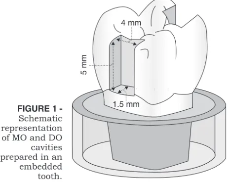
Twenty sound human molars were extracted for orthodontic reasons and stored in distilled water at 37°C for a maximum post-extraction period of 90 days19
. Previously to cavity preparations, tooth apices were sealed using Super BonderÒ
gel (Locti-te, Itapevi, Brazil). After that, root portions were coated with 3 coats of nail polish (Nutra NailÒ
embedded in PVC tubes (Tigre, São Paulo, Brazil) using Epoxide polyester resin (Buhler, Lake Bluff, IL, USA), activated using butanox M 50 catalyst, code BUTA/100 (RedeleaseÒ
, S.S. White, Rio de Ja-neiro, Brazil), maintaining the cement/enamel junction at a distance of 2 mm from the embedding resin (Figure 1).
Two class II preparations (MO/DO) were cut in each tooth with the following dimensions: 4 mm of width, 5 mm of height and 1.5 mm of depth to-wards the pulp. Preparations were made using number 3100 diamond burs (KG Sorensen, Barue-ri, Brazil) and a high-speed handpiece under cons-tant water cooling. Cavity standardization was ob-tained by using a special aligning device that allowed controlled movements on the three spatial directions6. Burs were replaced at each five cavity
preparations6,23
.
After that, cavities were cleaned using pumice paste (S.S. White, Rio de Janeiro, Brazil) in a Rob-son brush (KG Sorensen, Barueri, Brazil). MO pre-parations were restored using ABF experimental (Kuraray Medical Inc., Osaka, Japan) and DO were restored using Single Bond (3M ESPE, St. Paul, USA). All materials were applied strictly following the manufacturers’ instructions. Restorations were made using Z250 composite resin shade C3 (3M ESPE, St. Paul, USA) placed in increments of approximately 1 mm, so that each one was light-cured for 20 s using an Optilux 500 light-curing unit (Demetron/Kerr, Danbury, CT, USA), whose readings were maintained in 750 mW/cm2
. After storage in distilled water at 37°C for 24 hours, all restorations were finished using polyes-ter grinding papers (3M ESPE, St. Paul, USA).
For group 1 (n = 10)20
, teeth were immediately submitted to thermal cycling in 700 cycles betwe-en water baths held at 5° and 55°C, 1 min dwell time and 3 s of interval between each bath15,19. For
group 2, teeth were again stored in distilled water at 37°C for a period of 3 months. After that, ther-mal cycling was carried out as described for group 1.
Upon completion, tooth surfaces were coated with 3 coats of nail polish, except for the restorati-on and a 1 mm rim of tooth structure. Specimens were immersed in 0.5% methylene blue solution (Fórmula & Ação, São Paulo, Brazil) for 4 hours (neutral pH)19
. Teeth were thoroughly washed un-der tap water and sectioned longitudinally using a diamond disc mounted on a Labcut 1010 machine (Extec Corp., Enfield, USA) under water cooling, so that 3 sections with approximately 1 mm of thick-ness were obtained for each restoration18,23.
Secti-ons were embedded in PVC tubes with 0.5 mm of height using chemically-activated acrylic resin (JET ClássicoÒ
, Artigos Odontológicos Clássico, São Paulo, Brazil) in a 1:1 ratio, to protect and faci-litate manipulation during microleakage assess-ments.
Visual analysis was performed by 3 previously calibrated examiners under a stereomicroscope with 25 X magnification (Baucsh & Lomb, Roches-ter, NY, USA) according to the microleakage scores demonstrated in Figure 215. After scores were
attri-buted, results were analyzed regarding examiner agreement.
The section that presented the highest microle-akage score determined the final result, and was also submitted to morphometric analysis. For this study, the ImageLab 200027
image segmentation program was employed to assess dye penetration. This system consisted of a Digital Sony camera (Hyper HAD, Sony Corporation, Tokio, Japan)
FIGURE 1
-Schematic representation of MO and DO cavities prepared in an embedded tooth.
adapted to a Citoval 2 stereomicroscope (Carl Zeiss Jena, Germany), which was connected to a Pavili-on microcomputer (HP, Palo Alto, California, USA) equipped with a digitalizer plate and the proces-sing software ImageLab 2000 (Canborough, Wel-landport, Ontario, Canada). After image scanning, linear measurements in mm could be obtained and recorded for each section.
Firstly, to verify if there was a correlation betwe-en methods of evaluation, in which parametric and non-parametric data should be obtained, scores were turned into millimeters proportionally to the cavity size. For example: a score 1 attributed to a cavity whose axial wall was 1.8 mm deep was tur-ned into 0.6 mm (one third of 1.8 mm); scores 0 were maintained as 0 mm, despite the cavity size. Results were submitted to Pearson’s regression analysis. Results regarding adhesive systems and storage periods were also submitted to ANOVA and Tukey’s test.
RESULTS
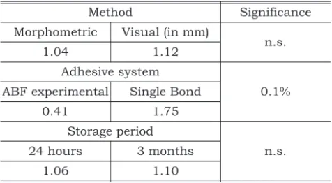
Regarding the two methods of evaluation, Pear-son’s regression analysis presented highly signifi-cant correlation (r = 0.978, p < 0.001) between vi-sual and morphometric methods (Graph 1), thus emphasizing that both can be reliably used and produce equivalent results.
ANOVA demonstrated no statistically signifi-cant difference between the two studied methods (p > 0.05). Regarding adhesive systems, the analy-sis demonstrated statistically significant differen-ces (p < 0.001), indicating that the ABF experimen-tal presented less microleakage when compared to the Single Bond adhesive system, either after the
immediate (24 hours) or the 3-month storage peri-od in distilled water (Table 1).
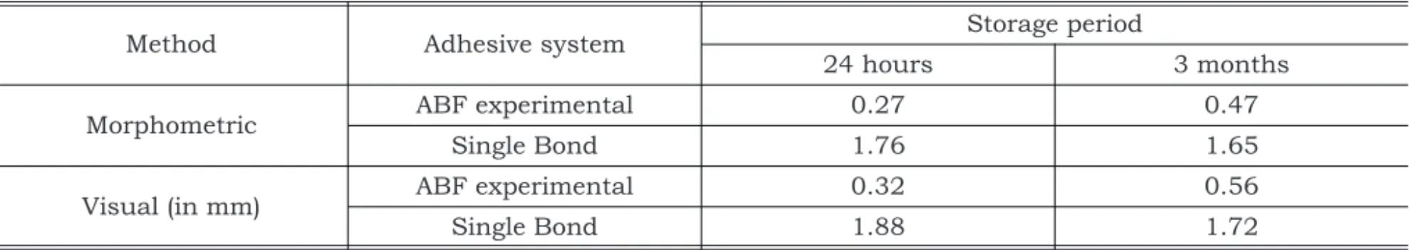
The storage period did not significantly influen-ce the results of microleakage (p > 0.05), showing that the storage period in distilled water for 3 months had no influence on the performance of the two tested adhesive systems (Table 2).
DISCUSSION
Pearson’s regression analysis demonstrated a highly significant correlation between the tested methods of evaluation. Moreover, it is possible to observe that the reliability interval at the level of 5% varied from 0.0023 to 0.1489, showing that va-lues obtained using both methods were very cohe-rent. B angular coefficient (5%) went from 0.9339 to 1.0745, and hence included value 1. This coeffi-cient indicates that a certain increase in the mor-phometric value corresponds to the same value ob-tained using score data, when we consider the cavity size.
Discrepancies were relatively higher when mi-croleakage values were very low to be visually de-tected and generally corresponded to score 0 (no dye penetration). In some of these cases, the mor-phometric method detected a small dye penetra-tion, which led to these discrepancies. Neverthe-less, statistical analysis proved that there is a highly significant correlation between the two methods. Therefore, when equipment is available, the morphometric analysis can be used with great reliability, although the visual method revealed to be practical and efficient to evaluate microleakage, also showing that the subjectivity attributed to it does not influence the results.
TABLE 1 -Mean values of microleakage (in mm) obtained for each experimental condition.
Method Significance
Morphometric Visual (in mm)
n.s.
1.04 1.12
Adhesive system
0.1% ABF experimental Single Bond
0.41 1.75
Storage period
n.s.
24 hours 3 months
1.06 1.10
n.s.: not significant.
Regarding adhesive systems, the ABF experi-mental (Table 1) showed a superior sealing ability than did Single Bond. Probably, bonding penetra-tion occurred at the same depth as the deminerali-zation obtained using acidic monomers11,25
, so that the risk of incomplete impregnation with the bon-ding resin does not exist, thus guarantying the marginal sealing observed in this study. As in pre-vious studies11
, Single Bond presented higher mi-croleakage when compared to the self-etching pri-mer systems, possibly as a result of incomplete monomer penetration in the demineralized dentin zone obtained following phosphoric acid et-ching3,11,12,27
.
The greater microleakage obtained using Single Bond can also be explained by the fact that its bon-ding mechanism is based on the total removal of the smear layer following etching with phosphoric acid, which leaves an exposed collagen network available for primer impregnation. Primer applica-tion must be made after slight drying, leaving den-tin sufficiently moist to prevent collapse of this col-lagen network22,25,27. Due to the difficulties in
verifying the ideal moisture to be left in dentin17,22,27,
marginal integrity could be compromised, resul-ting in higher levels of microleakage.
The idea of adhesives containing acidic mono-mers is very interesting as, in addition to obtaining excellent results, it renders the technique simpler and less sensitive. Clinically, the reduction of such a critical operative step as etching with phosphoric acid, which requires careful washing and drying, minimizes the possibility of post-operative sensiti-vity, as marginal sealing is improved1,3,11-13,27.
Once marginal sealing is obtained, it is expec-ted that it will be maintained for many years, gua-rantying the longevity of a restoration. However, the bonding interface is constantly in contact with saliva and consequently subjected to degradation in the oral cavity, especially in cases of incomplete monomer penetration through the demineralized
dentin9,11,21. Moreover, if incomplete polymerization
of resin monomers occurs, they can be washed out, leaving spaces in the polymeric matrix, which becomes soaked with water, and thus facilitates degradation2,7,9,11.
Some studies have already reported that bond strength decreases following long periods of stora-ge in water2,7,16
, and that the collagen layer presents higher porosity when analyzed using scanning electron microscopy, even after 6 months21
. Consi-dering this possible degradation, it is important to evaluate the performance of restorative materials after different storage periods. As in other stud-ies11,16, the period evaluated in the present study (3
months) was not enough to allow bonding degra-dation and consequently higher microleakage (Ta-ble 2). Li et al.11
(2001) found differences on micro-leakage results only after a storage period of 6 months. Hence, it is evident that more studies are necessary with longer storage periods to evaluate the longevity of these materials.
CONCLUSIONS
Based on the results obtained in this study, the following conclusions can be drawn:
1. The visual method of evaluation using calibra-ted examiners and data transformation allowed us to obtain the same conclusions as with the morphometric method, which is technologically more sophisticated.
2. The ABF experimental self-etching primer ad-hesive system presented significantly lower va-lues of microleakage than did the Single Bond one-bottle system, either in the immediate as-sessment (24 hours) or after 3 months of stora-ge in water.
3. The storage period of 3 months produced micro-leakage results similar to those produced after the 24 hour period, thus pointing out that the former is not enough to allow degradation of the bonding interface.
TABLE 2 -Mean values of microleakage (in mm) corresponding to the method versus adhesive system versus storage period interaction.
Method Adhesive system Storage period
24 hours 3 months
Morphometric ABF experimental 0.27 0.47
Single Bond 1.76 1.65
Visual (in mm) ABF experimental 0.32 0.56
ACKNOWLEDGEMENTS
The authors gratefully acknowledge Paulo San-tos for the preliminary drawings, and KG Soren-sen, Kuraray Medical Inc., Japan and 3M ESPE for the supply of materials. We also wish to thank the Laboratory of Informatics for the Development of Dentistry (Laboratório de Informática para
Desen-volvimento da Odontologia - LIDO), School of Den-tistry, University of São Paulo, and the Nucleus for Supporting Research in Dental Materials (Núcleo de Apoio à Pesquisa em Materiais Dentários -NAPEM).
REFERENCES
1. Bouillaguet S, Gysi P, Wataha JC, Ciucchi B, Cattani M, Godin C, et al. Bond strength of composite to dentin using conventional, one-step, and self-etching adhesive systems. J Dent 2001;29:55-61.
2. Burrow MF, Satoh M, Tagami J. Dentin bonding durability after three years using a dentin bonding agent with and without priming. Dent Mater 1996;12:302-7.
3. Christensen GJ. Self-etching primers are here. J Am Dent Assoc 2001;132:1041-3.
4. Déjou J, Sindres V, Camps J. Influence of criteria on the re-sults of in vitro evaluation of microleakage. Dent Mater 1996;12:342-49.
5. Ferrari M. Sealing ability of last generation of adhesive-re-storative materials placed on vital teeth. Trans Acad Dent Mater 1998;12:75-102.
6. Formolo E, Sartori A, Demarco F. Infiltração marginal em cavidades de classe V com o uso de diferentes materiais adesivos. RPG Rev Pos Grad 2001;8:306-12.
7. Gwinnett AJ, Yu S. Effect of long-term water storage on dentin bonding. Am J Dent 1995;8:109-11.
8. Hilton TJ. Can modern restorative procedures and materi-als reliably seal cavities?In vitroinvestigations. Trans Acad Dent Mater 1998;12:21-71.
9. Kitasako Y, Burrow MF, Nikaido T, Tagami J.Long-term tensile bond durability of two different 4-META containing resin cements to dentin. Dent Mater 2002;18:276-80. 10. Koibuchi H, Yasuda N, Nakabayashi N. Bonding to dentin
with a self-etching primer: the effect of smear layers. Dent Mater 2001;17:122-6.
11. Li HP, Burrow MF, Tyas MJ. The effect of long-term storage on nanoleakage. Oper Dent 2001;26:609-16.
12. Lucena-Martín C, González-Rodriguez MP, Ferrer-Luque CM, Robles-Gijón V, Navajas JM. Influence of time and thermocycling on marginal sealing of several dentin adhe-sive systems. Oper Dent 2001;26:550-5.
13. Manhart J, Chen HY, Mehl A, Weber K, Hickel R.Marginal quality and microleakage of adhesive class V restorations. J Dent 2001;29:123-30.
14. Milia E, Lallai MR, Garcia-Godoy F. In vivo effect of a self-etching primer on dentin. Am J Dent 1999;12:167-71. 15. Miranda Jr WG. Avaliaçãoin vitroda infiltração nas caixas
proximais de pré-molares humanos restaurados com dife-rentes adesivos universais e resinas compostas. [Tese de
Doutorado] São Paulo: Faculdade de Odontologia da USP; 1994.
16. Okuda M, Pereira PNR, Nakajima M, Tagami J. Relation-ship between nanoleakage and long-term durability of dentin bonds. Oper Dent 2001;26:482-90.
17. Pereira GDS, Paulillo LAMS, De Goes MF, Dias CTS. How wet should dentin be? Comparison of methods to remove excess water during moist bonding. J Adhes Dent 2001; 3:257-64.
18. Raskin A. Reliability of dye penetration for microleakage evaluation: influence of sections number. Trans Acad Dent Mater 1998;12:212.
19. Raskin A, D’Hoore W, Gonthier S, Degrange M, Déjou J.
Reliability ofin vitromicroleakage tests: a literature review. J Adhes Dent 2001;4:295-8.
20. Sano H, Takatsu T, Ciucchi, B, Horner JA, Matthews WG, Pashley DH.Nanoleakage: leakage within the hybrid layer. Oper Dent 1995;20:18-25.
21. Sano H, Toshikawa T, Pereira PNR, Kanemura N, Morigami M, Tagami J, et al. Long-term durability of dentin bonds made with a self-etching primer, in vivo. J Dent Res 1999;78:906-11.
22. Santini A, Mitchell S. Effect of wet and dry bonding tech-niques on marginal leakage. Am J Dent 1998;11:219-24. 23. Tapety CMC. Influência da interposição de bases na
infiltração marginal em cavidades classe II (MOD), res-tauradas com resinas compostas para dentes posteriores. [Dissertação de Mestrado] Bauru: Faculdade de Odon-tologia da USP; 2000.
24. Tay FR, Gwinnett AJ, Wei SH. The overwet phenomenon: a scanning electron microscopic study of the surface mois-ture in the acid-conditioned, resin-dentin interface. Am J Dent 1996;9:109-14.
25. Tay FR, Pashley DH. Aggressiveness of contemporary self-etching systems. I: Depth of penetration beyond dentin smear layers. Dent Mater 2001;17:296-308.
26. Ulson RCB. Avaliação da microinfiltração, utilizando di-versos sistemas adesivos, em restaurações de molares decíduos. [Dissertação de Mestrado] São Paulo: Faculdade de Odontologia da USP; 2000.
27. Unemori M, Matsuya Y, Akashi A, Goto Y, Akamine A.
Composite resin restoration and postoperative sensitivity: clinical follow-up in an undergraduate program. J Dent 2001;29:7-13.