Nautilus-shaped dynamic craniotomy: a new surgical
technique and preliminary results
Nautilus: craniotomia dinâmica – nova técnica cirúrgica e resultados preliminares
ABSTRACT
Introduction: Considering that craniosynostosis is a suture-related condition, the main challenge for its treatment is the fact that the brain is located in a closed compartment that does not have the required adaptability to accommodate its growth. The goal of treat ment is to restore stenotic suture adaptability and correct the compensatory cranial deformity. This paper proposes the combined use of spiral osteotomy with distraction osteogenesis by the use of distracting springs to remodel craniofacial defects caused by craniosynostosis. Methods: Between July 2010 and July 2012, 10 patients with cranio-synostosis were treated: 5 with oxycephaly, 3 with scaphocephaly, 1 with turricephaly, and 1 with trigonocephaly. The treatment consisted of the application of Lauritzen springs to correct the primary craniosynostosis defect in combination with a nautilus-shaped spiral craniotomy at the secondary deformation sites without dural detachment. Results:
Resolution of cranial deformity and remission of the clinical signs of intracranial hyper-tension were observed. None of the patients had complications such as cerebrospinal fluid fistula, local infection, seroma, or hematoma. Conclusions: The combined use of spiral osteotomy with spring-mediated distraction or contraction enables active reshaping of the skull and facilitates accommodation of the brain by the cranial cavity. (clinicaltrials.gov identifier: NCT01672619)
Keywords: Craniosynostoses. Osteogenesis, distraction. Craniotomy.
RESUMO
Introdução: Considerando-se que as craniossinostoses são afecções basicamente suturais, o fato de o cérebro estar aprisionado em um compartimento fechado, que não possui a
com-placência necessária para acompanhar seu crescimento, se constitui no desaio principal de
seu tratamento. O objetivo do tratamento é restabelecer a complacência da sutura estenótica e corrigir a deformidade craniana compensatória. Este trabalho propõe a associação de osteotomia helicoide à distração osteogênica proporcionada pelo uso das molas distratoras para remodelar defeitos craniofaciais causados por craniossinostoses. Método: Entre julho de 2010 e julho de 2012, foram tratados 10 pacientes portadores de craniossinostoses, sendo 5 oxicefalias, 3 escafocefalias, 1 turricefalia e 1 trigonocefalia. O tratamento consistiu na aplicação de molas de Lauritzen, para corrigir a deformidade primária da craniossinostose, com a associação de craniotomia helicoide em forma de Nautilus nos sítios de deformação secundária do crânio, sem descolamento dural. Resultados: Foi observada resolução da deformidade craniana e remissão dos sinais clínicos de hipertensão intracraniana. Nenhum This study was performed at the
Beneicência Portuguesa Hospital of São Paulo, São Paulo, SP, Brazil. Submitted to SGP (Sistema de Gestão de Publicações/Manager Publications System) of RBCP (Revista Brasileira de Cirurgia Plástica/Brazilian Journal of Plastic Surgery).
Article received: November 1, 2012 Article accepted: January 20, 2013
1. Full Member of the Sociedade Brasileira de Cirurgia Plástica/Brazilian Society of Plastic Surgery (SBCP), PhD in General Surgery by the Faculty of Medical Sciences of Santa Casa de São Paulo, Coordinator of the Advanced Plastic Surgery Center (NPA), São Paulo, SP, Brazil.
2. Full Member of the SBCP, Assistant Plastic Surgeon at the NPA, São Paulo, SP, Brazil.
3. Full Member of the SBCP, MSc in Plastic Surgery by the University of São Paulo, Assistant Plastic Surgeon at the NPA, São Paulo, SP, Brazil 4. Associate Member of the SBCP, Assistant Plastic Surgeon at the NPA, São Paulo, SP, Brazil.
5. Full Member of the Brazilian Society of Neurosurgery, MSc and PhD in Neurosurgery by the Federal University of São Paulo, Pediatric Neurosurgeon, Coordinator of the Center for Pediatric Neurosurgery (CENEPE), São Paulo, SP, Brazil.
Vera Lucia Nocchi cardim1 aLessaNdrados saNtos
siLVa2 roLf Lucas saLomoNs2 rodrigo fariado VaLLe
paciente apresentou complicações, como fístula liquórica, infecção local, seroma ou he-matoma. Conclusões: A associação da osteotomia helicoide com a distração ou contração promovida pelas molas permitiu remodelar ativamente o crânio, facilitando a acomodação
do conteúdo cerebral no continente craniano. Clinical trials.gov Identiier: NCT01672619. Descritores: Craniossinostoses. Osteogênese por distração. Craniotomia.
INTRODUCTION
Craniosynostoses, known and described since the times
of ancient Greece, were so clearly studied and classiied by
Virchow that his concepts remain as the basis of all unders-tanding of this disease to this day1,2.
Since the irst attempts to treat skulls deformed by cranio -synostoses, restoration of the stenotic line expandability (pri mary defect) and remodeling the areas of compensatory deformation (secondary defect) have been the main concerns. In the evolutionary history of the treatment of craniosy -nostoses, we reviewed extensive and morbid craniectomies and suturectomies that possibly involved the interposition of materials or caustic substances in an attempt to inhibit the recurrence of early suture closure3,4 through distraction osteo-genesis using external distractors5 and skullcap re mo deling1. Such remodeling, the preferred practice still to day, follows various types of osteotomies, but all involve craniotomy using dural detachment, back table remodeling of the osteotomized
cap, and its return as a graft ixed with varied materials.
However, these procedures have high morbidity, with the possibility of several complications related to dead space between the dura mater and cap grafts6.Furthermore, static remodeling causes cavity expansion that does not always adequately meet the actual requirement. Therefore, Lauritzen et al.7 proposed dynamic cranial remodeling using expan der springs in 1998. They applied the principles of osteotomy and interposition of metal springs, without detaching the dura, by inserting expansive forces on the stenotic lines that are distributed throughout the dura around the whole brain
content/cavity to reshape the skull7.
Because of the ideal characteristics of the spiral shape for modeling convex surfaces, Salyer and Bardach8 proposed its use in modeling osteotomies in scaphocephaly. They advo-cated extensive posterior biparietal craniotomy in which they performed a back-table spiral osteotomy of the cap; after being remodeled, the cap is attached to the skull as a graft. Tullous et al.9 and Solís-Salgado and Anaya-Jara10 advocated the use of spiral osteotomies for scaphocephaly treatment but performed them bilaterally to the sagittal line. Back-table remodeling was performed for the 2 large craniotomy plates through spiral osteotomies. The expanded form was secured with absorbable plates, and the pieces were returned to the skull as a graft.
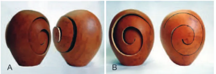
As demonstrated by the authors cited above, the curved shape of the spiral adapts perfectly to the remodeling of a surface curve like the skullcap. However, in the absence of fixation, the spiral shape of the osteotomy can turn bone into a spring that is able to expand or contract according to the direction of the force it receives (Figure 1). This form of osteotomy was therefore chosen by the authors to induce compliance in the areas of bone with secondary defects, which must indirectly compress or expand during the process of dynamic remodeling of the primary defect using springs.
The objective of this study is to demonstrate the use -fulness of nautilus-shaped spiral osteotomy for inducing indirect remodeling of skulls deformed by craniosynostoses associated with dynamic remodeling using implantable springs.
METHODS
Patients treated at the Plastic Surgery Department of the Advanced Plastic Surgery Center from July 2010 to July
2012 with a diagnosis of craniosynostosis were eligible to participate in this prospective study.
The inclusion criteria for the study were presence of residual cranial deformity after previous treatment, complex (involving more than one suture) or syndromic craniosynos-tosis, and trigonocephaly. Furthermore, patients with cranial deformities that required complex surgery involving cranial remodeling were included. The study excluded patients with simple craniosynostosis or for which treatment could be
Figure 1 - Representation of the shape of the osteotomy and its possibilities for expansion (A) or contraction (B) using the
fruit of Lagenaria siceraria (“gourd”) as a model of rigid structure.
achieved through suturectomies, expansion with springs, or video-assisted minimally invasive surgery.
Ten cases of craniosynostosis were treated, including 5 of oxycephaly, 3 of scaphocephaly, 1 of turricephaly, and 1 of trigonocephaly. The patients were aged 5-160 months (Table 1). Each patient underwent preoperative clinical, laboratory, and imaging testing. All procedures were per -formed with the permission and consent of the patients’ pa rents or guardians. The study protocol was approved by the Ethics and Re search Council of the Hospital da Real e
Benmérita Associa ção Portuguesa of Sao Paulo (Protocol number 777-12 dated March 30, 2012), the hospital where
the surgeries were performed.
All patients underwent surgery under general anesthesia, received intraoperative blood transfusion to replace blood loss (decreases in hemoglobin of 3-5 points), mean blood pressure control, and immediate postoperative follow-up in
the intensive care unit (ICU). The surgeries were performed
by collaborating pediatric neurosurgery and craniofacial surgery teams.
As described by Lauritzen et al.7, the procedure involved
2 surgical steps: irst, spring placement for nautilus-shaped
dynamic osteotomy; and second, spring removal performed 6-12 months after the initial procedure. The timing of spring removal was determined according to the degree of
ossi-ication of the osteotomy and evaluated by 3-dimensional
tomography of the skull. If consolidated osteotomies were observed, the springs were removed; presence of open osteo-tomies signaled ongoing remodeling and resulted in delay of spring removal. Spring removal was not delayed only in the presence of complications such as infection or exposure of the spring through the skin or scalp.
The cranial shape was evaluated postoperatively by 4 in dependent surgeons who used the following scale to grade
the results: insuficient, no attenuation of the pre-operative
deformity existed; partial, correction did occur but did not include all features of the deformity; and appropriate, total remission of the cranial deformity was obtained.
Surgical Technique
A broken-line coronal incision was made in the temporal
regions with subperiosteal detachment of the scalp laps.
The springs were used in osteotomies performed to cor -rect the primary defect (caused by sutural stenosis), whereas nautilus-shaped spiral osteotomies were used in the areas with secondary defects (caused by indirect remodeling).
In areas containing a compensatory hump, a nautilussha ped osteotomy was performed to enable collapsing, and the osteotomy traces were widened with a wear drill to decrease surface area. Gains in intracranial space were induced by the expansion springs applied according to craniosynostosis type.
In areas of compensatory fossa (lattening), a
nautilussha ped osteotomy was performed for expansion, and a cen -trifugal bevel was used to induce progressive increase in surface area. The combined action of the contracting springs to reduce excessively elongated regions was responsible for the ejection of the contents toward the area of fragility crea -ted by the expander.
If the area to be expanded included the insertion of the greater wing of the sphenoid bone, this was released. A cra niotomy hole was created using a wearing drill after the zygomaticofrontal suture. This enabled osteotomy of the wing of the sphenoid bone using a curved chisel without re -quiring craniotomy cap detachment and with minimal dural detachment.
All osteotomies were performed with minimal dural de -tachment without the removal of any skullcap segment for back-table modeling.
RESULTS
In the irst 12 postoperative hours, all patients had mo
-derate pain that was controlled by non-opioid analgesics. Edema of the operated region, including major eyelid expan-sion and chemosis, peaked on the second day postoperatively, and full resolution was seen in 8-15 days.
Length of ICU stay was 13 days, and the overall hospita
-lization time was 3-6 days. In the second stage of the proce-dure, the patients were hospitalized for 1 day only, and the
use of an ICU bed or blood transfusion was unnecessary.
All patients had an uneventful postoperative period, and those who showed neuropsychomotor impairment related to intracranial hypertension had signs suggestive of clinical
Table 1 – Characteristics of the study population.
Patient Sex Age Type of
craniosynostosis Syndrome
1 M 67 months Scaphocephaly No
2 M 69 months Oxycephaly Apert
3 F 87 months Oxycephaly No
4 F 15 months Oxycephaly Crouzon
5 M 72 months Scaphocephaly No
6 M 103 months Oxycephaly Crouzon
7 M 71 months Scaphocephaly Crouzon
8 F 160 months Turricephaly No
9 M 5 months Oxycephaly No
10 M 8 months Trigonocephaly No
improvement (parental reports of improvement in concen-tration, gain of motor skills, and remission of headache). No
patients experienced a CSF istula, seroma, hematoma, signs
of infection, spring externalization, or other complications. The desired cranial remodeling was obtained in all cases (Figures 2-8). The 4 surgeons who evaluated the postopera-tive cranial shape considered the results partial in 40% of cases and appropriate in 60%.
DISCUSSION
Realizing the compensatory effect of skull growth in the presence of stenosis, Virchow postulated that the direction of growth of the cranial vault is always parallel to the axis of the compromised suture by compensatory expansion of sutures perpendicular to the stenosis1.2. Therefore, deformation of the skull occurs because of not only growth cessation in the area of stenosis but also compensatory growth of the unaffected
sutures, which do not always fulill the function of providing suficient intracranial space for undamaged brain growth. This indirect compensatory remodeling determines the inal
shape of skulls with stenosis.
These concepts by Virchow led us to conclude that the skulls affected by craniosynostosis have 2 types of defects: primary, the synostosis itself; and secondary, the deforma -tion or compensatory remodeling of the skull. Therefore, there are 2 methods for classifying craniosynostoses: one that refers to the affected suture and pointing toward the primary defect, such as sagittal craniosynostosis and coronal cra
-niosynostosis; and the other referring to the inal skull shape,
i.e., pointing toward the secondary defect (scaphocephaly or turricephaly).
Because craniosynostosis is a suture-related condition, the main challenge in its treatment is the fact that the brain is located inside a closed compartment that does not have the required adaptability to accompany brain growth. All techniques proposed and performed since the dawn of medical science to this day have aimed to provide space for
A B C
Figure 2 - Patient with turricephaly. In A, lateral preoperative computed tomography of the skull. In B, intraoperative detail. In C, computed tomography of the skull performed in the
seventh postoperative month revealed remission of the “beaten silver sign.”
A
C
E
B
D
F
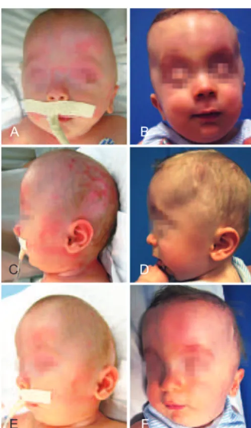
Figure 3 - Patient with oxycephaly. In A, C, and E, preoperative appearance on frontal, lateral, and left oblique views. In B, D, and F, appearance in the ifth postoperative month:
frontal view in frontal, lateral, and left oblique views.
A
C
B
D
Figure 4 - Oxycephaly. In A and C, preoperative computed tomography (CT) of the skull. In B and D, CT of the skull in the
ifth postoperative month revealing remission of the ingerprint-like patterns observed
A
D
G
B
E
H
C
F
I
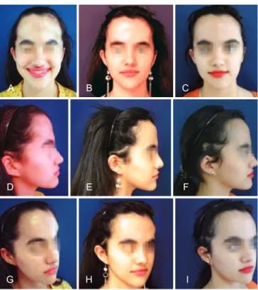
Figure 5 - Patient with turricephaly. In A, D,G, preoperative appearance from the frontal, right lateral, and right oblique views.
In B, E, H, appearance in the eighth postoperative month from the frontal, lateral right, and right oblique views. In C, F, and I,
appearance in the irst month after removal of springs in the frontal, right lateral, and right oblique views.
A
C
B
D
Figure 6 - Turricephaly. In A and C, preoperative computed tomography (CT) of the skull. In B and D, CT of the
skull in the eighth postoperative month revealing remission of ingerlike prints observed on
preoperative CT.
A
A B
Figure 7 - Scaphocephaly. In A, preoperative 3-dimensional skull computed tomography (CT), lateral view. In B, postoperative
3-dimensional skull CT.
A B
Figure 8 - Scaphocephaly. In A and B, preoperative computed tomography of the skull of the same patient as in Figure 7.
Axial section at the level of the lateral ventricles showing enlargement of the lateral-lateral diameter
and anterior posterior shortening.
the expansion of the brain in order to prevent or minimize compression damage11.
The consensus is that treatment should be performed as early as possible because the longer it occurs after the
irst year of life, the greater are the neurological sequelae
observed12-14. It is believed that after 2 years of age, the
brain no longer needs sutural expansion to inish its growth
because it can be achieved by simple cap remodeling alone. However, this hypothesis is not supported by clinical
indings: a group of children with craniosynostosis who were treated for decompression within the irst year of life
unexpanding and ossiied skull cap that does not respond to indirect or secondary remodeling in cranial areas not affected by surgical remodeling.
This necessity leads to another very common problem in craniofacial surgery, i.e., the progressive stabilization of dural elasticity with age. The longer surgery is delayed after
the irst 2 years of life, the more dificult it is for brain tissue
and the dural sheath to occupy the space offered, leading to the maintenance of dead space, seroma, infection, osteomye-litis, and loss of the cap’s remodeled area15.
The treatment of older skulls (after 2 years of age) through distraction osteogenesis with expansion springs allows for dynamic and progressive remodeling since it maintains adherence of the dura to the moving cap and the cap need not be removed to be reused as a graft. The bone remains vascu-larized and inserted, drastically decreasing trans- and
posto-perative morbidity. Moreover, expansion is not controlled
by external forces but rather by spring elasticity, and the need for brain expansion causes cessation of the expansion
to be modulated by the cap ossiication and the homeostasis
between pressure and intracranial space. Offering the tissues the opportunity to occupy the required space is a safer and more precise technique than just creating a pre-designed and static gain in space as in conventional remodeling16.
A nautilus-shaped spiral osteotomy in the areas of secon-dary defect, where an indirect response to compression or spring-induced distraction is sought, produces great plasti-city in the bone of the cap and keeps it adhered to the dura mater because there is no detachment or removal of the cap from its position. The bone spring created by spiral osteo-tomy allows the cap to respond to expansion within skulls
that were being contracted by springs and with lattening
(retraction) in skulls that were being dynamically expanded
with springs (Figure 1). Maintaining continuity between the
cap and the dura guarantees the absence of dead space. This extends the indication of cranial remodeling for any age, even in adults, where dural inextensibility does not usually allow for detachment and increases to the cavity without the burden of collections of dead space.
The minimally invasive feature of the procedure described here provides greater safety and lower morbidity, opening a wide range of treatment options for craniosynostosis. The only drawback is the need for 2 surgical procedures (one for spring placement and another for spring removal), even considering that the former is much less invasive than conventional surgery and that the latter is of very short dura-tion and can be performed in an outpatient setting.
The remission of indirect signs of cranial hypertension,
such as the disappearance of ingerlike impressions of the
skull that are present on CT intraoperatively and the impro-vement in symptoms (headache), suggests that osteotomy effectively reduced intracranial pressure. However, as this
variable was not evaluated preoperatively (for example, determination of papilledema), the extension of this study could be a future perspective.
The limitations of the short postoperative follow-up and the small number of patients in the present study are offset by the novelty of the nautilus-shaped dynamic osteotomy. The spiral shape of these osteotomies adds elasticity to the bone and transforms the cap itself into a spring to enable global reshaping of the skull with low morbidity, thus enabling the use of dynamic expansion at any age.
Spring forces act and spread throughout the dura mater
to interfere with the content/cavity ratio of the brain. There -fore, spiral osteotomies provide indirect skull remodeling by inducing retraction of the secondary humps associated with the use of expanding springs and inducing expansion of the
zones of lattening associated with the use of contracting
springs (Figures 7 and 8).
CONCLUSIONS
We successfully induced indirect remodeling of synos-totic skulls that were subjected to dynamic remodeling with implantable springs using nautilus-shaped spiral osteotomies.
REFERENCES
1. Persing JA, Jane JA, Shaffrey M. Virchow and the pathogenesis of cra -niosynostosis: a translation of his original work. Plast Reconstr Surg. 1989;83(4):738-42.
2. Mark ES, Persing JA, Christopher I, Shaffrey JAJ. Craniosynostosis. In: Rengachary SS, Wilkins RH, eds. Principles of neurosurgery. 2nd ed. London: Wolfe; 2004. p.8.2-8.15.
3. Guimarães-Ferreira J, Miguéns J, Lauritzen C. Advances in craniosy -nostosis research and management. Adv Tech Stand Neurosurg. 2004; 29:23-83.
4. Greensmith AL, Holmes AD, Lo P, Maxiner W, Heggie AA, Meara JG. Complete correction of severe scaphocephaly: the Melbourne method of total vault remodeling. Plast Reconstr Surg. 2008;121(4): 1300-10.
5. Cardim VLN, Dornelles RFV. Craniossinostoses. In: Carreirão S, Car -neiro Jr. LVF, eds. Cirurgia plástica para a formação do especialista. São Paulo: Atheneu; 2011. p.297-308.
6. Ortiz-Monasterio F, del Campo AF, Carrillo A. Advancement of the orbits and the midface in one piece, combined with frontal repositio-ning, for the correction of Crouzon’s deformities. Plast Reconstr Surg. 1978;61(4):507-16.
7. Lauritzen C, Sugawara Y, Kocabalkan O, Olsson R. Spring mediated dynamic craniofacial reshaping. Case report. Scand J Plast Reconstr Surg Hand Surg. 1998;32(3):331-8.
8. Salyer KE, Bardach J. Salyer & Bardach’s atlas of craniofacial & cleft surgery. Philadelphia: Lippincott-Raven; 1999.
9. Tullous MW, Henry MN, Wang PTH, Vollmer DG, Auber AE, Mancuso PA. Multiple-revolution spiral osteotomy for cranial reconstruction. Technical note. J Neurosurg. 2001;94(4):671-6.
11. Panchal J, Uttchin V. Management of craniosynostosis. Plast Reconstr Surg. 2003;111(6):2032-48.
12. Renier D. Intracranial pressure in craniosynostosis pre and postoperative recordings: correlation with functional results. In: Persing JA, Edger -ton MT, Jane JA, eds. Scientiic foundation and surgical treatment of craniosynostosis. Baltimore: Williams & Wilkins; 1989. p.263-9. 13. Arnaud E, Meneses P, Lajeunie E, Thorne JA, Marchac D, Renier D.
Postoperative mental and morphological outcome for nonsyndromic
brachycephaly. Plast Reconstr Surg. 2002;110(1):6-12.
14. Cohen MMJ. Craniosynostosis: diagnosis, evaluation and management. New York: Raven Press; 1986.
15. David DJ, Sheen R. Surgical correction of Crouzon syndrome. Plast Reconstr Surg. 1990;85(3):344-54.
16. Cardim VLN, Silva AS, Salomons RL, Dornelles RFV, Lima e Silva A, Blom JOS. Remodelagem de crânios maduros utilizando molas expansoras.Rev Bras Cir Craniomaxilofac. 2012;15(2):57-63.
Correspondence to Vera Lucia Nocchi Cardim