O
RIGINALA
RTICLE Revista Brasileira de FisioterapiaEvaluation of a neuropathic ulcers prevention
program for patients with diabetes
Avaliação de um programa para prevenção de úlceras neuropáticas em
portadores de diabetes
Lígia L. Cisneros
Abstract
Background:Neuropathic foot ulcers are among the major health problems faced by patients with diabetes mellitus. Objective: To evaluate the preventive efficacy of a therapeutic education and protective footwear program in the incidence and recurrence of neuropathic ulcers due to diabetes. Methods: Fifty-three patients with diabetes and neuropathy from a public healthcare unit in Porto Alegre, Rio Grande do Sul, took part in a clinical trial for two years. The participants were randomly allocated to an intervention group (n=30) or a control group (n=23). Therapeutic education was provided in group sessions, and protective footwear was supplied in accordance with individual prescriptions. The nonparametric Mann-Whitney test was used to determine differences in incidence and recurrence of ulceration between the groups. Life-table analysis and the Kaplan-Meier method were used to measure the duration of ulcer-free survival. Results: In the intervention group, the ulcer incidence rate was 38.1% compared to 51.1% in the control group. Among the participants who presented ulcers, 83% were in the control group and 16.7% in the intervention group. After one year, the participants in the intervention group had a 75% chance of being ulcer-free, compared with 61% in the control group, and these percentages reduced to 60% and 52% respectively after two years. There was a tendency toward shorter survival among the control group participants. Conclusion:Although the proposed program lowered recurrence rates and increased the duration of ulcer-free survival, it was unable to prevent occurrence and recurrence of neuropathic ulcers due to diabetes.
Identification number in the Australian New Zealand Clinical Trials Registry ACTRN 12609000693224
Key words: diabetic foot; primary prevention; health education; shoes.
Resumo
Contextualização:Úlceras neuropáticas nos pés são um dos grandes problemas de saúde enfrentados por portadores de diabetes
mellitus. Objetivo: Avaliar a eficácia preventiva de programa de educação terapêutica e de calçados para proteção dos pés quanto à incidência e recorrência de úlceras neuropáticas por diabetes. Métodos: Um total de 53 pacientes de uma unidade de saúde pública de Porto Alegre/RS, portadores de diabetes e neuropatia, participaram de um ensaio clínico durante dois anos. Os sujeitos foram alocados aleatoriamente em grupo de intervenção (GI) (n=30) ou controle (GC) (n=23). A educação terapêutica foi realizada em grupo, e o calçado para proteção fornecido conforme prescrição individual. Utilizou-se o teste não paramétrico de Mann Whitney para determinar a diferença de incidência e recorrência de ulceração entre os grupos. A análise da tábua de vida e o método de Kaplan-Meier foram usados para medir o tempo de sobrevida sem úlcera. Resultados: A incidência de lesão no GI foi de 38,1% versus 57,1% no GC. Dos sujeitos que apresentaram úlcera, 83% pertenciam ao GC e 16,7% ao GI. Em um ano, os participantes do GI o mostraram 75% de probabilidade de se encontrarem sem lesão, contra 61% do GC, reduzindo para 60% e 52%, respectivamente, em dois anos. Há uma tendência de menor sobrevida em participantes do GC. Conclusão: Embora com índices menores de recorrência e maior sobrevida sem lesão, o programa proposto não foi capaz de prevenir a ocorrência e recorrência de úlceras neuropáticas por diabetes.
Número de identificação de Registro de Ensaios Clínicos (Australian New Zealand Clinical Trials Registry): ACTRN12609000693224
Palavras-chave: pé diabético; prevenção primária; educação em saúde; sapatos.
Received: 31/07/2008 – Revised: 17/12/2008 – Accepted: 30/06/2009
1 Department of Physical Therapy, Universidade Federal de Minas Gerais (UFMG), Belo Horizonte (MG), Brazil
Correspondence to: Ligia de Loiola Cisneros ,UFMG, Departamento de Fisioterapia Escola de Educação Física, Fisioterapia e Terapia Ocupacional, Avenida Antônio Carlos, 6.627 Pampulha CEP 31270-901, Belo Horizonte (MG), Brazil, e-mail: [email protected]
32
Introduction
Diabetes mellitus (DM) is a metabolic disorder of multiple etiologies characterized by chronic hyperglycemia resulting from impaired production and/or use of insulin. he disease can be classiied into two major groups: type 1 diabetes (au-toimmune or idiopathic) and type 2 diabetes, which is charac-terized by a defect in insulin secretion and action1. A serious
health problem in diabetic patients is foot ulcers. An otherwise simple lesion can lead to functional losses2,3 and culminate in
loss of the limb or even death4. Several factors are involved in
the development of foot ulcers in diabetic patients: neuropathy, peripheral vascular disease, limitation of joint motion, trophic skin disorders and abnormal distribution of mechanical forces in the feet5-7. Among them, the most important etiologic factor
is diabetic peripheral neuropathy5,8-10.
he International Consensus on the Diabetic Foot rein-forced reports of several studies on the amputation of diabet-ics by recommending multi-professional actions to achieve the 50% reduction in amputations proposed in the St. Vincent Declaration11: inspection of patients’ feet during clinic visits,
use of appropriate footwear, education for self-care and con-tinuous follow-up of those who have already had foot injuries12.
herefore, the role of the physical therapist within the multi-professional team is to educate the diabetes patients and to prescribe and follow-up the use of orthoses13.
herapeutic education and foot protection with footwear are two of the ive crucial points deined by the Consensus12
for the care of diabetic patients at risk of neuropathic injuries due to insensitivity. hese interventions, which are comple-mentary, have been identiied as strategies that can reduce the incidence and recurrence of neuropathic injuries in diabetic patients14-18. he Consensus12 recommends that these
interven-tions be targeted speciically at patients at high risk of injury, however as essentially preventive actions, they should be also targeted at patients with less severe neuropathy because it is a complication that worsens with the development of diabetes5, 9,10,16. herefore, the purpose of the present study was to evaluate
the efectiveness of a program of injury prevention for patients with diabetic neuropathy, consisting of patient education for self-care and use of special protective footwear.
Methods
his was an experimental study performed through a clini-cal trial in a convenience sample with duration of two years. he participants were selected from a unit of the National Health System (SUS) in Porto Alegre, Rio Grande do Sul. At the time, the unit ofered follow-up consultations and non-systematic
instruction on the prevention of diabetic foot. An initial sam-ple of 563 patients was tracked to identify those who were at risk of foot injury due to neuropathy. To identify the degree of risk, monoilament testing was performed using the monoila-ment Semmes-Weinstein 5.07 (10g)19,20. Fifty-three individuals
that were followed-up clinically and through laboratory test-ing were identiied with the condition of interest: neuropathy caused exclusively by DM. he ethical approval for this study was obtained from the Municipal Health Department of Porto Alegre, Rio Grande do Sul (approval number 1279/00), and the study was conducted according to the human research guide-lines put forward by Resolution 196/96 of the National Health Council.
Procedures
Monofilament Testing
Cutaneous sensitivity was evaluated using the Semmes-Weinstein 5.07 monoilament (GWLHDC, Carville, Louisiana, USA). he test was conducted with the participant in the su-pine position, after familiarization with the test. he test sites were: the digital pulp of the hallux and the head of the irst and ifth metatarsal12. he forced-choice protocol described by
Boulton et al.21 was followed. he inability to feel the ilament
in two of the three evaluated points was considered an indica-tion of risk of ulceraindica-tion.
Plantar pressure measurement
he dynamic footprints were obtained in the standing position with bare feet and semi-weight bearing, using the Harris and Beath footprinting mat22 (Apex Foot Products
Cor-poration, Englewood, NJ). For the measurement of the natural stride length and for familiarization with the equipment, the participant walked along a regular ive-meter path three times before the test. he result was used to identify the presence of overpressure and as a reference for determining the size of the footwear that would be delivered to the participant.
33
tibial arteries) and deformities (claw toe, hammer toe or bone protuberances). hese data, together with the results of the monoilament testing and the plantar pressure measurement were used to classify the risk of foot injury23, namely: risk 1
(insensitivity), 2 (insensitivity and plantar overpressure or de-formity), 3 (insensitivity and previous ulcers), 4 (insensitivity, previous ulcers and plantar overpressure) and 5 (neuropathic fracture).
he participants in the control group maintained the rou-tine care assistance ofered by the unit where the study was conducted, and those in the intervention group underwent the prevention program. Both groups were monitored by the researcher through foot inspection to survey the incidence and recurrence of neuropathic injury. his dichotomous in-formation was used to evaluate the efectiveness of the pro-posed program. Individual consultations were held quarterly in the irst 18 months, with a total of seven consultations referred to as “times” ( from 0 to 6). At the end of the two-year study period, the participants were evaluated for the last time (time 7). he control group received instructions on foot care and use of footwear when requested during individual consultations with the researcher. he participants who had neuropathic injuries during the study received medical and nursing care and instructions on how to reduce loads on the afected limb.
Intervention
he intervention consisted of a preventive program, implemented by the researcher, composed of therapeutic education (weekly group meetings) and provision of two pairs of special protective shoes. he therapeutic education was conducted in four meetings of 90 minutes in groups of up to eight participants. he focus group technique was employed to address and discuss issues that are suggested internation-ally for prevention programs of diabetic foot complications: DM complications, disease treatments, inspection and foot hygiene or choice and use of footwear14. Specially prepared
games were used as teaching aids24, with questions on the
issue at the end of each meeting. he participants received the footwear only after the completion of the educational program, one pair at the beginning of the study and another pair after the fourth re-evaluation (time 4), with the recom-mendation of daily use. he researcher monitored the irst time the shoe was worn and the two-week adaptation phase. he footwear was designed to meet the demands of this study, respecting the characteristics of a therapeutic shoe18,25,26. Two
models, one open and one closed, were created in three dif-ferent widths and didif-ferent colors, using the Brazilian refer-ence for standard footwear measures (French point), and the
values of the perimeter of a section of the shoe were resized to proportional increases in height and width, following the same logic used in the American point. he width of the shoe was deined by the distance from the head of the irst to the head of the ifth metatarsal, and the size, by the greatest lon-gitudinal length of the foot. he participants could choose the color and model (open or closed). Adherence to the footwear was evaluated by its use: not daily, daily use for up to six hours a day, or for more than six hours a day.
Statistical analysis
Descriptive analysis and normality tests (Shapiro-Wilk) were performed using the statistical package SPSS for Win-dows, version 14.0 (Chicago Illinois Software). A level of 5% (α value=0.05) of statistical signiicance was considered in all tests. For comparison between groups, the nonparametric Mann-Whitney U test was used for the quantitative variables. We chose to use nonparametric tests due to the size of some samples and the asymmetric nature of the tested variables. Categorical variables, because they are proportions, were com-pared using the Pearson chi-square test or Fisher’s exact test for samples with low frequency.
To study the time until the occurrence of injuries and compare it between groups, we used the method of survival analysis27, employed when one wants to study the time until
the occurrence of events of interest (neuropathic injury). his method allows the inclusion of information contained in the censored data. Censures (loss due to incomplete observation time) occurred due to withdrawal, death or completion of the study before the occurrence of injury. he life table technique was used to obtain an estimation of the survival function, and the Kaplan-Meier method was used to construct the survival curve. To test the diference in survival time between groups, the log-rank test was used. A signiicance level of 5% was considered.
Results
he descriptive data of the study population are shown ac-cording to group in Tables 1 and 2. Of the 53 participants of the total sample, 51 (96.2%) had type 2 DM. he time of diagnosis of DM was more than 10 years (mean 14.5±10.2). he mean age was 62 years, and 33 (62.3%) participants were male. he diferences between groups were not statistically signiicant (p<0.05) in any of the demographic and clinical variables col-lected at the beginning of the evaluation (time 0).
34
into the lower risk categories (risk 1 and 2) in both groups, 21 (70%) in the intervention group and 17 (74%) in the control group. Among the participants classiied as risk 4, six (66.7%) were in the intervention group. he diferences between the
two groups, regarding the distribution into categories of risk of injury, were not signiicant (p=0.256).
he censure (loss of follow-up) in the total sample was 14 participants, seven of each group. herefore, the 24 months of follow-up were completed by 21 participants in the interven-tion group and 14 in the control group. One participant in the intervention group withdrew from the study before complet-ing the educational program and, therefore, did not receive the footwear. he other 29 participants completed the therapeutic education program, received the shoes and wore them: 34.5% daily for up to six hours, alternating with other shoes and 37.9% daily for more than six hours. he others (27.6%) did not wear the shoes daily. Table 3 shows data of the occurrence ( irst episode) and recurrence of neuropathic foot injuries recorded in the two groups at the end of the study. Sixteen participants had the irst episode of neuropathic injury, 38.1% from the in-tervention group and 57.1% from the control group, which was not a signiicant diference (p=0.317). Of these 16 participants, 12 (75%) had high risk for injury (risk 3 or 4). Of those who were at risk 4, the occurrence of injury was detected in two participants (100%) in the control group and two (50%) in the intervention group. Of the six participants who had recurrence of injury during the study, ive (83.3%) belonged to the control group and one (16.7%) to the intervention group; the diference was not signiicant (p=0.119).
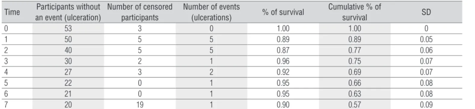
Table 4 shows the survival data of the total sample, consid-ering the censures since the start of the study (time 0) until the time of occurrence of the event (neuropathic injury). he per-centage of survival concerns each moment (time) of follow-up, and the cumulative percentage of survival is the time without the event over the length the study. At the end of the study (time 7), 19 participants of the total sample remained free of injuries. Of these, 13 participants belonged to the intervention group and six to the control group, therefore the cumulative survival was 60% and 52% respectively.
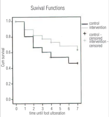
Figure 1 shows the graph of the survival function of both groups, with a trend toward shorter survival time, i.e. shorter
Variable
Group
p-value Intervention
(n=30)
Control (n=23)
Diabetes diagnosis (years) 14±10 15±10.5 0.602**
Type of Diabetes n (%)
1 1(50) 1(50) 0.999*
2 29(56.9) 22(43.1)
Male n (%) 21(63.6) 12(36.4) 0.255*
Age (years) 64.4±9.2 59.8±9.0 0.074**
Table 1. Characteristics of the studied population.
Data are means ± SD; * Significant difference at p<0.05 (Fisher’s Exact Test); ** Signifi-cant difference at p<0.05 (Mann-Whitney Test).
Intervention n (%) Control n (%) p-value*
Risk 1 6(37.5) 10(62.5) 0.256
Risk 2 15(68.2) 7(31.8)
Risk 3 3(50) 3(50)
Risk 4 6(66.7) 3(33.3)
Table 2. Foot risk categories in the two groups.
* Significant difference at p< 0.05 (Fisher’s Exact Test).
Foot
ulceration Intervention n(%) Control n (%) p-value*
Occurrence
No 13(61.9) 6(42.9) 0.317
Yes 8(38.1) 8(57.1)
Recurrence
No 7(70) 3(30) 0.119
Yes 1(16.7) 5(83.3)
Table 3. Occurrence and recurrence of neuropathic foot ulcerations in the two groups after 24-month follow-up.
* Significant difference at p<0.05 (Fisher’s Exact Test).
Time Participants without an event (ulceration)
Number of censored participants
Number of events
(ulcerations) % of survival
Cumulative % of
survival SD
0 53 3 0 1.00 1.00 0
1 50 5 5 0.89 0.89 0.05
2 40 5 5 0.87 0.77 0.06
3 30 2 1 0.96 0.75 0.07
4 27 3 2 0.92 0.69 0.07
5 22 0 1 0.95 0.66 0.08
6 21 0 1 0.95 0.63 0.08
7 20 19 1 0.90 0.57 0.09
Table 4. Survival time in the population studied: time until foot ulceration.
35
time until the occurrence of the event (neuropathic injury) among participants in the control group. However, the test for comparison of the curves of the two groups did not provide a signiicant diference (p=0.362).
Discussion
he present study included participants with insensitivity only (risk 1) up to larger losses (risk 4) caused by diabetic neu-ropathy. he success in preventing the recurrence of ulcers in di-abetic patients, in response to the combination of footwear and therapeutic education, was discussed by Maciejewski et al.18 in
a review of the literature published between 1980 and 2003. he authors emphasized that these multifactorial interventions are directed to patients at high risk of injury due to neuropathy and ischemia caused by diabetes. According to the International Consensus on Diabetic Foot12, the interventions of therapeutic
education and foot protection should be directed especially to high-risk patients, given that they are more likely to have com-plications. However, those with a milder neuropathy condition should not be excluded, due to the risk of deterioration. he data from the present study conirm the priority of assistance to patients at higher risk. Of the participants with injuries, 75% were risk 3 or 4, which is equivalent to 80% of the initial sample of participants at risk 3 and 4. Of the 38 participants classiied as risk 1 and 2, only four (10.5%) had the event. Considering the indings of Calle-Pascual et al.16, it can be inferred that the
proile of the sample may have inluenced the results of this study. hese authors studied DM patients at diferent stages of neuropathy undergoing a prevention program and found that the reduction in the incidence of neuropathic injury is lower in patients with milder neuropathy. Calle-Pascual et al.16 studied
the patients for 4.6 years; therefore, to verify preventive efects in patients with mild neuropathy even, with deterioration, it is necessary to monitor them for longer period than those moni-tored by Calle-Pascual et al.16.
he follow-up losses by withdrawal or death were similar in both groups, indicating that the proposed procedures did not interfere with the adherence to the study. hese high levels of sample loss often occur in studies on patients with DM. Polon-sky28 attributes this to the diiculties and frustrations
regard-ing the management of chronic diseases such as diabetes. he author describes this behavior as diabetes burnout.
he adherence to the proposed intervention is a positive point of the present study, considering its importance to the result of preventive actions16,18. Protective footwear was given
only to the participants who completed the therapeutic edu-cation program. Of the 30 participants, 29 completed the pro-gram. Of the participants who received the shoes, 72.4% wore
them daily, alternating with or without other footwear. his is a good result, considering the diiculties shown by Johnson, Newton and Goyder29 in their study on the prospects of the
dia-betes patients in relation to therapeutic footwear. According to these authors, patient involvement in choosing the model and color of the footwear may motivate the use, which could ex-plain the good adherence observed in this study. his informa-tion on adherence was not used for analysis of associainforma-tion with the occurrence of injuries, because the intent of the study was to evaluate the two interventions, education and protection of the feet, applied in conjunction. To evaluate individual efects of each intervention, a design with two other groups would be required to isolate the efects of therapeutic education or of the protective footwear.
he size of the sample (39, at the end of the study) may have afected the results of this study because it is insuicient to evaluate a prevention program. he magnitude of efect of pre-ventive interventions, such as education of patients, is small. According to a systematic review by Valk, Kriegsman and As-sendelft14, a sample of at least 430 participants is required to
detect clinically relevant diferences between groups. For n = 39 (sample at time 7), considering 20% of efect, the statistical power is 0.2430. his value indicates a high probability of type II
error in the results obtained in the present study. To conirm the null hypothesis of diferences between the groups, a larger sample would be required.
Figure 1. Survival function using Kaplan-Meier analysis to compare groups.
0 1 2 3 4 5 6 7
1.0
Suvival Functions
time until foot ultceration
control
control -censored
censored intervention
intervention -0.8
0.6
Cum sur
vival
0.4
0.2
36
References
Conclusion
Despite lower rates of recurrence of injury and a greater probability of remaining without injury, no signiicant difer-ence was found as a result of the implementation of the thera-peutic education program combined with the use of protective footwear in diabetic patients.
Acknowledgements
To Dr. Cristina Rolim Neumann, endocrinologist at the City Hall of Porto Alegre, Rio Grande do Sul, and to Prof. Carmen Lúcia Bezerra Machado, Department of Basic Studies, Faculty of Education, Universidade Federal do Rio Grande do Sul, for the technical assistance and advice.
1. Brasileiro Filho G. Bogliolo Patologia. 6ª ed. Rio de Janeiro: Guanabara Koogan; 2000.
2. Andreassen CS, Jakobsen J, Andersen H. Muscle weakness: a progressive late complication in diabetic distal symmetric polyneuropathy. Diabetes. 2006;55(3):806-12.
3. Bruce DG, Davis WA, Davis TM. Longitudinal predictors of reduced mobility and physical disability in patients with type 2 diabetes: the fremantle diabetes study. Diabetes Care. 2005;28(10):2441-7.
4. Ghanassia E, Villon L, Thuan Dit Dieudonné JF, Boegner C, Avignon A, Sultan A. Long-term outcome and disability of diabetic patients hospitalized for diabetic foot ulcers: a 6.5-year follow-up study. Diabetes Care. 2008;31(7):1288-92.
5. Boulton AJ. The diabetic foot: grand overview, epidemiology and pathogenesis. Diabetes Metab Res Rev. 2008;24 Suppl 1:S3-6.
6. Giacomozzi C, D’Ambrogi E, Cesinaro S, Macellari V, Uccioli L. Muscle performance and ankle joint mobility in long-term patients with diabetes. BMC Musculoskelet Disord. 2008;9:99.
7. Lavery LA, Peters EJ, Armstrong DG. What are the most effective interventions in preventing diabetic foot ulcers? Int Wound J. 2008;5(3): 425-33.
8. Zimny S, Schatz H, Pfohl M. The role of limited joint mobility in diabetic patients with an at-risk foot.Diabetes Care. 2004;27(4):942- 6.
9. Gershater MA, Löndahl M, Nyberg P, Larsson J, Thörne J, Eneroth M, et al. Complexity of factors related to outcome of neuropathic and neuroischaemic/ischaemic diabetic foot ulcers: a cohort study. Diabetologia. 2009;52(3):398-407.
10. Nather A, Bee CS, Huak CY, Chew JL, Lin CB, Neo S, et al. Epidemiology of diabetic foot problems and predictive factors for limb loss. J Diabetes Complications. 2008;22(2):77-82.
11. World Health Organization. International Diabetes Federation. Diabetes care and research in Europe: the Saint Vincent declaration. Diabet Med. 1990;7(4):360.
12. Grupo de Trabalho Internacional Sobre Pé Diabético. Consenso internacional sobre pé diabético. Brasília: Ministério da Saúde; 2001.
13. Krishnan S, Nash F, Baker N, Fowler D, Rayman G. Reduction in diabetic amputations over 11 years in a defined U.K. population: benefits of
multidisciplinary team work and continuous prospective audit. Diabetes Care. 2008;31(1):99-101.
14. Valk GD, Kriegsman DM, Assendelft WJ. Patient education for preventing diabetic foot ulceration. Cochrane Database Syst Rev. 2005;25(1):CD001488.
15. Lott DJ, Hastings MK, Commean PK, Smith KE, Mueller MJ. Effect of footwear and orthotic devices on stress reduction and soft tissue strain of the neuropathic foot. Clin Biomech (Bristol, Avon). 2007;22(3):352-9.
16. Calle-Pascual AL, Durán A, Benedí A, Calvo MI, Charro A, Diaz JA, et al. A preventative foot care programme for people with diabetes with different stages of neuropathy. Diabetes Res Clin Pract. 2002;57(2):111-7.
17. Bus SA, Valk GD, van Deursen RW, Armstrong DG, Caravaggi C, Hlavácek P, et al. The effectiveness of footwear and offloading interventions to prevent and heal foot ulcers and reduce plantar pressure in diabetes: a systematic review. Diabetes Metab Res Rev. 2008;24 Suppl 1:S162-80.
18. Maciejewski ML, Reiber GE, Smith DG, Wallace C, Hayes S, Boyko EJ. Effectiveness of diabetic therapeutic footwear in preventing reulceration. Diabetes Care. 2004;27(7):1774-82.
19. Pham H, Armstrong DG, Harvey C, Harkless LB, Giurini JM, Veves A. Screening techniques to identify people at high risk for diabetic foot ulceration: a prospective multicenter trial. Diabetes Care. 2000;23(5):606-11.
20. Crawford F, Inkster M, Kleijnen J, Fahey T. Predicting foot ulcers in patients with diabetes: a systematic review and meta-analysis. QJM. 2007;100(2):65-86.
21. Boulton AJ, Armstrong DG, Albert SF, Frykberg RG, Hellman R, Kirkman MS, et al. Comprehensive foot examination and risk assessment. A report of the task force of the foot care interest group of the American Diabetes Association, with endorsement by the American Association of Clinical Endocrinologists. Phys Ther. 2008;88(11):1436-43.
22. Pedrosa HC, Leme LAP, Novaes C, Saigg M, Sena F, Gomes EB, et al. The diabetic foot in south America: progress with the brazilian save the diabetic foot project. International. Diabetes Monitor. 2004;16(4):17-24.
23. Birke JA, Sims DS. The insensitive foot. In: Hunt GC, McPoil TG (editors).
Physical therapy of the foot and ankle. 2nd ed. New York: Churchill
Livingstone; 1995; p.159-206.
37 25. Dahmen R, Haspels R, Koomen B, Hoeksma AF. Therapeutic footwear for
the neuropathic foot: an algorithm. Diabetes Care. 2001;24(4):705-9.
26. Loiola LV, Schmid H. Os pés dos pacientes com diabetes. In: Braga, WRC (editor). Clínica médica – diabetes mellitus. Porto Alegre: MEDSI; 2001. p.577-95.
27. Bustamante-Teixeira MT, Faerstein E, Latorre MR. Técnicas de análise de sobrevida. Cad Saúde Pública. 2002;18(3):579-94.
28. Polonsky WH. Emotional and quality-of-life aspects of diabetes management. Curr Diab Rep. 2002;2(2):153-9.
29. Johnson M, Newton P, Goyder E. Patient and professional perspectives on prescribed therapeutic footwear for people with diabetes: a vignette study. Patient Educ Couns. 2006;64(1-3):167-72.
30. Portney LG, Watkins MP. Foundations of clinical research: applications to