From the Radiology Institute, Hospital das Clínicas, Faculty of Medicine, University of São Paulo – São Paulo/SP, Brazil.
E-mail: [email protected]
Received for publication on October 24, 2003. Accepted for publication on April 02, 2004.
ORIGINAL RESEARCH
RETROSPECTIVE EVALUATION OF BONE PAIN
PALLIATION AFTER SAMARIUM-153-EDTMP
THERAPY
Marcelo Tatit Sapienza, Carla Rachel Ono, Maria Inês Cury Guimarães, Tomoco Watanabe, Paulo Aguirre Costa and Carlos Alberto Buchpiguel
SAPIENZA MT et al. Retrospective evaluation of bone pain palliation after samarium-153-EDTMP therapy. Rev. Hosp.
Clín. Fac. Med. S. Paulo 59(6):321-328, 2004.
PURPOSE: The aim of this study was to evaluate the degree of metastatic bone pain palliation and medullar toxicity
associated with samarium-153-EDTMP treatment.
METHODS: Seventy-three patients with metastatic bone pain having previously undergone therapy with
samarium-153-EDTMP (1 mCi/kg) were retrospectively evaluated. Routine follow-up included pain evaluation and blood counts for 2 months after treatment. Pain was evaluated using a subjective scale (from 0 to 10) before and for 8 weeks after the treatment. Blood counts were obtained before treatment and once a week for 2 months during follow-up. Dosimetry, based upon the urinary excretion of the isotope, was estimated in 41 individuals, and the resulting radiation absorbed doses were correlated with hematological data.
RESULTS: Reduction in pain scores of 75% to 100% was obtained in 36 patients (49%), with a decrease of 50% to
75%, 25% to 50%, and 0% to 25% in, respectively, 20 (27%), 10 (14%), and 7 (10%) patients. There was no significant relationship between the pain response and location of the primary tumor (breast or prostate cancer). Mild to moderate myelosuppression was noted in 75.3% of patients, usually with hematological recovery at 8 weeks. The mean bone marrow dose was 347 ± 65 cGy, and only a weak correlation was found between absorbed dose and myelosuppression (Pearson coefficient = .4).
CONCLUSIONS: Samarium-153-EDTMP is a valuable method for metastatic bone pain palliation. A mild to moderate
and transitory myelosuppression is the main toxicity observed after samarium therapy, showing a weak correlation with dosimetric measures.
KEYWORDS: Samarium. Pain. Scintigraphy. Palliative. Care. Neoplasm. Metastasis.
Bone metastases are common in the progression of various tumors such as prostate, breast, and lung carcinoma, and they often entail an occurrence of progressive pain.1 Control of pain and its consequences (eg, depression, movement constraint, and dependence) are important goals in oncological treatment requiring a multidisciplinary approach to the patient.
The use of ionizing radiation is one of the alternatives for pain treat-ment, together with the administration of drugs (eg, anti-inflammatories,
opioids, chemotherapeutic drugs, receptor blockades, phosphonates) and surgical interventions. Local radio-therapy results in a good clinical re-sponse in approximately 80% of the cases (complete in 30% to 60%) last-ing for over 4 months, although with an effect confined to the irradiated
site.2,3 Hemibody radiotherapy (iso-lated or sequential) may be used in pa-tients with widespread metastases, re-sulting in a complete response in ap-proximately 20% of the cases, and a partial one for 50% to 100%. Radia-tion on wide areas is limited by hematopoietic, pulmonary, and gastrointestinal toxicity produced by the high radiation absorbed dose.2,3
bone metastases. Beta radiation energy is almost completely delivered to the specific site of uptake, with a minimal effect on neighboring tissues. The main radioisotopes used for palliation of bone pain due to metastatic malig-nancies are strontium-89 and samar-ium-153, with response rates between 60% and 80%.2,4 The isotope most fre-quently used in Brazil is samarium-153 due to its wide availability and low cost (150 dollars per treatment, compared to approximately 2,000 dol-lars for strontium-89).
S a m a r i u m - 1 5 3 – l a b e l e d ethylenediaminetetramethylene phosphonic acid (153Sm-EDTMP) is re-tained in the mineral phase of bone, probably by adsorption to hydroxya-patite crystals, similarly to the mecha-nism involved in the uptake of radiopharmaceuticals used in bone scans. Treatment with 153Sm-EDTMP was first described in 1989,5 and it was first approved for clinical use in the USA in 1997. In Brazil, samarium-153 is produced by the Institute for Ener-getic and Nuclear Researches (IPEN -Instituto de Pesquisas Energéticas e Nucleares – São Paulo) and has been used clinically since 1995, initially as part of an international program coor-dinated by the International Atomic Energy Agency (IAEA – Vienna), and later as a routine procedure covered by the Brazilian healthcare system ( SUS-Sistema Único de Saúde).
Clinical response to treatment with 153Sm-EDTMP (pain reduction) is
de-scribed in 55% to 88% of the cases, beginning 5 to 7 days after injection, and lasting for 2 to 17 weeks.6,7,8 The shorter half-life of samarium-153 com-pared to strontium-89 (2 versus 50 days) may be related to the smaller in-terval for the response start (5 to 7 ver-sus 10 to 20 days) and to its shorter duration (2 to 3 versus 3 to 4 months). Bone marrow toxicity is the main adverse reaction observed after treat-ment with samarium-153, generally
occurring in a mild to moderate de-gree, reaching a peak between 3 and 4 weeks and subsiding after 6 to 8 weeks. Grade III toxicity is described in approximately 10% of the patients treated with doses of 1 mCi/kg,9,10,11,12 and grade IV toxicity occurs in less than 1% of cases. In addition to beta-particle emission (energy = 810 keV), samarium-153 emits gamma radiation (energy = 103 keV), which may be used for biodistribution and dosimetric measures. A bone uptake of 55% to 75% of the injected dose is confirmed in these measurements,13,14 resulting in a radiation absorbed dose of approxi-mately 7 cGy/mCi in normal bone and 42 cGy/mCi in osteoblastic bone le-sions.2
To the best of our knowledge, clinical results of the treatment with 153Sm-EDTMP in Brazil have not yet
been described, even though there is almost one decade of experience.
The objectives of the current study were:
1- to evaluate the clinical response (pain intensity variation) and the marrow toxicity after treatment with 153Sm-EDTMP;
2- to correlate the degree of marrow toxicity with the radiation ab-sorbed dose of the bone marrow.
METHODS
Patients
Medical records of 178 patients were retrospectively reviewed. Patients had been sent for treatment with 153 Sm-EDTMP to the Nuclear Medicine Serv-ice of our Institute between 1995 and 2002. Patient records were considered adequate if there were data for at least 75% of the routine clinical and labo-ratory fields (daily pain evaluation and weekly blood counts for 8 weeks). As part of the treatment planning, all patients had a consultation with a
nuclear medicine physician to confirm the indications and to exclude even-tual contraindications to the treatment, and all signed an informed consent. Indications for treatment were pain due to bone metastases requiring increas-ing analgesic doses, clinically uncon-trolled pain, or recurring pain in a pre-viously irradiated site. An additional criterion was the detection of multiple osteoblastic lesions in a recent bone scan (less than a 3-month interval) showing moderate to outstanding ra-diopharmaceutical uptake. Treatment was contraindicated for patients with one or more of the following criteria: severely disabled patients, leukopenia (<2,500/mm3) or thrombocytopenia (<100,000 mm3) in a recent blood test (less than 1 week), progressive drop of the platelet count, less than 1 month interval from chemotherapy or 2 months from radiotherapy involving more than 20% of the bone marrow, or pregnancy. Patients with pain due to a suspected pathological fracture, nerve compression, or soft tissue invasion were directed to complementary inves-tigation and treatment with other modalities because treatment with 153Sm-EDTMP was not indicated.
Treatment and follow-up
Pain intensity before treatment was subjectively assessed by the patients themselves using a 0-to-10 scale (0 = no pain, 10 = maximum pain). Anal-gesics used, their doses, and their use intervals were also recorded. Outpa-tient treatment was performed by the intravenous administration of 153 Sm-EDTMP (IPEN - São Paulo, Brazil) through an upper limb access.
grouped according to their pain inten-sity drop, that is, 0% to 25%, 25.1% to 50%, 50.1% to 75%, and 75.1% to 100%. Pain flare, a transitory worsen-ing of pain after treatment,4,15 was de-fined by a greater-than-2-point increase in the pain scale within 5 days after the administration of 153Sm-EDTMP.
Blood counts were also obtained weekly during the same 8-week period, and the corresponding leukocyte count, platelet count, and hemoglobin concentration were recorded. Absolute and percent variations of these param-eters were calculated compared to ba-sal values (obtained at 1 week or less pretreatment). Medullary toxicity, based on the lowest leukocyte and platelet values observed during follow-up, was classified in accordance with the National Cancer Institute Com-mon Toxicity Criteria - Version 1.
Dosimetric calculations
After the administration of 153 Sm-EDTMP, 41 of the 73 patients under-went dosimetric calculation. Urinary excretion of samarium-153 by these 41 patients was measured hourly for 6 hours, and an activity retention curve was drawn. Previously described mod-els16,17 were used to determine the ac-tivity distribution in the different in-tervals. These data were used to calcu-late the accrued activity integral in the cortical and trabecular bone using equation 1, where A = accrued activ-ity, A(t) = retained activity in each structure in the various times, … = in-termediate terms between 1 and 24
hours, and λ = the samarium-153 dis-integration constant.
The radiation absorbed dose of the hematopoietic marrow resulting from the emission of beta particles from the isotope retained in the cortical and in the trabecular bone was calculated us-ing equation 2, where S = radiation ab-sorbed dose per accrued activity unit,
cor = cortical bone, trab = trabecular bone. The marrow radiation absorbed dose was corrected according to the patient’s weight, based on the 70 kg and 170 cm reference-man (ICRP 23).
Statistical evaluation
Mean and standard deviation of the initial pain intensity, clinical response, and medullary toxicity were estimated for the whole group of patients. The Student t test was used to compare pain intensity prior to and at the end of the eighth week after treatment (1-tailed paired t test, P <.05), as well as to com-pare the treatment response between patients with primary breast or prostate cancer (2-tailed t test, P <.05). The re-lationship between the decrease in platelet and leukocyte counts and the radiation absorbed dose of the bone marrow was evaluated using Pearson’s linear correlation coefficient.
RESULTS
Medical records were considered appropriate for analysis in 73 patients (37 males and 36 females; age range, 31 to 89 years, mean ± SD, 60 ± 15
years) who had the following as a pri-mary tumor: prostate carcinoma (n = 36), breast carcinoma (n = 29), lung carcinoma (n = 5), colorectal carci-noma (n = 1), parathyroid carcicarci-noma (n = 1), or unknown primary tumor (n = 1). Patient records were considered inappropriate for analysis in the re-maining 105 patients (62 followed up at Hospital das Clínicas de São Paulo; 43 at other institutions).
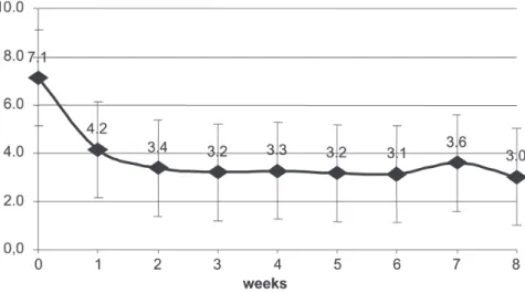
The mean value for pain intensity dropped from 7.1 ± 2.8 to 3.4 ± 2.8 in the first 2 weeks after treatment, hold-ing at 3.0 ± 2.3 in the eighth week af-ter treatment (Figure 1). Pain intensity in the eighth week after treatment was significantly lower than pain before treatment (P <.001).
During follow-up, a maximum pain intensity reduction of 5.83 ± 2.13 points with respect to the initial value (percent reduction: 70.5% ± 27.7%) was observed. A 75% to 100% pain re-duction occurred in 36 patients (49%), and a 50% to 75% reduction occurred in 20 cases (27%). Pain reduction was less than 50% in the other cases (25% to 50% in 10 cases and 0% to 25% in 7 cases) (Figure 2). No statistical dif-ference was observed in pain intensity reduction either in absolute scale (P = .983) or in percent scale (P = .069) when patients with prostate and breast carcinoma were compared.
In 7 patients, a transitory worsen-ing of pain was observed durworsen-ing the first week after treatment, with a mean increase of 3.76 ± 0.78 in the pain scale. The response subsequently ob-served in this subset of patients was similar to that seen in the others (re-duction of 70% ± 35% compared to the initial pain intensity).
Pretreatment blood count values were as follows: hemoglobin = 11.5 ± 2.0 g/dL, leukocytes = 6,820 ± 3,480/ mm3, and platelets = 237,200 ± 95,000/mm3. 75.3% of the patients ex-perienced leukopenia and/or thrombo-cytopenia during follow-up: 71.2% of Equation 2
the patients had leukopenia (grade I toxicity = 26.0%, grade II = 28.8%, grade III = 16.4%); and 53.4% had thrombocytopenia (grade I toxicity = 15.1%, grade II = 17.8%, grade III = 19.2%, grade IV = 1.4%) (Figure 3). Leukopenia and thrombocytopenia occurred simultaneously in 36 cases, while isolated leukopenia or thrombo-cytopenia occurred, respectively, in 16 and 3 patients. Platelet count variation was only weakly correlated with leukocyte count variation (Pearson’s coefficient r = .41). The correlation be-tween the minimum and basal values was also low for both leukocyte and platelet counts (r =.30 and .62).
The platelet and leukocyte nadir occurred between 4 and 5 weeks after
treatment, with recovery to 78% of ba-sal leukocyte values (average ± SD = 4,910 ± 1,830/mm3) and 88% of basal platelet values (187,700 ± 81,500/ mm3) at the end of the eighth week (Figure 4). Persistent leukopenia was still present between the seventh and the eighth weeks in 26% of the cases (4% had between 1,000 and 2,000 leukocytes/mm3, 8% had between 2,000 and 3,000 leukocytes/mm3, and 14% had between 3,000 and 4,000 leukocytes/mm3). Thrombocytopenia was still present in 16% of cases (4% had between 25 and 50 thousand platelets/mm3, 4% had between 50 and 75 thousand platelets/mm3, and 8% had between 75 and 100 thousand platelets/mm3).
The radiation absorbed dose of bone marrow varied from 195 to 468 cGy (mean ± SD = 347 ± 65 cGy) in the 41 patients. The radiation ab-sorbed dose had a low correlation with the maximum decrease in leukocyte counts during follow-up (Pearson’s co-efficient r = .40), as well as with the maximum drop in platelet counts (Pearson’s coefficient r = .48).
DISCUSSION
Intravenous injection of strontium-89 and samarium-153 for the treatment of pain due to osteoblastic metastasis has been widely described in the lit-erature, mainly for patients with hor-mone-refractory prostate carcinoma or with breast carcinoma, with reported responses between 55% and 83% of patients.1,5,6,7,15,18,19-22 The clinical re-sponse observed in this study was similar to that described in literature, the pain having been reduced to less than 50% of basal levels in 76% of cases, with no distinction as to the pri-mary tumor (breast or prostate). Pain improvement after treatment is prob-ably related to a reduction in both in-flammatory infiltrate and cytokine re-lease, together with a change in osteo-clastic and osteoblastic function. Pain increase during the first week after treatment (a phenomenon known as “pain flare” and probably related to cytokine release) was observed in 10% of patients, a value comparable to that described in the literature.15
Our findings of bone marrow tox-icity were also similar to those de-scribed in the literature, with leukopenia in 71.2% of patients and thrombocytopenia in 53.4% of our pa-tients. Concentration of leucocytes and platelets reached a nadir between 4 and 5 weeks after treatment. There was a good recovery in blood counts dur-ing follow-up, reachdur-ing 78% of initial leukocyte count and 88% of initial
Figure 1 - Pain intensity before and for 8 weeks after treatment with 153Sm-EDTMP.
platelet count. In addition to the risks of leukopenia and thrombocytopenia, long-lasting marrow depression is a reason for concern because it may cause difficulty in future treatments with cytostatic agents. Even though leukopenia persisted in 26% of pa-tients and thrombocytopenia in 16%, it is difficult to determine if medullary function was permanently jeopardized, since further improvement in labora-tory parameters may have occurred af-ter the 8-week follow-up. On the other hand, bone marrow infiltration by the primary disease and the effect of pre-vious chemotherapy could also con-tribute to the nonreversion of the med-ullary depression.
The usual activity for treatment with samarium-153-EDTMP is 1 mCi/ kg.10,11,23 With this activity, various au-thors describe the radiation absorbed dose in the marrow to be between 230 and 590 cGy16,18,21,24 (a range that in-cludes the results of this study, ie, 347 ± 65 cGy). Administration of doses above 1 mCi/kg seems not to lead to better pain control,9 even though it re-sults in higher marrow toxicity.9,12,15,24 Though other studies have shown that administration of higher activities in-creases the risk of bone marrow toxic-ity,9,24,25 the current study did not show a direct correlation between toxicity and individual dosimetric measures. The low correlation observed suggests that other factors not evaluated in this study (such as previous chemotherapy, radiotherapy, and the extent of bone metastasis) could have reduced the bone marrow reserve, thus increasing the radiopharmaceutical toxicity.
Retrospective analysis imposed several restrictions on the current study, some of which were due to the fact that the follow-up file card had been designed approximately 8 years ago. We particularly regret that fields were not included for the clinical find-ings of medullary toxicity (febrile neu-tropenia, bleeding, and transfusions)
Figure 3 - a) Number of patients with medullary toxicity after treatment with 153Sm EDTMP
(characterized by thrombocytopenia)*. b) - Number of patients with medullary toxicity after treatment with 153Sm-EDTMP (characterized by leucopenia)*.
and for the objective evaluation of clinical parameters (for instance, Karnofsky’s index of clinical perform-ance). The comparison of average weekly pain intensity with the pretreat-ment value had the purpose of reduc-ing the interference of natural oscilla-tions on pain intensity. Additionally, it should be noted that the initial pain score was based on an isolated evalu-ation; thus, a bias may have occurred due to a return to the average during follow-up.
The possibility of a selection bias must also be considered since it is more probable that patients with an unsatis-factory response to treatment would fail to complete follow-up. Though these data were not included in the current study, we observed that patients with incomplete information were prone to higher pain intensity in the basal evalu-ation (8.64 ± 2.0 points) and to a lower response intensity (average reduction of 4.4 ± 2.7 points).
In addition to the difficulties pecu-liar to retrospective data collections
and the lack of a control group, this study also had limitations inherent to the type of clinical situation evaluated, including the possible simultaneous or sequential use of other therapies and the interference of patients’ activities according to pain intensity. We consid-ered patients’ basal situations as a ref-erence for the evaluation of clinical re-sponse and of adverse reactions during follow-up, and it is our experience that other treatments are generally intro-duced before the indication for samar-ium-153-EDTMP. It should be noted that there was no overlapping of chemotherapy, introduction of ster-oids, or hormone therapy in the period comprising the previous month and the 2 follow-up months after samar-ium-153-EDTMP administration—fac-tors that could interfere in the response and adverse reactions analysis. There were analgesic consumption changes during the follow-up period, with a tendency towards reduction in the dose and frequency of use among pa-tients with a pain reduction higher
than 50%; this suggests that the ob-served response had effectively origi-nated from use of samarium-153-EDTMP. However, analgesic types, doses, and use intervals were incom-pletely reported in almost all cases and therefore could not be included in the results.
CONCLUSIONS
Treatment with 153Sm-EDTMP re-sulted in a good clinical response with a reduction in the subjective appraisal of pain to less than 50% of the basal intensity in 76% of the patients.
The main adverse effect observed during follow-up was a transitory med-ullary depression to a mild to moder-ate degree that was only weakly cor-related with dosimetric measures. Leukopenia was observed in 71.2% of the patients and thrombocytopenia in 53.4%; however, most of the patients had recovered at the end of the eighth week.
RESUMO
SAPIENZA MT e col. Avaliação retros-pectiva do tratamento da dor óssea metastática com Samário-153-EDTMP. Rev. Hosp. Clín. Fac. Med. S. Paulo 59(6):321-328, 2004.
OBJETIVO: O presente trabalho teve por objetivo avaliar o efeito pali-ativo da dor e a toxicidade medular as-sociados ao tratamento com Samário-153-EDTMP em pacientes com metás-tases ósseas.
MÉTODOS: O estudo foi realiza-do de forma retrospectiva, a partir realiza-do levantamento de prontuário de 178
pacientes submetidos a tratamento com 1mCi/kg de 153Sm-EDTMP devi-do à devi-dor por metástases ósseas. Os prontuários de 73 pacientes foram con-siderados adequados para análise dos parâmetros clínicos (intensidade da dor) e laboratoriais (hemograma). A in-tensidade da dor foi avaliada em esca-la de 0 a 10 pelo próprio paciente, an-tes e durante 8 semanas após o trata-mento. Hemograma completo foi rea-lizado antes do tratamento e a cada se-mana nas 8 sese-manas seguintes. Estu-dos de Estu-dosimetria foram realizaEstu-dos em 41 dos 73 pacientes, baseados na excreção urinária e retenção do
radio-isótopo, sendo a dose de radiação ab-sorvida correlacionada à toxicidade medular.
plaque-topenia), em geral de grau leve a mo-derado e com recuperação ao término da 8o semana. A dose média de medu-la foi de 347±65 cGy, havendo baixa correlação entre a dosimetria medular e a queda da contagem de leucócitos (coeficiente de correlação linear de 0,40) ou de plaquetas (coeficiente de
correlação linear = 0,48).
CONCLUSÕES: O tratamento
com Samário-153-EDTMP permitiu um adequado controle da dor por metástases ósseas, com significativa redução na intensidade da dor. A toxicidade medular transitória foi a principal reação adversa observada,
em geral de grau leve a moderado, apresentando baixa correlação com as medidas dosimétricas.
UNITERMOS: Samário. Dor. Cin-tilografia. Assistência paliativa. Metástase.
REFERENCES
1. Coleman RE. Skeletal complications of malignancy. Cancer 1997;80:1588-94.
2. Mcewan AJ. Use of radionuclides for the palliation of bone metastases. Semin Radiat Oncol 2000;10:103-14.
3. Saarto T, Janes R, Tenhunen M, Kouri M. Palliative radiotherapy in the treatment of skeletal metastases. Eur J Pain 2002;6:323-30.
4. Silberstein EB, Eugene L, Saenger SR. Painful osteoblastic metastases: the role of nuclear medicine. Oncology (Huntingt) 2001;15:157-63.
5. Turner JH, Claringbold PG, Hetherington EL, Sorby P, Martindale AA. A phase I study of samarium-153 ethylene-diaminetetramethylene phosphonate therapy for disseminated skeletal metastases. J Clin Oncol 1989;7:1926-31.
6. Collins C, Eary JF, Donaldson G, Vernon C, Bush NE, Petersdorf S, et al. Samarium-153-EDTMP in bone metastases of hormone refractory prostate carcinoma: a phase I/II trial. J Nucl Med 1993;34:1839-44.
7. Sandeman TF, Budd RS, Martin JJ. Samarium-153-labelled EDTMP for bone metastases from cancer of the prostate. Clin Oncol (R Coll Radiol ) 1992;4:160-4.
8. Lovera C, Massardo T, Galleguillos MC, et al. [Analgesic response and secondary effects in patients with osteoblastic metastasis, treated with Samarium-153 ethylenediaminotetramethylene-phosphate]. Rev Med Chil 1998;126:963-71.
9. Alberts AS, Smit BJ, Louw WK, van Rensburg AJ, van Beek A, Kritzinger V, et al. Dose response relationship and multiple dose efficacy and toxicity of samarium-153-EDTMP in metastatic cancer to bone. Radiother Oncol 1997;43:175-9. 10. Serafini AN, Houston SJ, Resche I, Quick DP, Grund FM, Ell PJ,
et al. Palliation of pain associated with metastatic bone cancer using samarium-153 lexidronam: a double-blind placebo-controlled clinical trial. J Clin Oncol 1998;16:1574-81. 11. Resche I, Chatal JF, Pecking A, Ell P, Duchesne G, Rubens R, et al.
A dose-controlled study of 153Sm-ethylenediaminete-tramethylenephosphonate (EDTMP) in the treatment of patients with painful bone metastases. Eur J Cancer 1997; 33:1583-91.
12. Tian JH, Zhang JM, Hou QT, Oyang QH, Wang JM, Luan ZS, et al. Multicentre trial on the efficacy and toxicity of single-dose samarium-153-ethylene diamine tetramethylene phosphonate as a palliative treatment for painful skeletal metastases in China. Eur J Nucl Med 1999;26:2-7.
13. Singh A, Holmes RA, Farhangi M, Volkert WA, Williams A, Stringham LM, et al. Human pharmacokinetics of samarium-153 EDTMP in metastatic cancer. J Nucl Med 1989;30:1814-8.
14. Bartlett ML, Webb M, Durrant S, Morton AJ, Allison R, Macfarlane DJ. Dosimetry and toxicity of Quadramet for bone marrow ablation in multiple myeloma and other haematological malignancies. Eur J Nucl Med Mol Imaging 2002;29:1470-7. 15. Farhanghi M, Holmes RA, Volkert WA, Logan KW, Singh A. Samarium-153-EDTMP: pharmacokinetic, toxicity and pain response using an escalating dose schedule in treatment of metastatic bone cancer. J Nucl Med 1992;33:1451-8. 16. Logan KW, Volkert WA, Holmes RA. Radiation dose calculations
in persons receiving injection of samarium-153 EDTMP. J Nucl Med 1987;28:505-9.
17. Turner JH, Martindale AA, Sorby P, Hetherington EL, Fleay RF, Hoffman RF, et al. Samarium-153 EDTMP therapy of disseminated skeletal metastasis. Eur J Nucl Med 1989;15:784-95.
18. Cameron PJ, Klemp PF, Martindale AA, Turner JH. Prospective 153Sm-EDTMP therapy dosimetry by whole-body scintigraphy. Nucl Med Commun 1999;20:609-15. 19. Friedland J. Local and systemic radiation for palliation of
metastatic disease. Urol Clin North Am 1999;26:391-402, x. 20. Altman GB, Lee CA. Strontium-89 for treatment of painful bone metastasis from prostate cancer. Oncol Nurs Forum 1996;23:523-7.
21. Eary JF, Collins C, Stabin M, Vernon C, Petersdorf S, Baker M, et al. Samarium-153-EDTMP biodistribution and dosimetry estimation. J Nucl Med 1993;34:1031-6.
23. Serafini AN. Systemic metabolic radiotherapy with samarium-153 EDTMP for the treatment of painful bone metastasis. Q J Nucl Med 2001;45:91-9.
24. Bayouth JE, Macey DJ, Kasi LP, Fossella FV. Dosimetry and toxicity of samarium-153-EDTMP administered for bone pain due to skeletal metastases. J Nucl Med 1994;35:63-9.