Article
J. Braz. Chem. Soc., Vol. 25, No. 10, 1832-1838, 2014. Printed in Brazil - ©2014 Sociedade Brasileira de Química 0103 - 5053 $6.00+0.00
A
http://dx.doi.org/10.5935/0103-5053.20140115*e-mail: [email protected]
Leishmanicidal Galloylquinic Acids are Noncompetitive Inhibitors of Arginase
Lorena R. F. de Sousa,a Suelem D. Ramalho,a João B. Fernandes,a
Maria Fátima das G. F. da Silva,a Mônica R. da C. Iemma,a Caroindes J. Corrêa,a
Dulce H. F. de Souza,a Maria I. S. Limab and Paulo C. Vieira*,a
aDepartamento de Química and bDepartamento de Botânica, Universidade Federal de São Carlos,
Rod. Washington Luís, km 235, 13565-905 São Carlos-SP, Brazil
Ácidos galoilquínicos, substâncias que têm apresentado atividade leishmanicida, foram isolados do extrato de acetato de etila da planta Byrsonima coccolobifolia. Estes compostos fenólicos
juntamente com o ácido gálico demonstraram ser uma nova classe de inibidores não-competitivos potentes em arginase (ARG) de Leishmaniaamazonensis (Ki variando de 0,10 a 0,68 µmol L-1). O ácido quínico não apresentou atividade inibitória significativa em ARG demonstrando que a unidade galoila tem características importantes que permitem a interação enzima-inibidor. A atividade inibitória significativa do ácido gálico frente à ARG pode ser uma indicação para o entendimento da resposta imune previamente observada em Leishmania donovani, uma vez que
a atividade enzimática da arginase está associada à diminuição dos níveis de NO no processo de infecção por Leishmania.
Leishmanicidal galloylquinic acids were isolated from the ethyl acetate extract of Byrsonima coccolobifolia. These phenols and gallic acid showed to be a new class of potent noncompetitive inhibitors of arginase ARG (Ki ranging from0.10 to 0.68 µmol L-1) from Leishmania amazonensis. Quinic acid did not exhibit significant inhibition of ARG, indicating that galloyl moiety has important features that allows the enzyme-inhibitor interactions. The significant inhibitory activity of gallic acid on ARG can be a clue to understand the immune response previously observed on L. donovani,
since ARG activity is associated with the decrease of the levels of NO in Leishmania infection.
Keywords: arginase, Leishmania, galloylquinic acids, Byrsonima coccolobifolia
Introduction
Leishmaniasis is a deadly infectious tropical disease
caused by the protozoan of the genus Leishmania,
which affects more than 12 million people of broad geographical distribution.1,2 The challenges for healthcare
of leishmaniasis found on available drugs such as, high toxicity, undesirable side effects, high cost and parasite resistance, reveal an urgent problem and the need for new efficient drugs.3-5
Exploring for novel therapeutic opportunities, new biochemical targets have been investigated, in particular
arginase (ARG) from Leishmania amazonensis have
been considered an attractive target in the search for new leishmanicidal agents. The biochemical pathway that this enzyme is involved is essential for the protozoa development in their life cycle.6-8 In addition, the crystal
structure of ARG from Leishmania mexicana was solved,9
making this protease more interesting to investigate for new antileishmanial compounds.
Arginase is a metalloenzyme with binuclear manganese center, which catalyzes the last step in the urea cycle in mammals, allowing hydrolyzes of L-arginine to L-ornithine and urea.10 In infected macrophages the
substrate L-arginine is used by ARG and by nitric oxide synthase (NOS) in two different metabolic pathways. The reaction catalyzed by ARG is carried out in the polyamines (PAs) metabolism, essential to preserve parasite viability, and the NOS pathway generate nitric oxide (NO) molecules, which production increases oxidative stress.11,12 The balance between NOS and
mammalian arginase is competitively regulated by TH1
and TH2 cytokines as protective response. Leishmania
protozoa explore the immune response of TH2 increasing
Sousa et al. 1833 Vol. 25, No. 10, 2014
Nω-hydroxy-L-arginine and
2(S)-amino-6-boronohexanoic acid are synthetic aminoacid derivatives, which inhibit in a competitive mode ARG from L. mexicana
with Ki values of 85 and 1.3 µmol L-1 respectively. However,
in an in vivo study these synthetic compounds could not
contain the infection completely.7,15 Flavonoids isolated
from plants have been reported as inhibitors of ARG from
L. amazonensis. The presence of hydroxy groups in their
structures showed to be important features for inhibitory activity.16-19
Quinic acid is a hydrated form of shikimic acid and together with galloylquinic acids (tannic acids) and gallic acid are derivatives from shikimate pathway, available and widespread in plant sources.20,21 Tannins have been
associated with many biological activities and to health beneficial effects.22-24 Additionally, tannic acids have
presented antileishmanial activity (EC50 = 2-38 µmol L-1),
and their leishmanicidal potency have been associated to the number of galloyl groups substituents in the shikimic acid moiety.25 Gallic acid singly reduces Leishmania donovani
amastigotes (EC50 = 4.4 µg mL-1) in murine macrophages
by activating leishmanicidal macrophage functions.26 Also,
tannins identified from fractions of cajazeira (Spondias mombin L.) showed leishmanicidal effect in vitro on
amastigotes of Leishmania chagasi, with IC50 in the range of
0.61 to 17.07 µg mL-1.27 However, so far, no study has been
reported on this class of secondary metabolites exploring their inhibitory activity on ARG from L. amazonensis.
Based on the usage of natural products as invaluable tools in the search for new drugs5,28 and in the antiprotozoal
activities previously found in Byrsonima species,29,30
we performed a phytochemical study of fractions of
Byrsonima coccolobifolia Kunth. (Malpighiaceae) extract
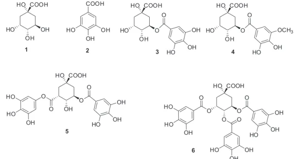
that significantly inhibited ARG enzyme. Gallic acid (2) and galloylquinic acids (3-6)were isolated from ethyl acetate extract from leaves and stems of B. coccolobifolia,
which were identified by 1D and 2D NMR spectra, and through comparison with data previously reported.31-35
Galloylquinic acids (3-6) and gallic acid (2) were potent inhibitors of ARG with high affinity. Quinic acid (1)36
isolated from Myrcia lingua (O. Berg) Mattos (Myrtaceae)
was also tested on ARG to compare inhibitory activity with those galloylquinic acid derivatives, but the inhibition of ARG found for this compound was not significant.
Experimental
General experimental procedures
The measurement of urea in the enzymatic assays was performed on a Beckman Coulter DU 800 spectrophotometer at 600 nm. The 1D and 2D nuclear magnetic resonance (NMR) data were acquired on a Bruker DRX-400 NMR spectrometer (1H: 400 MHz; 13C: 100 MHz) using D
2O and
MeOH-d4 as solvents.Silica gel 60 (Merck, 230-700 mesh)
and Sephadex LH-20 (Amersham Pharmacia Biotech AB) together with thin-layer chromatography (TLC) on pre-coated aluminum silica 60 F254 (Merck) were used to isolate the compounds. Compounds were visualized in TLC UV254/366 and by the usage of the stain sulfuric vanillin solution. The solvents ethanol (EtOH), methanol (MeOH), hexane, acetone, ethyl acetate (EtOAc) from Vetec were used to prepare the extracts and for chromatographic procedures.
Leishmanicidal Galloylquinic Acids are Noncompetitive Inhibitors of Arginase J. Braz. Chem. Soc. 1834
Plant material
B. coccolobifolia leaves and stems were collected in
the cerrado at Federal University of São Carlos (UFSCar), São Carlos - SP, Brazil. The plant material was identified by Dr Maria Inês Salgueiro Lima, and deposited at the Herbarium of the Botany Laboratory (HUFSCar) in UFSCar (voucher No. 8367).
Extraction and isolation
The crude extracts from the leaves (10.0 g) and stems (30.5 g) of B. coccolobifolia were liquid-liquid partitioned
leading to the EtOAc extracts (2.0 g of leaves and 10.0 g of stems), and both showed inhibitory activity against ARG. The procedure of extraction and some of initial chromatography steps were described previously by us.19 The subfraction F14IV (128.0 mg) from the EtOAc
extract from leaves (BcFA), after two chromatography columns using sephadex LH-20 (F14IV: 53.0 × 4.0 and
37.0 × 1.4 cm; MeOH isocratic) afforded 3,5-di-O
-galloylquinic acid (5) (2.0 mg). Further purification of the resulting fraction 8 by silica flash (0.8 × 14.0 cm; 6:4
acetone/hexane isocratic) provided gallic acid (2) (2.0 mg). The EtOAc extract from stems (BcCA) was chromatographed on a silica gel column (60-200 mesh, 12.0 × 5.0 cm; 1:10 acetone/hexane isocratic) yielding
four fractions. Fraction 4 (4.0 g) obtained from BcCA was then fractionated several times by sephadex LH-20 columns (50.0 × 4.5 cm; 7.0 × 5.0 cm; 1.3 × 36.0 cm;
and 1.5 × 52 cm; MeOH isocratic) to give the compounds
5-O-galloylquinic acid (3) (1.5 mg), 5-O
-(3-methylgalloyl)-quinic acid (4) (1.5 mg) and 3,4,5-tri-O-galloylquinic
acid (6) (3.0 mg). The structures of the known compounds
were determined by analysis of 1H and 13C NMR,
DEPT-135, HSQC, and HMBC spectra, and confirmed by comparison with the literature data.31-35
Chemicals
Quercitrin hydrate ≥ 78% (Sigma Q3001) was
purchased from Sigma-Aldrich and the natural quinic acid (1)36 was previously isolated from Myrcia lingua (O. Berg)
Mattos (Myrtaceae) in our laboratory. The Enzymatic Urea Kit was obtained from Biotécnica (Varginha, MG, Brasil).
Arginase assay
The recombinant arginase of L. amazonensis was
expressed and purified as described previously.19,36,37 The
kinetics measurements of ARG were performed as reported
before,10,19,39 resulting a K
m value of 22.6 ± 1.7 mmol L-1
(R2 = 0.996).Compounds were serially diluted, using at least
10 concentrations for IC50 determinations (quinic acid (1) was
diluted from 5000 to 1.22 µmol L-1 and compounds (2-6), the
concentrations were between 250 and 0.024 µmol L-1). Mix I
was prepared using 50 µL of CHES buffer solution at pH 9.6, 8 µL of arginase solution and 292 µL of water. A volume of 5 µL sample of each concentration of inhibitor was added to 35 µL of mix I, and the reaction mixture was incubated for 10 min at 37 °C. Then 10 µL of L-arginine solution was added to the reaction giving 50 mmol L-1 substrate and 50 mmol L-1
of CHES buffer at pH 9.6 in a final assay volume, which was incubated again for 10 min at 37°C. The urea was measured spectrophotometrically at 600 nm by an enzymatic colorimetric assay,40 using a commercially available kit
(Biotécnica, Brazil). A volume of 10 µL of the enzymatic reaction was added to 500 µL of reagent 1 previously prepared (50 mmol L-1 phosphate buffer, pH 6.7, 60 mmol L-1
salicylate, 3.2 mmol L-1 sodium nitroprusside, and 30000 IU
urease) and incubated at 37°C for 10 min. After incubation, 500 µL of reagent 2 (140 mmol L-1 NaOCl and 150 mmol L-1
NaOH) was added and incubated at 37°C for 10 min. The enzymatic assay was performed in duplicate and a negative control and a positive control (quercitrin)16 were used. Type
of inhibition of active compounds was evaluated using the same procedure, but increasing substrate concentrations in the range of 6.25-72.0 mmol L-1. K
is (affinity constant of
enzyme-inhibitor) and Kii (affinity constant of inhibitor with enzyme-substrate complex) for compounds (2-6) were found by double-reciprocal (Lineweaver-Burk) plots, using slopes and ordinate intercepts plotted versus the respective inhibitor
concentrations in the abscissa. The constants are resulting from the straight lines (linear regression), which the abscissa intercepts leads to –Kii and –Kis. The equations used are
Sl = Km/V + [I] Km/KisV, slope (Sl) and Or = 1/V + [I]/KiiV,
intercept (Or), derived from Lineweaver-Burk equation,
following bellow.41-43 In addition, Dixon plots of 1/velocity versus inhibitor concentration data was also used to find
Kis values (1/V = [1 – (Kis/Kii)]/Vmax , [I] = −Kis). All data
analyses were carried out with the SigmaPlot 12.0 and GraFit5 software.
Results and Discussion
Sousa et al. 1835 Vol. 25, No. 10, 2014
inhibited ARG (Table 1). However, quinic acid (1) did not show expressive inhibition on ARG. Furthermore, high activity was demonstrated by compounds with large number of phenolic hydroxy groups. Comparing the IC50 values for compounds (2-6) the potency of gallic acid (2) is more significant, but increasing the galloyl groups in the quinic acid moiety (3-6) did not show a considerable change in the IC50 values, as observed previously for leishmanicidal potency in tannic acids.25
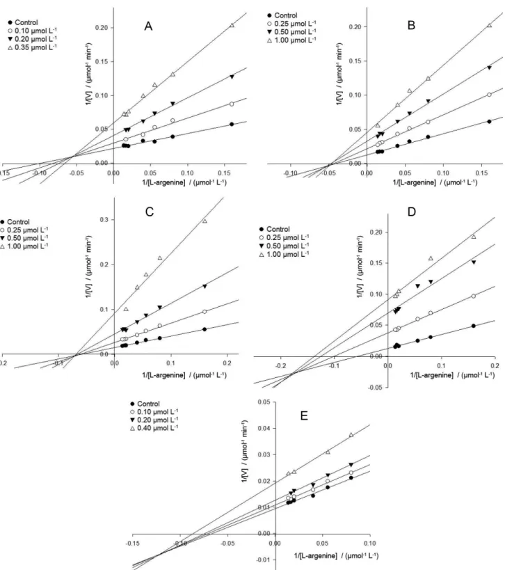
Kinetics studies of compounds (2-6) showed that these inhibitors shared a common behavior acting as noncompetitive inhibitors. For compounds 2, 5 and 6
the noncompetitive inhibition is also frequently known as mixed type, while inhibitors 3 and 4 are designated as pure noncompetitive inhibitors. Recently, gallic acid (Ki = 7.2 ± 1.4 µmol L-1) and epigallocatechin-3-gallate
(EGCG) (Ki = 4.0 ± 0.5 µmol L-1) showed potent ARG
inhibitory activity with a noncompetitive and mixed type, respectively.44
Analysis of Lineweaver-Burk plot (Figure 2A) for gallic acid (2) showed the intersecting lines converging to the left of the y-axis and above the abscissa (Kii > Kis), which means the inhibitor 2 binds with higher affinity to the free enzyme than to the enzyme-substrate complex. Additionally, when quinic acid is substituted in the position 5 by a galloyl (compound 3) or a methoxy galloyl moieties (compound 4), this behavior changes a little, each inhibitor (3 and 4) now binds with the same affinity to both, free enzyme and enzyme-substrate complex (Kii = Kis). This can also be seen by the double reciprocal plots (Figures 2B and 2C), which the nest of lines converge nearly at the x-axis. On the other hand, for the substituted quinic acid with two or more galloyl moieties (compound
5 and 6), the bind affinity is higher to the enzyme-substrate complex than to the free enzyme (Kii < Kis), and the line
intercept is now located below the abscissa in both plots (Figures 2D and 2E). The calculated values of affinity constants (Table 1) support the findings.
Gallic acid and derivatives have been reported as responsible for benefic effects and a number of chemopreventive properties provided by green tea and wine consumption.45,46 Also, other therapeutic effects have
been described, such as anti-cancer, antimicrobial and antiviral activity.47-50 Additionally, some gallotannins and
tannic acids have showed inhibitory potential on enzymes like α-glucosidase, fatty acid synthase (FAS) and HIV-1 protease. 24,51,52 This is contradictory with previous screening
for enzyme inhibitors that showed that tannins and lauryl gallate nonspecifically inhibited enzymes through the formation of aggregates, acting as promiscuous protein binders.53-55 The aggregate-based promiscuous inhibition
could be related to the large reactive functional groups of these compounds.55
We have identified the inhibition mechanism of the quinic acid esters (3-6) and gallic acid (2) as noncompetitive and mixed inhibitors, suggesting that the inhibitory activity of ARG would not be related to the formation of aggregates. In addition, some of these phenols have already been reported as leishmanicidal compounds. Previous study showed that gallic acid increases the expression of nitric oxide synthase (iNOS) and cytokine genes in Leishmania major parasitised
RAW 264.7 cells, as properties to combat the infection.56
This phenol demonstrated in in vitro study on L. donovani,
a nonspecific immune response recognized by release of tumour necrosis factor (TNF) and elevated nitric oxide (NO) concentration.26
Leishmania stimulate the PAs production for their
development and decrease the level of NO prompted by NOS.7,8,57 Thus, ARG inhibition could turn on the
production of NO, instead of PAs preventing the infection.
Conclusions
Gallic acid and derivatives isolated from B. coccolobifolia
showed to be novel noncompetitive and mixed inhibitors of ARG from L. amazonensis, with Ki values in the low
Table 1. IC50 and Ki values for secondary metabolites active on ARG
Compound IC50 / (µmol L-1)b Kis / (µmol L-1)b Kii / (µmol L-1)b Type of inhibition
Positive controla 12.20 ± 1.83 7.20 ± 0.90a 7.20 ± 0.90a Noncompetitivea
1 147.48 ± 15.44 – – –
2 0.13 ± 0.01 0.10 ± 0.03 0.18 ± 0.004 Mixed
3 0.44 ± 0.04 0.43 ± 0.014 0.41 ± 0.003 Noncompetitive
4 0.49 ± 0.04 0.16 ± 0.01 0.14 ± 0.01 Noncompetitive
5 0.46 ± 0.04 0.57 ± 0.03 0.23 ± 0.01 Mixed
6 0.31 ± 0.02 0.68 ± 0.02 0.35 ± 0.002 Mixed
aQuercitrin is a noncompetitive inhibitor used as a positive control (K
ii and Kis obtained from the reference 18); bdata values represent means of individual
Leishmanicidal Galloylquinic Acids are Noncompetitive Inhibitors of Arginase J. Braz. Chem. Soc. 1836
µmol L-1 range. In conclusion, our results suggest that the
ability of galloyl moiety in induce immunological response demonstrated earlier may be also a result of the inhibition of ARG activity. In this view, the galloylquinic acids should be considered as promising hits in the investigation of leishmanicidal compounds.
Supplementary Information
Supplementary data (1H, 13C NMR and Dixon plots of
acids) are available free of charge at http://jbcs.sbq.org.br as PDF file.
Figure 2. Lineweaver-Burk plots of gallic acid 2 (A), 5-O-galloylquinic acid 3 (B), 5-O-(3-methylgalloyl)-quinic acid 4 (C), 3,5-di-O-galloylquinic acid
Sousa et al. 1837 Vol. 25, No. 10, 2014
Acknowledgments
This research was supported by the State of São Paulo Research Foundation (FAPESP, Fundação de Amparo à Pesquisa do Estado de São Paulo) Proc. 2010-52326-9, INBEQMEdi and the National Council for Scientific and Technological Development (CNPq, Conselho Nacional de Pesquisa e Desenvolvimento), Brazil.
References
1. Chawla, B.; Madhubala, R.; J. Parasit. Dis.2010, 34, 1. 2. http://www.who.int/leishmaniasis/en/, accessed in May 2014. 3. Amato, V. S.; Padilha, A. R. S.; Nicodemo, A. C.; Duarte,
M. I. S.; Valentini, M.; Uip, D. E.; Boulos, M.; Amato Neto, V.; Int. J. Infect. Dis. 2000, 4, 153.
4. Gil, E. S.; Paula, J. R.; Nascimento, F. R. F.; Bezerra, J. C. B.; J. Basic Appl. Pharm. Sci. 2008, 29, 223.
5. López, R. E. S. Quim. Nova 2010, 33, 1541.
6. da Silva, M. F. L.; Zampieri, R. A.; Muxel, S. M.; Beverley, S. M.; Floeter-Winter, L. M.; PLoS One2012, 7,e34022. 7. Iniesta, V.; Gómez-Nieto, L. C.; Corraliza, I.; J. Exp. Med.2001,
193, 777.
8. Roberts, S. C.; Tancer, M. J.; Polinsky, M. R.; Gibson, K. M.; Heby, O.; Ullman, B.; J. Biol. Chem.2004, 279, 23668. 9. D’Antonio, E. L.; Ullman, B.; Roberts, S. C.; Dixit, U. G.;
Wilson, M. E.; Hai, Y.; Christianson, D. W.; Arch. Biochem. Biophys.2013, 535, 163.
10. da Silva, E. R.; da Silva, M. F.; Fischer, H.; Mortara, R. A.; Mayer, M. G.; Framesqui, K.; Silber, A. M.; Floeter-Winter, L. M.; Mol. Biochem. Parasitol.2008, 159, 104.
11. Balaña-Fouce, R.; Calvo-Álvarez, E.; Álvarez-Velilla, R.; Prada, C. F.; Pérez-Pertejo, Y.; Reguera, R. M.; Mol. Biochem. Parasitol.2012, 181, 85.
12. Colotti, G.; Ilari, A.; Amino Acids 2011, 40, 269.
13. Kropf, P.; Herath, S.; Weber, V.; Modolell, M.; Müller, I.; Parasite Immunol. 2003, 25, 439.
14. Kropf, P.; Fuentes, J. M.; Fähnrich, E.; Arpa, L.; Herath, S.; Weber, V.; Soler, G.; Celada, A.; Modolell, M.; Müller, I.; FASEB J.FJ Express2005, 19, 1000.
15. Riley, E.; Roberts, S. C.; Ullman, B.; J. Parasitol.2011, 41, 545.
16. da Silva, E. R.; Maquiaveli, C. C.; Magalhães, P. P.; Exp. Parasitol.2012, 130, 183.
17. Cruz, E. de M.; da Silva, E. R.; Maquiaveli, C. C.; Alves, E. S.; Lucon Jr., J. F.; dos Reis, M. B.; de Toledo, C. E.; Cruz, F. G.; Vannier-Santos, M. A.; Phytochemistry 2013, 89, 71. 18. Manjolin, L. C.; dos Reis, M. B. G.; Maquiaveli, C. C.;
Santos-Filho, O. A.; da Silva, E. R.; Food Chem. 2013, 141, 2253. 19. de Sousa, L. R. F.; Ramalho, S. D.; Burger, M. C. M.; Nebo, L.;
Fernandes, J. B.; da Silva, M. F. G. F.; Iemma, M. R. C.; Corrêa,
C. J.; de Souza, D. H. F; Lima, M. I. S.; Vieira, P. C.; J. Nat. Prod. 2014, 77, 392.
20. Dewick, P. M.; Medicinal Natural Products: a Biosynthetic Approach, 2nd ed.; John Wiley & Sons: England, 2001.
21. Rawat, G.; Tripathi, P. R. K.; Appl. Microbiol. Biotechnol.2013, 97, 4277.
22. Nepka, C.; Asprodini, E.; Kouretas, D.; Eur. J. Drug Metab. Pharmacokinet. 1999, 24, 183.
23. Crozier, A.; Jaganath, I. B.; Clifford, M. N.; Nat. Prod. Rep.
2009, 26, 1001.
24. Fan, H.; Wu, D.; Tian, W.; Ma, X.; Biochim. Biophys. Acta 2013, 1831, 1260.
25. Kolodziej, H.; Kiderlen, A. F.; Phytochemistry2005, 66, 2056. 26. Kayser, O.; Kolodziej, H.; Kiderlen, A. F.; Phytother. Res. 2001,
15, 122.
27. Accioly, M. P.; Bevilaqua, C. M. L.; Rondon, F. C. M.; de Morais, S. M.; Machado, L. K. A.; Almeida, C. A.; de Andrade Jr., H. F.; Cardoso, R. P. A.; Vet. Parasitol. 2012, 187, 79.
28. Clardy, J.; Walsh, C.; Nature 2004, 432, 829.
29. Guilhon-Simplicio, F.; Pereira, M. M.; Quim. Nova 2011, 34, 1032.
30. Sannomiya, M.; Fonseca, V. B.; da Silva, M. A.; Rocha, L. R. M.; dos Santos, L. C.; Hiruma-Lima, C. A.; Souza Brito, A. R. M.; Vilegas, W.; J. Ethnopharmacol.2005, 97, 1. 31. de Almeida, S. C. X.; de Lemos, T. L. G.; Silveira, E. R.; Pessoa,
O. D. L.; Quim. Nova2005, 28, 57.
32. Pawlowska, A. M.; De Leo, M.; Braca, A.; J. Agric. Food Chem.
2006, 54, 10234.
33. Itoh, A.; Tanaka, Y.; Nagakura, N.; Nishi, T.; Tanahashi, T.; J. Nat. Med.2006, 60, 146.
34. Bouchet, N.; Levesque, J.; Blond, A.; Bodo, B.; Pousset, J. L.; Phytochemistry 1996, 42, 189.
35. Moore, J. P.; Westall, K. L.; Ravenscroft, N.; Farrant, J. M.; Lindsey, G. G.; Brandt, W. F.; Biochem. J.2005, 385, 301. 36. Pauli, G. F.; Poetsch, F.; Nahrstedt, A.; Phytochem. Anal. 1998,
9, 177.
37. Camacho, M. D. R.; Phillipson, J. D.; Croft, S. L.; Marley, D.; Kirby, G. C.; Warhurst, D. C.; J. Nat. Prod. 2002, 65, 1457. 38. Bradford, M. M.; Anal. Biochem.1976, 72, 248.
39. da Silva, E. R.; Floeter-Winter, L. M.; Exp. Parasitol.2010, 125, 152.
40. Fawcett, J. K.; Scott, J. E.; J. Clin. Pathol.1960, 13, 156. 41. Bisswanger, H.; Enzyme Kinetics: Principles and Methods, 2nd
ed.; WILEY-VCH Verlag GmbH & Co. KGaA: Germany, 2008. 42. Dixon, M.; Biochem. J.1953, 55, 170.
43. Krupka, R. M.; Biochem. J. 1963, 2, 76.
44. dos Reis, M. B. G.; Manjolin, L. C.; Maquiaveli, C. C.; Santos-Filho, O. A.; da Silva, E. R.; Plos One 2013, 8, e78387.
45. Okuda, T.; Yoshida, T.; Hatano, T.; Fortschr. Chem. Org. Naturst.
Leishmanicidal Galloylquinic Acids are Noncompetitive Inhibitors of Arginase J. Braz. Chem. Soc. 1838
46. Paixão, N.; Pereira, V.; Marques, J. C.; Camara, J. S.; J. Sep. Sci. 2008, 31, 2189.
47. Liu, X.; Kim, J.; Li, Y.; Li, J.; Liu, F.; Chen, X.; J. Nutr.2005, 135,165.
48. Ximenes, V. F.; Lopes, M. G.; Petronio, M. S.; Regasini, L. O.; Silva, D. H.; da Fonseca, L. M.; J. Agric. Food Chem. 2010, 58, 5355.
49. Giftson, J. S.; Jayanthi, S.; Nalini, N.; Invest. New Drugs 2010, 28, 251.
50. Morais, M. C.; Luqman, S.; Kondratyuk, T. P.; Petronio, M. S.; Regasini, L. O.; Silva, D. H.; Bolzani, V. S.; Soares, C. P.; Pezzuto, J. M.; Nat. Prod. Res. 2010, 24, 1758.
51. Flausino Jr., O. A.; Dufaua, L.; Regasini, L. O.; Petrônio, M. S.; Silva, D. H. S.; Rosec, T.; Bolzani, V. S.; Reboud-Ravauxa, M.; Curr. Med. Chem. 2012, 19, 4534.
52. Wana, C.; Yuan, T.; Li, L.; Kandhi, V.; Cech, N. B.; Xie, M.; Seeram, N. P.; Bioorg. Med. Chem. Lett. 2012,22, 597. 53. Pohjala, L.; Tammela, P.; Molecules 2012, 17, 10774. 54. Coan, K. E.; Shoichet, B. K.; Mol. Biosyst. 2007, 3, 208. 55. Coan, K. E.; Maltby, D. A.; Burlingame, A. L.; Shoichet, B. K.;
J. Med. Chem. 2009, 52, 2067.
56. Radtke, O. A.; Kiderlen, A. F.; Kayser, O.; Kolodziej, H.; Planta Med.2004, 70, 924.
57. Gaur, U.; Roberts, S. C.; Dalvi, R. P.; Corraliza, I.; Ullman, B.; Wilson, M. E.; J. Immunol. 2007, 179, 8446.
Submitted on: March 10, 2014
Published online: May 23, 2014
Supplementary Information
S
I
J. Braz. Chem. Soc., Vol. 25, No. 10, S1-S9, 2014.Printed in Brazil - ©2014 Sociedade Brasileira de Química 0103 - 5053 $6.00+0.00
*e-mail: [email protected]
Leishmanicidal Galloylquinic Acids are Noncompetitive Inhibitors of Arginase
Lorena R. F. de Sousa,a Suelem D. Ramalho,a João B. Fernandes,a
Maria Fátima das G. F. da Silva,a Mônica R. da C. Iemma,a Caroindes J. Corrêa,a
Dulce H. F. de Souza,a Maria I. S. Limab and Paulo C. Vieira*,a
aDepartamento de Química and bDepartamento de Botânica, Universidade Federal de São Carlos,
Rod. Washington Luís, km 235, 13565-905 São Carlos-SP, Brazil
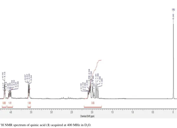
Figure S2.13C NMR spectrum of quinic acid (1) acquired at 100 MHz in D 2O. Figure S1.1H NMR spectrum of quinic acid (1) acquired at 400 MHz in D
Leishmanicidal Galloylquinic Acids are Noncompetitive Inhibitors of Arginase J. Braz. Chem. Soc.
S2
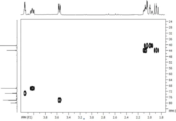
Figure S3. HSQC of quinic acid (1) acquired at 400 MHz in D2O.
Sousa et al. S3 Vol. 25, No. 10, 2014
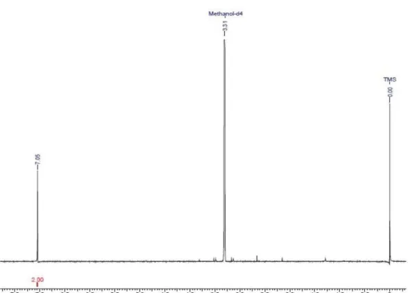
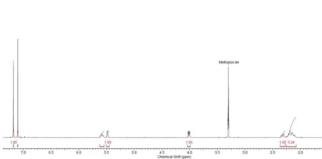
Figure S6.1H NMR spectrum of 5-O-galloylquinic acid (3) acquired at 400 MHz in MeOH- d 4. Figure S5.1H NMR spectrum of gallic acid (2) acquired at 400 MHz in MeOH-d
Leishmanicidal Galloylquinic Acids are Noncompetitive Inhibitors of Arginase J. Braz. Chem. Soc.
S4
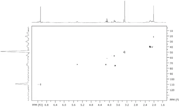
Figure S7. HSQC of 5-O-galloylquinic acid (3) acquired at 400 MHz in MeOH- d4.
Sousa et al. S5 Vol. 25, No. 10, 2014
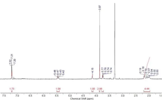
Figure S9.1H NMR spectrum of 5-O-(3-methylgalloyl)-quinic acid(4) acquired at 400 MHz in MeOH- d 4.
Leishmanicidal Galloylquinic Acids are Noncompetitive Inhibitors of Arginase J. Braz. Chem. Soc.
S6
Figure S11. HMBC of 5-O-(3-methylgalloyl)-quinic acid (4) acquired at 400 MHz in MeOH- d4.
Sousa et al. S7 Vol. 25, No. 10, 2014
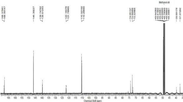
Figure S13.13C NMR spectrum of 3,5-di-O-galloylquinic acid (5) acquired at 100 MHz in MeOH- d 4.
Leishmanicidal Galloylquinic Acids are Noncompetitive Inhibitors of Arginase J. Braz. Chem. Soc.
S8
Figure S15. HMBC of 3,5-di-O-galloylquinic acid (5) acquired at 400 MHz in MeOH- d4.
Sousa et al. S9 Vol. 25, No. 10, 2014
Figure S17. HSQC of 3,4,5-tri-O-galloylquinic acid (6) acquired at 400 MHz in MeOH-d4.