O R I G I N A L A R T I C L E UDC: 616.831-073-71:616.8-053.31-036 DOI: 10.2298/VSP1206492V
The prognostic value of amplitude-integrated electroencephalography
in neonates with hypoxic-ischemic encephalopathy
Prognosti
þ
ka vrednost elektroencefalografije integrisanih amplituda kod
novoro
ÿ
en
þ
adi sa hipoksi
þ
ko-ishemijskom encefalopatijom
Brankica Vasiljeviü*, Svjetlana Maglajliü-Djukiü†, Miroslava Gojniü*
*Institute of Gynecology and Obstetrics, Clinical Center of Serbia, Belgrade, Serbia,
†
University Children's Hospital, Belgrade, Serbia
Abstract
Background/Ǎim. Diagnosis of perinatal hypoxic-ische-mic encephalopathy (HIE) and early prediction neurologi-cal outcome is important and difficult. The aim of this study was to determine the prognostic value of amplitude-integrated electroencephalography (aEEG) for abnormal neurodevelopment outcome in a neonate with HIE. Methods. A total of 90 neonates > 32 gestational age (GA) with HIE were enrolled prospectively. All neonates with HIE were categorized into three grades according to the Sarnat and Sarnat clinical scoring system (mild HIE, moderate HIE and severe HIE). aEEG traces were re-corded with a cerebral function monitor (CFM) during the first 72 h of life. The neurodevelopment outcome was as-sessed at 12 months of age of corrected gestational age. Results. The pattern of aEEG correlated with the severity of HIE (p < 0.0001) and subsequent neurodevelopment outcome (p < 0.001). We found that aEEG background patterns exhibited superior prediction of abnormal out-comes at 12 months of age (sensitivity of 91.7% and speci-ficity of 94.3%, positive predictive value of 78.6% and negative predictive value of 98.1%) when compared to aEEG seizure (sensitivity of 94% and specificity of 48%, positive predictive value of 57% and a negative predictive value of 92%). Electroclinical dissociation seizure was de-tected in 28% of the neonates with HIE. Conclusions. Our results confirm that aEEG is simple and accurate bedside diagnostic method for assessing extension of hy-poxic-ischemic brain damage and early identification of neonates with perinatal HIE who are at high risk of neu-rodevelopmental impairment.
Key words:
elektroencephalography; intensive care units, neonatal; hypoxia-ishemia, brain; infant, premature; infant, newborn; prognosis.
Apstrakt
Uvod/Cilj. Dijagnoza perinatalne hipoksiÿno-ishemiÿke encefalopatije (HIE) i prognoza kasnijeg neurološkog ishoda je važna i, istovremeno, vema teška. Cilj ove studije bio je da se utvrdi prognostiÿki znaÿaj elektroencefalograma integrisanih amplituda (aEEG) za utvrĀivanje neurološke prognoze kod novoroĀenÿeta sa HIE. Metode. Ovom prospektivnom studijom bilo je obuhvaýeno 90 novoroĀ en-ÿadi > 32 gestacijske nedelje (GN) sa HIE. Sva ispitivana novoroĀenÿad bila su neurološki procenjivana prema Sarnat i Sarnat kliniÿkom skoru i podeljena u 3 grupe (blaga HIE, srednje teška HIE i teška HIE). Snimanje aEEG vršeno je cerebralnim funkcionim monitorom (CFM) tokom prva 72 sata života. Neurološka procena ispitivane dece sprovedena je u uzrastu od 12 meseci i korigovana je prema gestacijskoj zrelosti na roĀenju. Rezultati. Registrovana aEEG aktiv-nost bila je u korelaciji sa težinom HIE (p < 0,0001) i kasni-jim razvojem neuroloških sekvela (p < 0,001). Naši rezultati ukazuju da registrovana osnovna aEEG aktivnost ima višu prediktivnu vrednost za loš neurološki ishod u izrastu od 12 meseci (senzitivnost 91,7%, specifiÿnost 84,3%, pozitivna prediktivna vrednost 78,6% i negativna prediktivna vrednost 98,1%) od registrovanih aEEG konvulzija (senzitivnost 94%, specifiÿnost 48%, pozitivna prediktivna vrednost 57% i negativna prediktivna vrednost 92%). Elektrokliniÿka diso-cijacija konvulzija je registrovana kod 28% novoroĀenÿadi sa HIE. Zakljuÿak. Naši rezultati ukazuju da je aEEG jed-nostavna i precizna dijagnostiÿka metoda za procenu ek-stenzivnosti hipoksiÿno-ishemiÿkog moždanog ošteýenja i ranu identifikaciju novoroĀenÿadi sa HIE kod kojih je pri-sutan visok rizik od kasnijeg nastanka neuroloških sekvela.
Kljuÿne reÿi:
Introduction
Perinatal hypoxic-ischemic encephalopathy (HIE) is a common cause of neonatal morbidity and mortality and neu-rological disabilities among survivors 1. Each year 1.2 mil-lion neonates die and about 1 milmil-lion infants have permanent neurological disability caused by HIE 2. Determination of se-verity of perinatal HIE by the clinical criteria and appropriate neuroimaging or neurophysiologic techniques remains the main prognostic tool.
Neonates with mild HIE have a uniformly good prog-nosis and those with severe HIE a very high risk for early death or severe disabilities. Thus, especially for the neonates with moderate HIE, neuroimaging and neurophysiological examinations have a great value. Unfortunately, the predic-tive values of neuroimaging techniques (i.e. magnetic reso-nance imaging, computerized tomography and ultrasound) are limited during the first days of life 3. In contrast, neuro-physiological examinations of functional integrity of the brain have been shown to be useful also during the first days of life. High predictive values have been reported for evoked potentials (somatosensory, visual and auditory), conventional encephalography (EEG) and amplitude-integrated encephalography (aEEG) 4. Both serial registra-tion of convenregistra-tional EEG and measurement of evoked po-tentials in a neonatal intensive care unit (NICU) require considerable technical skill, time and expertise in interpre-tation and may not be rapidly available in most hospitals. An alternative technique is aEEG recorded with a cerebral function monitor (CFM), designed for long-term monitoring brain activity at bedside 5. CFM is a simplified single- or two-channel electroencephalogram monitor 6. A signal is obtained from a single pair of electrodes placed at the P3 and P4 position of the 10–20 International System, i.e. in the left and right parietal region. A guard or reference elec-trode positioned anterior to the vertex was also used to re-duce the effects of electrical interference. The use of two channels (two pair of bilateral frontoparietal electrodes) has the advantage of defining laterality in unilateral lesions, not available in the single channel devices. The signal is ampli-fied and passed through an asymmetrical band filter which attenuates activity below 2 Hz and above 15 Hz in order to minimize artefacts from muscle activity and electrical inter-ference. Additional processing includes semi logarithmic amplitude compression, rectification and time compression. A signal is presented electronically on the device monitor and can be recorded on paper with a semi-logarithmic scale at slow speed (6 cm/hr). A second trace continuously rec-ords the electrode impedance. EEG waveform can also be displayed on the monitor. The bandwidth in aEEG traces re-flects variations in upper and lower margins of activity or patterns of aEEG, both of which depend on the maturity and severity of the illness of a newborn infant 7.
In this study we prospectively evaluated the prognostic value of aEEG for assessing extension of hypoxic-ischemic brain damage and early identification of neonates with peri-natal HIE who are at high risk of neurodevelopmental im-pairment.
Methods
Our study was performed from January 2007 to January 2009 and was approved by the Ethical Committee for Medi-cal Research of the MediMedi-cal Faculty at the University of Bel-grade. Our institute serves as a referral center for high-risk pregnancies, with delivery numbers of 7,000–7,500 per year. We studied 90 neonates under 32 weeks gestational age (GA) with perinatal HIE admitted to neonatal intensive care units (NICU) at the Institute of Gynecology and Obstetrics, Clinical Center of Serbia, Belgrade. Written consent was obtained from all parents. Perinatal HIE was diagnosed if fetal distress (meconium staining of liquor or abnormal fetal heart rate), metabolic acidosis [pH < 7.20, base excess (BE)
10 mmol/L and lactate > 3mmol/L in arterial cord blood within 60 min of birth], immediate neonatal depression Ap-gar score (AS) 6 at 5 min and/or delayed spontaneous res-piration, necessitating artificial ventilation at 5 min, and early neonatal encephalopathy (within the first 24 of life) were presented. All the neonates were resuscitated according to the guidelines of the Newborn Resuscitation Program of the American Academy of Pediatrics and American Heart Association 8, 9.
A complete obstetrical history and physical examina-tions were obtained on admission. Perinatal HIE was catego-rised into three stages according to the Sarnat and Sarnat clinical scoring system 10. Head sonograms were performed on all the neonates before enrolment.
aEEG recordings started after initial stabilization in all 90 neonates with HIE. aEEG was recorded during the first 72 h of life using the Cerebral Function Monitor Olympic 6000 (Olympic Biomedical, USA) from biparietal adhesive elec-trodes and displayed on the integral printer at 6 cm/h. Han-dling of the neonates, observed clinical seizures, and admini-stration of anticonvulsants or sedatives were recorded by the nursing staff. We excluded any aEEG records within 30 minutes of anticonvulsant administration. The CFM also re-corded the impedance across the electrodes which was al-ways below 10 k.
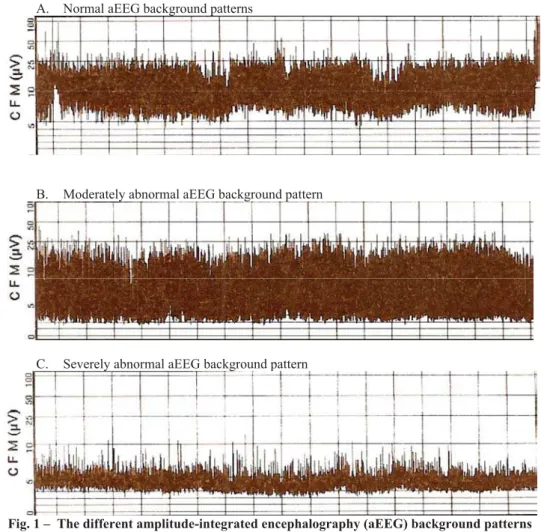
Evaluation of aEEG recording should begin with the background pattern and then proceed to the presence or ab-sence of seizures. aEEGs were described with either voltage criteria, using the upper and lower margins of activity or patterns of aEEG.
less than 10 ȝV and the lower margin is usually less than 5
ȝV. A severely abnormal trace is characterized by a general suppression of amplitude and this pattern may be accompanied by brief bursts of higher voltage spikes which appear as single spikes above the background activity – “burst suppression”. A severely abnormal trace is usually seen with severe encepha-lopathy and is often accompanied by seizure activity).
In addition, any of these three groups could be accom-panied by seizures. Seizures were manifested as periods of sudden increase in voltage accompanied by narrowing of the band of aEEG activity and followed by a brief period of sup-pression (Figure 2). Arousal during care procedures may be misinterpreted as seizures. It is therefore important that all the procedures are documented to facilitate correct interpre-tation of aEEG. Administration of anticonvulsants or seda-tives or handling of the neonates was recorded by the nursing staff. Status epilepticus often looks like a ‘saw tooth’ pattern
but a continuously raised background may also be seen. Cor-rect interpretation is only possible when simultaneous raw EEG is also available.
Neurodevelopment outcome was assessed at 12 months of corrected gestational age by the neonatologist and pediat-ric neurologist using the Denver Developmental Screening Test (DDST). Neurodevelopment outcomes were classified as normal, mild motor abnormality (slight abnormality in muscular tone or mild delayed of motor development) and severe adverse outcome [cerebral palsy (CP), epilepsy or if died in follow-up period].
We excluded neonates with congenital malformations, chromosomal abnormalities, inherited metabolic disorders, congenital or acquired neonatal infections and maternal drug addiction.
Statistical Package for Social Science software 11.5.1 for Windows (SPSS Inc., Chicago, IL, U.S.A.) was used for A. Normal aEEG background patterns
B. Moderately abnormal aEEG background pattern
C. Severely abnormal aEEG background pattern
Fig. 1 – The different amplitude-integrated encephalography (aEEG) background patterns
data management and statistical analysis. Values of p < 0.05 were considered statistically significant. All values were ex-pressed as mean value with standard deviation (SD) or as percentages (%) for descriptive purposes unless otherwise stated. Group comparisons were performed with the Fisher exact test or Kruskall-Wallis test and Wilcoxon rank sum test. The predictive value of aEEG for determining neurode-velopmental prognosis was assessed by calculation of sensi-tivity and specificity and positive and negative predictive values (PPV and NPV, respectively). Primary analysis was made by assessing the predictive value of abnormal aEEG background activity alone and/or seizure aEEG activity.
Results
We studied 90 neonates (> 32 GA) with perinatal HIE. The majority of neonates in our study developed mild HIE (Table 1). Birth weight (BW) and gestational age were simi-lar, while gender distribution was different among the neo-nates with mild and advanced clinical stage of HIE (p < 0.05) (Table 1). At term, the incidence of HIE ranged from 3 to 4 per 1,000 live births during our study period. Predictive ca-pacities AS at 1 and 5 min and arterial blood cord values of pH, BE and lactate for severity of HIE and abnormal
out-come are shown in Table 1. Apgar scores at 1 min (W = 166;
p < 0.05) and AS at 5 min (W = 181; p < 0.01) and pH val-ues of arterial blood cord (W = 184; p < 0.001), BE (W = 167; p < 0.05) and lactate (W = 51; p < 0.05) correlated well with severity of HIE. Arterial cord blood pH had best pre-dictive capacities for the severity of HIE and subsequent ab-normal neurological outcome (W = 916; p < 0.001). We de-tected no significant difference among arterial blood cord pH
values, BE and lactate or AS at 1min and 5 min in preterm and term neonates.
A relation between aEEG patterns and severity of HIE and neurologic outcome is summarized in Figure 3. There was a close relationship between the findings on aEEG pat-terns and severity of HIE (p < 0.0001) (Figure 3). The aEEG patterns in the first 72 h of life also had high predictive value for abnormal outcome (p < 0.001) (Figure 3). All neonates with normal aEEG patterns had good outcome while neo-nates with severely abnormal aEEG patterns (low voltage or inactive isoelectric background activity) either died or sub-sequently had an abnormal neurologic outcome. All the three HIE groups could be accompanied by seizures (Figure 3). Electrographic seizures without clinical correlates were de-tected on the CFM in 28% of the neonates with HIE (Figure 4). In the neonates with seizures, we found that the interictal aEEG activity correlated with the subsequent outcome. Se-verely abnormal aEEG patterns accompanied with seizures had a poor outcome. We detected no significant difference among aEEG patterns in preterm and term neonates.
Neurodevelopment outcome at 12 months of cor-rected gestational age corresponded well with severity of HIE (p < 0.01) and gestation age (p < 0.05) (Table 2). All the infants with severe HIE developed neurological
se-quels (cerebral palsy or epilepsy). Two preterm neonates with severe HIE died, one within the early neonatal period because of multiorgan failure, the other later because of respiratory dysfunction. In determining the prognostic value of aEEG, we included death and neurodevelopmen-tal impairment as a single outcome group. Incidences of neurological sequels were significantly higher in preterm infants (p < 0.05). We found that aEEG background
pat-Table 1 Clinical and biochemical characteristics at birth in neonates with different stage of hypoxic-ischemic encephalopathy
Parameters HIE HIE I HIE II HIE III NS
GA 36.6 ± 2.6 36.4 ± 2.5 37.4 ± 2.6 36.2 ± 3.1 35.8 ± 2.7
BW (g) 2711 ± 810 2699 ± 775 2889 ± 796 2311 ± 1000 2450 ± 93
M /F(%) 62/38 54/46 67/33* 100/0* 67/33*
AS (1min) 3.4 ± 1.4 4.1 ± 1.1 2.6 ± 1.3 1.3 ± 0.7* 2.5 ± 1.6*
AS (5min) 5.5 ± 1.7 5.6 ± 0.5 4.3 ± 1.2 2.7 ± 1.0* 3.8 ± 1.8** pH 7.09 ± 0.11 7.15 ± 0.05 7.03 ± 0.11** 6.88 ± 0.09*** 6.97 ± 0.14*** BE (mmol/L) -13.1 ± 4.2 -11.0 ± 1.7 -15.5 ± 4.5* -19.7 ± 3.9** -16.5 ± 5.1* Lactate(mmol/L) 7.4 ± 4.4 5.4 ± 2.9 9.6 ± 4.6* 13.9 ± 3. 9* 11.0 ± 5.1*
N 90 57 24 9 15
Values are expressed as mean± standard deviation (SD) or percentages (%). Kruskal-Wallis F2 test and Wilcoxon rank sum test with continuity
correction. Significance *p < 0.05 or **p < 0.01 or ***p < 0.001. BW = birth weight: AGA = appropriate BW for gestation age: SGA = small BW for gestation age and LGA = large BW for gestation age; GA = gestational age; M = male and F = female; AS = Apgar score; BE = base excess;
N = numbers of neonates; HIE = hypoxic-ischemic encephalopathy (HIE stage I, HIE stage II and HIE stage III); NS = neurological sequels.
Table 2 Neurodevelopment outcome (NDO) in neonates with different stages of hypoxic-ischemic
encephalopathy and different gestation age at birth
NDO HIE HIE I HIE II HIE III PRETERM TERM
Normal NDO (%) 73.9 91.3** 54.2** 0** 62.7 84.4*
M (%) 12.5 7 29.2** 0* 16.3 * 8.9
CP (%) 10.2 1.7** 12.5 71.4** 14* 6.7
EPI (%) 3.5 0 4.1 28.6** 13.3 * 0
Values are expressed as percentages (%); Fisher exact test significance; *p < 0.05 or ** p < 0.01;
terns exhibited superior prediction of abnormal outcomes at 12 months of age (sensitivity of 91.7% and specificity of 94.3%, positive predictive value of 78.6% and negative predictive value of 98.1%) when compared to aEEG sei-zure (sensitivity of 94% and specificity of 48%, positive predictive value of 57% and negative predictive value of 92%) (Table 3).
Discussion
Outcome prediction after perinatal asphyxia is impor-tant and difficult. Despite vast advances in neonatal intensive care the incidence of HIE continues to have a significant im-pact on the perinatal morbidity and mortality. The incidence of perinatal HIE varies at different gestational ages. At term,
the incidence ranges from 3 to 5 per 1,000 live births and an incidence approaching 60% in premature newborns 12. In our study the incidence of perinatal HIE was similar. The age-dependent vulnerability to hypoxic-ischemic insults seen in the immature brain can be explained by the high density of N-methyl-d-aspartate (NMDA) receptors and neuronal nitrie oxide synthase (nNOS)-positive cells 13, 14. The immature
brain is especially sensitive to oxidative damage relative to the mature brain are poor antioxidant capabilities and a high concentration of free iron and lipids.
In our study males had higher incidence of long term developmental disabilities than females. A recent analysis of a European database of 4,500 children with CP found that the incidence of CP was 30% higher in males than females.
Fig. 3 – The differentamplitude-integrated encephalography (aEEG) characteristics in neonates with different stages of hy-poxic-ischemic encephalopathy and subsequent neurologic outcome
N-aEEG = normal aEEG background patterns; S-aEEG- = moderately abnormal aEEG background patterns; T-aEEG- severely abnormal aEEG background pat-ternsM; K-aEEG = seizures aEEG activity; HIE = hypoxic-ischemic encephalopathy (HIE stage I, HIE stage II and HIE stage III); NS = neurological sequels.
Fig. 4 – Electroclinical dissociation seizure in neonates with different stage of hypoxic-ischemic encephalopathy and subse-quent neurologic outcome
HIE = hypoxic-ischemic encephalopathy (HIE stage I, HIE stage II and HIE stage III); NS = neurological sequels
Table 3 The predictive value of amplitude-integrated electroencephalography (aEEG) background activity and seizures aEEG
activity for determining neurodevelopmental prognosis in neonates with hypoxic-ischemic encephalopathy
aEEG activity Sensitivity (%)
Specificity (%)
PPV (%)
NPV (%)
Background 91.7 94.3 78.6 98.1
Seizures 94 48 57 92
Basic research of the causes of CP has revealed that gender may influence the pathogenesis of developmental brain in-jury. Sex differences in the immature brain appear to be strongly influenced by intrinsic differences between male and female cells and this is influenced by sex chromosomes and sex hormones 15, 16.
The Apgar scoring system, first devised in 1952, has been used to assess a newborn’s condition and to reflect the need for resuscitation 17. Over time, the Apgar score has also been used to define asphyxia, which is inappropriate, as many other conditions (e.g., congenital anomalies, prematur-ity, maternal drug administration) can result in low scores that are not reflective of asphyxia 18. On the other hand, an-other study stated that low Apgar scores at 5 min are associ-ated with death or cerebral palsy, and this association in-creased if both Apgar scores at 1 and 5 minute were low 19. Our results also showed that the Apgar score at 5 minute re-mained a valid predictor of neonatal mortality, but using it alone to predict long-term outcome was inappropriate.
The incidence of long-term complications depends on the severity of hypoxic-ischemic encephalopathy which is in concordance with our results. In adults, neuronal necrosis and apoptosis after global ischemia are slow, and last for several hours to several days. Studies in neonatal experi-mental model suggest a quicker cellular destruction and en-ergy substrates in the neonatal brain continue to run down for 12 h to 48 h after perinatal hypoxia 20. Therefore, neuropro-tective intervention might be effective 6 h to 8 h after peri-natal hypoxic-ischemic insult 18. Intervention and treatment following perinatal asphyxia may not be free of risk.
A CFM could be useful for selecting those neonates who might benefit from early intervention after perinatal asphyxia, avoid unnecessary risks related to treatment for those neonates that do not need intervention 21, 22. A CFM provides a continu-ous real-time display of cerebral electrical activity at bedside to assist the clinician in making immediate treatment decisions and in identifying high-risk neonates, which requires closer attention. The benefit of CFM in a NeuroCare Unit (NCU) setting is its simplicity and ease of interpretation. It can be ap-plied rapidly by nursing staff at any time of the day. We showed that aEEG correlates closely with neurologic outcome in neonates with HIE. The ability of aEEG background ab-normalities to predict abnormal outcome has been previously studied in asphyxiated neonates 23. Our study showed that neonates with normal aEEG patterns within the first 72 h of life were likely to survive without sequelaes, by contrast, neo-nates with severely abnormal aEEG patterns or worse pattern were at a risk of death or neurological sequela. These findings are in accordance with previous studies 24. Our data also show that normalization of abnormal background patterns is associ-ated with normal neurologic outcomes, whereas severely ab-normal aEEG patterns that persisted beyond the age of 72 h are associated with adverse outcomes. These findings are in accordance with previous studies 25.
Diagnosis of seizures in a neonate has been based on clinical recognition of repetitive, stereotypic motor activity or behavioural phenomena. All neonates with either clinical or silent seizures were treated with antiepileptic drugs
(phe-nobarbital as drug of first choice). Treatment with antiepi-leptic drugs has never changed a normal pattern into a se-verely abnormal one, although in some infants, the pattern became transiently more discontinuous than before, for 30 to 60 min. By excluding from analysis the parts of the records that were associated with drug administration or handling of the neonates we minimized possible confounding effects. A CFM aids in the detection of seizures and displays their se-verity, duration and frequency in real time 26. In neonates with seizures, we found that the interictal aEEG activity cor-related with a subsequent outcome. The neonates with sei-zures and a normal-amplitude aEEG had a normal outcome, whereas the neonates with seizures and moderately abnormal or suppressed amplitude aEEG had a poor outcome. A CFM also helps to evaluate response to anticonvulsive therapies and to identify subclinical seizures. Electrographic seizures without clinical correlates are common in neonates with HIE. From previous studies, it is estimated that > 50% of seizures identified on EEG or aEEG in neonates may be silent 27. However, there are studies with the opposite results, report-ing a rate of electroclinical dissociation seizure of approxi-mately < 30%–50% in neonates with HIE, which is in accor-dance with our findings. Moreover, once anticonvulsant therapy is initiated, electrographic seizures may persist well after cessation of clinical seizure activity 28. The importance of electrographic seizures is underscored by recent data showing that electrographic seizures with or without clinical correlates may have detrimental effects on the neonatal brain. Animal and human data indicate that seizures in the developing brain may be harmful, at least in the short term, considering disturbances in cerebral blood flow, energy me-tabolism, and excitotoxic amino acids. This suggests that an-ticonvulsant therapy that suppresses clinical but not elec-trographic seizures may not be fully effective in preventing brain injury and that an appropriate goal of anticonvulsant therapy is to suppress both clinical and electrographic sei-zures. Brief seizure activity may be missed, and neonatolo-gists with limited experience in reading aEEGs may misin-terpret the presence or absence of seizures 29. These studies stress the fact that experience is required to be able to inter-pret aEEG traces, and one should also be aware of the limi-tations of the technology. The long duration of aEEG re-cording outweighs the limitations of obtaining detailed in-formation during much shorter, 30–40 min of full montage EEG recording. Newer systems provide access to raw EEG, and may offer 2 to 4 channels of recording. The use of these two modalities in conjunction is likely to provide the best in-formation at the current state of the technology 30.
very unlikely that immaturity influenced our results. Our study shows that when CFM is used in combination with standard neurological examination, it enhances the clini-cian’s ability to identify neonates at risk for poor long-term neurodevelopment outcome. Just like monitoring of respira-tion, heart rate, and saturation is routine in neonatal intensive care settings for high risk neonates, continuous aEEG re-cording to monitor brain function may be appropriate for neonates with HIE and may be considered a standard of care.
Conclusion
In conclusion, our findings confirm that continuous aEEG is a simple and accurate bedside diagnostic method for assessing extension of hypoxic-ischemic brain damage and
early identification of neonates with perinatal HIE who are at high risk of developmental delay. aEEG improves our ability to detect neonates at risk of hypoxic-ischemic brain injury at an earlier stage, when the window for therapeutic action is still open, optimize timing and assessment of neuroprotective treatment at the same time.
Acknowledgements
The authors are thankful to Dusica Gavrilovic, Institute for Radiology and Oncology, Department of Medical Educa-tion for her statistical help.
We declare that none of the authors has any competing interests with regard to the manuscript.
R E F E R E N C E S
1. Vannucci RC. Mechanisms of perinatal hypoxic-ischemic brain
damage. Semin Perinatol 1993; 17(5): 330î7.
2. Lincetto O. Birth asphyxia. In: World Health Organisation, editor.
Symmary of the revious meeting and protocol overview. Geneve: World Health Organisation; 2007. p. 3–35.
3. Vasiljeviý B, Maglajliý-Djukiý S, Stankoviý S, Lutovac D, Gojniý M.
Predictive value of color Doppler neuro-sonography for the development of neurological sequels in newborn infants with hypoxic ischemic encephalopathy. Vojnosanit Pregl 2011; 68(10): 825–31. (Serbian)
4. Blankenberg FG, Loh NN, Bracci P, D'Arceuil HE, Rhine WD,
Norbash AM, et al. Sonography, CT, and MR imaging: a
pro-spective comparison of neonates with suspected intracranial ischemia and hemorrhage. AJNR Am J Neuroradiol 2000; 21(1): 213î8.
5. Eken P, Toet MC, Groenendaal F, de Vries LS. Predictive value of
early neuroimaging, pulsed Doppler and neurophysiology in full term infants with hypoxic-ischaemic encephalopathy. Arch Dis Child Fetal Neonatal Ed 1995; 73(2): F75î80.
6. Rennie JM, Chorley G, Boylan GB, Pressler R, Nguyen Y, Hooper R.
Non-expert use of the cerebral function monitor for neonatal seizure detection. Arch Dis Child Fetal Neonatal Ed 2004;89(1):F37-40.
7. Burdjalov VF, Baumgart S, Spitzer AR. Cerebral function
monitor-ing: a new scoring system for the evaluation of brain maturation in neonates. Pediatrics 2003; 112(4): 855î61.
8. International Liaison Committee on Resuscitation. 2005 International
Consensus on Cardiopulmonary Resuscitation and Emergency Cardiovascular Care Science with Treatment Recommenda-tions. Part 7: Neonatal resuscitation. Resuscitation 2005; 67(2î3): 293î303.
9. Finer N, Leone T. Oxygen saturation monitoring for the
pre-term infant: the evidence basis for current practice. Pediatr Res 2009; 65(4): 375î80.
10. Sarnat HB, Sarnat MS. Neonatal encephalopathy following fetal
distress. A clinical and electroencephalographic study. Arch Neurol 1976; 33(10): 696î705.
11. Al Naqeeb N, Edwards AD, Cowan FM, Azzopardi D.
Assess-ment of neonatal encephalopathy by amplitude-integrated electroencephalography. Pediatrics 1999; 103(6): 1263î71.
12. Vannucci SJ, Hagberg H. Hypoxia-ischemia in the immature
brain. J Exp Biol 2004; 207(Pt 18): 3149î54.
13. Ikonomidou C, Bosch F, Miksa M, Bittigau P, Vöckler J, Dikranian
K, et al. Blockade of NMDA receptors and apoptotic neurode-generation in the developing brain. Science 1999; 283(5398): 70î4.
14. Bal-Price A, Brown GC. Inflammatory neurodegeneration
medi-ated by nitric oxide from activmedi-ated glia-inhibiting neuronal res-piration, causing glutamate release and excitotoxicity. J Neuro-sci 2001; 21(17): 6480î91.
15.Johnston MV, Hagberg H. Sex and the pathogenesis of cerebral
palsy. Dev Med Child Neurol 2007; 49(1): 74î8.
16.Wang X, Zhu C, Hagberg H, Korhonen L, Sandberg M, Lindholm D,
et al. X-linked inhibitor of apoptosis (XIAP) protein protects against caspase activation and tissue loss after neonatal hy-poxia-ischemia.Neurobiol Dis 2004;16(1): 179î89.
17.Apgar V, Holaday DA, James LS, Weisbrot IM, Berrien C.
Evaluation of the newborn infant; second report.J Am Med Assoc 1958; 168(15): 1985î8.
18.Pinheiro JM. The Apgar cycle: a new view of a familiar scoring
system. Arch Dis Child Fetal Neonatal Ed 2009; 94(1): F70î2.
19.American Academy of Pediatrics, Committee on Fetus and Newborn;
American College of Obstetricians and Gynecologists and Committee on
Obstetric Practice. The Apgar score. Pediatrics 2006; 117(4):
1444î7.
20.Shah PS, Beyene J, To T, Ohlsson A, Perlman M. Postasphyxial
hypoxic-ischemic encephalopathy in neonates: outcome pre-diction rule within 4 hours of birth.Arch Pediatr Adolesc Med 2006; 160(7): 729î36.
21.Perlman JM. Intervention strategies for neonatal
hypoxic-ischemic cerebral injury. Clin Ther 2006; 28(9): 1353î65.
22.Toet MC, Hellström-Westas L, Groenendaal F, Eken P, de Vries LS.
Amplitude integrated EEG 3 and 6 hours after birth in full term neonates with hypoxic-ischaemic encephalopathy. Arch Dis Child Fetal Neonatal Ed 1999; 81(1): F19î23.
23.Schulzke SM, Rao S, Patole SK. A systematic review of cooling
for neuroprotection in neonates with hypoxic ischemic en-cephalopathy - are we there yet? BMC Pediatr 2007; 7: 30.
24.Spitzmiller RE, Phillips T, Meinzen-Derr J, Hoath SB.
Amplitude-integrated EEG is useful in predicting neurodevelopmental outcome in full-term infants with hypoxic-ischemic encepha-lopathy: a meta-analysis. J Child Neurol 2007; 22(9): 1069î78.
25.Hellström-Westas L, Rosén I, Svenningsen NW. Predictive value of
early continuous amplitude integrated EEG recordings on outcome after severe birth asphyxia in full term infants.Arch Dis Child Fetal Neonatal Ed 1995; 72(1): F34î8.
26.Van Rooij LG, Toet MC, Osredkar D, van Huffelen AC,
Groenendaal F, de Vries LS. Recovery of amplitude integrated
27.Boylan GB, Pressler RM, Rennie JM, Morton M, Leow PL, Hughes R, et al. Outcome of electroclinical, electrographic, and clinical seizures in the newborn infant.Dev Med Child Neurol 1999; 41(12): 819î25.
28.Hahn CD, RivielloJJ. Neonatal Seizures and EEG:
Electroclini-cal Dissociation and Uncoupling. Neoreviews 2004; 5(8): 350î5.
29.Scher MS, Alvin J, Gaus L, Minnigh B, Painter MJ. Uncoupling of
EEG-clinical neonatal seizures after antiepileptic drug use. Pediatr Neurol 2003; 28(4): 277î80.
30.Hellström-Westas L, Rosén I, De Vries LS, Greisen G.
Amplitude-integrated EEG Classification and Interpretation in Preterm and Term Infants. Neoreviews2006; 7(2): 76î87.
31.Degos V, Loron G, Mantz J, Gressens P. Neuroprotective
strate-gies for the neonatal brain. Anesth Analg 2008; 106(6): 1670î80.
32.Massaro AN, Kadom N, Chang T, Glass P, Nelson K, Baumgart S.
Quantitative analysis of magnetic resonance images and neu-rological outcome in encephalopathic neonates treated with whole-body hypothermia. J Perinatol 2010; 30(9): 596î603.
33.Olischar M, Klebermass K, Kuhle S, Hulek M, Kohlhauser C,
Rück-linger E, et al. Reference values for amplitude-integrated
elec-troencephalographic activity in preterm infants younger than 30 weeks' gestational age. Pediatrics 2004; 113(1 Pt 1): 61î6.