Screening for carbohydrate-binding
proteins in extracts of Uruguayan plants
1Cátedra de Bioquímica, Facultad de Química, and 2Cátedra de Botánica, Departamento de Química Orgánica,
Facultad de Química, Montevideo, Uruguay A. Plá1, E. Alonso2,
F. Batista-Viera1 and
L. Franco Fraguas1
Abstract
The presence of carbohydrate-binding proteins, namely lectins, ß-galactosidases and amylases, was determined in aqueous extracts of plants collected in Uruguay. Twenty-six extracts were prepared from 15 Uruguayan plants belonging to 12 Phanerogam families. Among them, 18 extracts caused hemagglutination (HAG) that was inhibited by mono- and disaccharides in 13 cases, indicating the presence of lectins. The other 8 extracts did not cause any HAG with the four systems used to detect HAG activity (rabbit and mouse red cells, trypsin-treated rabbit and mouse red cells). For the extracts prepared from Solanum commersonii, HAG activity and HAG inhibition were similar for those prepared from tubers, leaves and fruits, with the chitocompounds being responsible for all the inhibitions. Purification of the S. commersonii tuber lectin was carried out by affinity chroma-tography on asialofetuin-Sepharose, and SDS-PAGE under reducing conditions gave a single band of Mr of approximately 80 kDa. The
monomer N-acetylglucosamine did not inhibit HAG induced by the purified lectin, but chitobiose inhibited HAG at 24 mM and chitotriose inhibited it at 1 mM. ß-Galactosidase activity was detected in leaves and stems of Cayaponia martiana, and in seeds from Datura ferox. Only traces of amylase activity were detected in some of the extracts analyzed. The present screening increases knowledge about the occur-rence of carbohydrate-binding proteins present in regional plants.
Correspondence L. Franco Fraguas Cátedra de Bioquímica Facultad de Química CC 1157 Montevideo Uruguay
Fax: +598-2-924-1906 E-mail: [email protected]
Research supported by the International Foundation for Science (IFS, Project F-2834-2), Sweden (to L. Franco Fraguas), Program for the Development of Basic Sciences (PEDECIBA-QUIMICA), Uruguay, and International Program in the Chemical Sciences (IPICS, Project URU: 02), Uppsala University, Sweden.
Received October 15, 2002 Accepted April 14, 2003
Key words
•Phanerogam families •Uruguayan plants •Asialofetuin-Sepharose •Carbohydrate-binding
proteins •Lectins
•ß-Galactosidases •Amylases
Introduction
Because of the essential role of protein-carbohydrate interactions in a wide variety of biological recognition processes, these interactions have been and still are a subject of permanent interest. Proteins that bind car-bohydrates are present in large numbers in all living cells and are involved in a myriad of important biological functions. Carbohy-drate-binding proteins include lectins and enzymes that bind carbohydrates. Lectins
soluble proteins and it has been suggested that they might function simply as storage proteins, although this postulate cannot an-swer the question of why lectins possess carbohydrate-binding sites of high specific-ity (2). Furthermore, due to their functional biological relevance, the design of high-af-finity reagents to occupy their carbohydrate recognition domains offers the prospect of a source of new drugs.
The enzymes ß-galactosidases and amy-lases are widely used in diagnostic tech-niques. ß-Galactosidases (EC 3.2.1.23) are widely distributed in the plant kingdom, al-though their precise role is not well under-stood. The interest in these enzymes has been focused on their in vivo functions con-cerning the degradation of such galactose-containing cell wall polysaccharides as ga-lactan-pectin polymers and xyloglucan in relation to cell growth, fruit ripening and seed germination (3). Amylases (EC 3.2.1.1) are enzymes catalyzing the hydrolysis of glycosidic α-1,4 links in polysaccharides.
There are a number of industrial and bio-technological applications of amylases, the largest volume being used for thinning starch and for the liquefaction process in the sugar, alcohol and brewing industries. Some of them are used in pharmaceuticals, in sewage treatment and in animal feed (4). Due to the great economic potential of these proteins and their biological activities, the search for new sources may reveal interesting new pro-teins with improved properties.
Uruguay possesses a phanerogamic flora consisting of approximately 2600 species belonging to 850 genera and 150 families (5) spontaneously distributed country-wide. This flora includes species which are widespread in the world and some others with a more restricted distribution area, but there are also a few other species which are endemic only to our region. In the present study we have explored for the first time the presence of carbohydrate-binding proteins, namely lec-tins, ß-galactosidases and amylases, in
aque-ous extracts prepared from plants belonging to the Phanerogam families, whose distribu-tion area is our country, Uruguay. As a result of this first screening, we purified a lectin from an endemic species of the region, Sola-num commersonii.
Material and Methods
Pre-packed PD-10 columns (Sephadex G-25) and CNBr-activated Sepharose 4B were from Pharmacia (Uppsala, Sweden). Polyvinylpolypyrrolidone (PVPP), dinitro-salicylic acid, o-nitrophenyl-ß-D-galactopy-ranoside (ONPG) and all the sugars were from Sigma (St. Louis, MO, USA). The bicinchoninic acid (BCA) protein assay kit was purchased from Pierce (Rockford, IL, USA).
Plant material
All plants were collected in rural places in Uruguay and identified, and a sample was registered and deposited at the Herbarium of the Facultad de Química, and the Herbarium of Eduardo Alonso Paz, General Flores 2124, Montevideo, Uruguay. The plants selected for study are indicated in Table 1.
Preparation of plant extracts
The fruits and flowers were washed with distilled water, processed with a blender and extracted with PBS buffer for 2 h at 4ºC. The suspensions were filtered through cheese cloth and then centrifuged at 9200 g for 30 min at 4ºC. The supernatant solutions were adjusted to 50% saturation with ammonium sulfate. The mixture was left to stand over-night and then centrifuged at 9200 g for 20 min at 4ºC. The precipitates were collected and resuspended in PBS buffer.
4ºC. The solution was precipitated with am-monium sulfate at 50% saturation, left to stand overnight and centrifuged as before. The precipitate was collected and resus-pended with 50 mM Tris-HCl buffer, pH 7.4. The leaves were washed with distilled water, treated in a mortar by abrasion with sand (acid and heat treated and washed with distilled water) and extracted with PBS buf-fer. The mixture was filtered through cheese cloth and then centrifuged at 9200 g for 30 min at 4ºC. The dark solutions were treated overnight with PVPP (15 mg/ml) and the mixtures were centrifuged as indicated above. The clear supernatants were precipitated with ammonium sulfate at 50% saturation and processed as above with PBS buffer.
The stems were washed with distilled water and processed with a food processor and the extract was prepared as described for the leaves.
The seeds were dried at room tempera-ture, washed with distilled water and ground to meal with a mill. The meal was suspended in 50 mM acetate buffer, pH 6.0, processed with a blender and extracted for 2 h at 4ºC. The suspension was treated as indicated above. The dark solution was treated with PVPP, the mixture was centrifuged as before and the treatment was repeated. The clear supernatant was then concentrated by pre-cipitation with ammonium sulfate at 50% saturation. The mixture was left to stand overnight and then centrifuged at 9200 g for 20 min at 4ºC; the precipitate was collected and resuspended in acetate buffer.
Protein determination
Protein was determined by the BCA method (6). An appropriate dilution of the sample (100 µl) was incubated with 2 ml BCA reagent for 15 min at 60ºC and the absorbance was read at 562 nm. A calibra-tion curve was prepared using pure bovine serum albumin (0.02-0.2 mg/ml) as stan-dard.
Amylase activity
Amylase activity was determined by the method of Bernfeld (7) using 1% soluble starch in 0.05 M acetate buffer, pH 5.0, as substrate. A calibration curve was constructed using glucose solution (0-1 mg/ml) as stan-dard. One enzyme unit (EU) was defined as the amount of enzyme catalyzing the pro-duction of 1 µmol of glucose per minute under the assay conditions. The specific amy-lase activity, EU/mg protein, is the ratio of the EU/ml to the protein concentration (mg/ml).
ß-Galactosidase activity
ß-Galactosidase activity was determined essentially by the method of Giacomini et al. (8) using ONPG as substrate and a molar extinction coefficient of 3.5 x 10³ M-1 cm-1
for the free o-nitrophenol. The EU was de-fined as the amount of enzyme hydrolyzing 1 µmol of ONPG per min under the assay conditions. Specific ß-galactosidase activ-ity, EU/mg protein, is the ratio of the EU/ml to the protein concentration (mg/ml).
Hemagglutination activity
The lectin activity was determined by meas-uring hemagglutination (HAG) by the method of Nowak et al. (9) using rabbit and mouse red cells and estimated by the two-fold serial dilu-tion assay. The erythrocytes were obtained from fresh blood collected in Alsever’s medi-um, washed four times with PBS buffer by centrifugation for 3 min at 1500 g and diluted to give a suspension of 4% red cells.
Trypsinization of red cells
ratio of LU/ml to protein concentration (mg/ml). Red cells from rabbits and mice (with and without trypsin treatment) were used to test HAG. The purification factor is the ratio of specific activity after to specific activity before affinity chromatography.
Sugar specificity
The saccharide specificity of lectin bind-ing to erythrocytes was determined by inhib-iting agglutination with 100 mM sugar solu-tions in 0.15 M NaCl. The lectin dilution used for the end point was the highest dilu-tion able to cause 50% HAG. The sugars used are given in Table 2 and/or mentioned in the Results section. Fetuin and asialofetuin were also tested at 8 mg/ml concentration. To determine the minimum concentrations required for HAG inhibition by these differ-ent carbohydrates, a two-fold serial dilution of the saccharide solutions was performed. The contents of the wells were mixed by gentle shaking and covered with plastic wrap, and the extent of HAG was detected visually after 30 min of incubation. These inhibition studies were performed with the extracts that were able to cause visible HAG under the conditions described.
Preparation of asialofetuin-Sepharose
Fetuin (300 mg) was dissolved in 25 ml of 0.2 N HCl and heated at 80ºC for 1 h. The solution was then cooled to 25ºC, neutral-ized with NaOH and dialyzed overnight against 0.2 M NaHCO3 buffer, pH 7.9, to
remove the free sialic acid. The resulting asialofetuin solution (31 ml) was adjusted to 0.1 M NaHCO3, pH 8.3, containing 0.5 M
NaCl and coupled to CNBr-activated Sepha-rose 4B (15 g of the freeze-dried powder was suspended in 1 mM HCl and washed for 15 min with 1 mM HCl on a sintered glass filter). The suspension was agitated slowly at room temperature for 2 h. The remaining active CNBr groups were blocked with 1 M
ethanolamine, pH 9.0, for 2 h at room tem-perature. The product was washed with three cycles of alternating pH. The adsorbent thus prepared contained 120.2 mg asialofetuin per gram of dry gel, as determined by total amino acid analysis.
Purification of the Solanum commersonii lectin
The precipitate obtained with ammonium sulfate at 50% saturation was resuspended in 50 mM Tris/HCl buffer, pH 7.4 (4.0 ml), and applied to a column (2.0 ml) packed with asialofetuin-Sepharose gel equilibrated with the same buffer. The column was eluted with 0.1 M Glc-NAc in the same buffer. The eluted fractions were pooled and concen-trated with an Amicon-10 ultrafilter. The concentrate was used for SDS-PAGE and to study HAG activity and HAG inhibition. Electrophoretic analysis was carried out with the PhastSystem equipment (Pharmacia, Uppsala, Sweden) using 12.5% homoge-neous Phast gels and 8-25 gradient Phast gels. SDS-PAGE was performed under re-ducing and nonrere-ducing conditions and pro-teins were stained with silver according to manufacturer instructions.
Results and Discussion
The botanical data, as well as the com-mon names of the Uruguayan plants used in this screening are reported in Table 1. We included the references where these species have been mentioned or described, indicat-ing their traditional uses by the local popula-tion. We used these materials to determine HAG, ß-galactosidase and α-amylase
rab-Table 1. Plant species studied in the present investigation.
Scientific name (Family) Herbarium number Common names References
Bidens laevis (Asteraceae) EAP N/N MVFQ 4164
-Cayaponia martiana (Cucurbitaceae) EAP 2872 Tayuyá 10
Sebastiania brasiliensis (Euphorbiaceae) EAP 2877 Blanquillo Sebastiania schottiana (Euphorbiaceae) EAP 2878 Sarandí negro
Sesbania virgata (Fabaceae) EAP 2968 Acacia mansa
Vigna luteola (Fabaceae) EAP N/N MVFQ 4166 Porotillo
Xylosma venosum (Flacourtiaceae) EAP 2879 Espina corona 11
Pavonia sepium (Malvaceae) EAP 2874 Malvavisco de cerco 12,13
Myrsine coriacea (Sw.) R. Br. Ex Roem & EAP 2875 Canelón Schult (Myrsinaceae)
Oxalis articulata (Oxalidaceae) Plá N/N MVFQ 4165 Macachín
Salix humboldtiana (Salicaceae) EAP 2876 Sauce criollo 14
Allophylus edulis (Sapindaceae) EAP 2870 Chal chal 11,14
Datura ferox (Solanaceae) EAP N/N MVFQ 4163 Chamico 11,14,15
Solanum commersonii (Solanaceae) EAP N/N MVFQ 4218 Batatilla purgante 15 Daphnopsis racemosa (Thymelaeaceae) EAP 2873 Envira
EAP, MVFQ and Plá: Eduardo Alonso Paz, Montevideo Faculdad de Química and A. Plá Herbaria, respectively. N/N, not numbered.
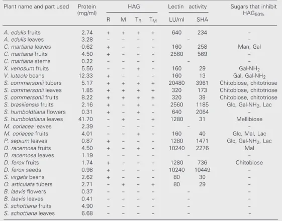
Table 2. Hemagglutination activity of extracts prepared from native and trypsin-treated red cells from Uruguayan plants.
Plant name and part used Protein HAG Lectin activity Sugars that inhibit
(mg/ml) HAG50%
R M TR TM LU/ml SHA
A. edulis fruits 2.74 + + + + 640 234
-A. edulis leaves 3.28 - - - - -
-C. martiana leaves 0.62 + - - - 160 258 Man, Gal
C. martiana fruits 4.50 + - - - 2560 569
-C. martiana stems 0.22 - - - - -
-X. venosum fruits 5.56 - - + - 160 29 Gal-NH2
V. luteola beans 12.33 + - - - 160 13 Gal, Gal-NH2
S. commersonii tubers 5.17 + + + + 20480 3961 Chitobiose, chitotriose
S. commersonii leaves 1.85 + + + + 320 173 Chitobiose, chitotriose
S. commersonii fruits 8.22 + + + + 320 39 Chitobiose, chitotriose
S. brasiliensis fruits 2.16 + - + - 2560 1185 Glc, Gal-NH2, Lac
S. humboldtiana flowers 0.31 + - + - 640 2064
-S. humboldtiana leaves 41.70 - + - + 1280 31 Mellibiose
M. coriacea leaves 2.39 - - - - -
-M. coriacea fruits 4.01 - - + - 160 40 Glc, Mal, Lac
P. sepium leaves 0.87 + - - - 1280 1471 Glc, Gal-NH2, Lac
D. racemosa fruits 4.50 + - + - 10240 2276 Mal
D. racemosa leaves 1.19 - - - - -
-D. ferox fruits 1.74 + - - - 1280 736 Chitobiose
D. ferox seeds 0.98 + - - - 10240 10449
-S. virgata beans 2.62 + - - - 80 30
-O. articulata tubers 2.71 - + - + 80 29
-B. laevis flowers 0.37 - - - - -
-B. laevis leaves 0.41 - - - - -
-S. schottiana fruits 4.90 - - - - -
-S. schottiana leaves 6.68 - - - -
-HAG: hemagglutination; HAG50%: the extract dilution one below the highest dilution not able to agglutinate
red blood cells; LU: lectin units, as defined in Material and Methods; M: mouse red cells; R: rabbit red cells; SHA: specific hemagglutination activity (LU/mg protein); TM: trypsin-treated mouse red cells;TR:
activity. The extract agglutinated untreated and trypsin-treated rabbit red cells but did not cause agglutination of untreated or tryp-sin-treated mouse red cells. HAG was inhib-ited by glucose (Table 2) but not by the other glucose-related sugars tested (2-amino-2-deoxy-D-glucose, Glc-NH2 and
N-acetyl-D-glucosamine, Glc-NAc). The 1-amino-1-deoxy-ß-D-galactose (Gal1-NH2) inhibited
HAG. Lactose and maltose were the only two disaccharides tested that caused HAG inhibition. Traces of ß-galactosidase activity were detected but no amylase activity was observed.
Solanum commersonii (Solanaceae)
The tuber, fruit and leaf extracts were all positive for HAG activity. The specific HAG activity was 23-fold higher in the tubers (3961 LU/mg) than in the leaves (173 LU/ mg) (Table 2). The extracts agglutinated un-treated and trypsin-un-treated rabbit and mouse red cells and HAG was inhibited by N,N’-diacetylchitobiose (chitobiose) and N,N’,N”-triacetylchitotriose (chitotriose). The mono-mer Glc-NAc did not inhibit HAG even at a concentration of 500 mM.
Cayaponia martiana (Cucurbitaceae)
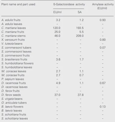
The leaf extract was positive for HAG activity. The extract agglutinated only rabbit red cells and the activity was inhibited by different sugars including mannose, galac-tose, trehalose and raffinose. The glycopro-tein Fetuin also inhibited HAG. The fruit extract was positive for HAG activity, agglu-tinating only the rabbit red cells and this activity was inhibited by Fetuin. The specif-ic HAG activity found in the fruit extract was 20-fold higher than the specific HAG activ-ity found in the leaf extract.
ß-Galactosidase activity was detected in all the plant parts analyzed, i.e., leaves, stems and fruits. The specific activities for the leaf and stem extracts were similar (193.5 and
Table 3. Distribution of the enzymatic activities analyzed in extracts from Uruguayan plants.
Plant name and part used ß-Galactosidase activity Amylase activity
(EU/ml)
EU/ml SA
A. edulis fruits 3.2 1.2 0.93
A. edulis leaves - -
-C. martiana leaves 120.0 193.5
-C. martiana fruits 25.0 5.5
-C. martiana stems 46.0 209.0
-X. venosum fruits - - 0.80
V. luteola beans - -
-S. commersonii tubers - - 0.07
S. commersonii leaves - -
-S. commersonii fruits - -
-S. brasiliensis fruits 3.6 1.7
-S. humboldtiana flowers - -
-S. humboldtiana leaves - -
-M. coriacea leaves 2.7 1.1
-M. coriacea fruits 2.7 0.7
-P. sepium leaves - -
-D. racemosa fruits 4.9 1.1 0.67
D. racemosa leaves - -
-D. ferox fruits - -
-D. ferox seeds 37.0 37.8
-S. virgata beans - -
-O. articulata tubers - -
-B. laevis flowers - - 0.13
B. laevis leaves - -
-S. schottiana fruits - -
-S. schottiana leaves - -
-SA: specific activity, defined as enzyme unit per mg of soluble extractable protein (EU/mg).
bit and mouse red cells.
In some cases, the activities were deter-mined in different parts of the same plant. Only in one case (Solanum commersonii) did we detect the same type of HAG activity and the same inhibitory behavior in extracts from tubers, fruits and leaves. The results of HAG inhibition by some of the sugars used in the present study are indicated in Table 2. ß-Galactosidase activity was found in some cases, and only traces of amylase activity were detected under the assay conditions used (Table 3).
Sebastiania brasiliensis (Euphorbiaceae)
209.0 EU/mg, respectively) and much higher than for the fruit extract (5.5 EU/mg).
Salix humboldtiana (Salicaceae)
The leaf and flower extracts were posi-tive for HAG activity. The leaf extract agglu-tinated untreated and trypsin-treated mouse red cells but did not agglutinate untreated or trypsin-treated rabbit red cells. HAG was inhibited only by mellibiose. In the flower extract, HAG was positive only for the un-treated rabbit red cells. The specific HAG activity for the leaf extract was 67-fold lower than the specific HAG activity for the flower extract and was not inhibited by any of the sugars assayed, indicating that the HAG, in this case, was nonspecific. Neither amylase nor ß-galactosidase activity was detected in the extracts.
Allophylus edulis (Sapindaceae)
The fruit extract was positive for HAG activity but was not inhibited by any of the sugars assayed, indicating that the HAG, in this case, was nonspecific and possibly due to extract components other than lectins. Traces of ß-galactosidase activity as well as amylase activity were detected in the fruit extract. The leaf extract was negative for the activities assayed.
Myrsine coriacea (Myrsinaceae)
The fruit extract was positive for HAG activity only for the trypsin-treated rabbit red cells and the activity was inhibited by glucose, maltose and lactose. The leaf ex-tract was negative for HAG. Traces of ß-galactosidase activity were detected in both fruit and leaf extracts, while no amylase activity was detected in either extract.
Datura ferox (Solanaceae)
The fruit extract was positive for HAG
activity, which was inhibited by chitobiose and also by Fetuin. The seed extract was positive for HAG activity but none of the sugars tested were able to inhibit HAG. ß-Galactosidase activity was detected (38 EU/ mg of soluble extractable proteins) but no amylase activity was found in the seed ex-tract.
Other plant extracts
The seed extract from Vigna luteola was positive for HAG with untreated rabbit red cells and the activity was inhibited by galac-tose and Gal1-NH2. The seed extract from Sesbania virgata was also positive for HAG with untreated rabbit red cells but in this case the activity was not inhibited by the tested sugars. The Oxalis articulata tubers were positive for HAG activity when using un-treated and trypsin-un-treated mouse red cells, although the HAG was not inhibited by the sugars tested. The leaf extract from Pavonia sepium was positive for HAG with untreated rabbit red cells and the activity was inhibited by glucose, Glc-NH2,
2-amino-2-deoxy-D-galactose (Gal2-NH2), maltose and lactose.
The fruit extract from Daphnopsis racemosa
was positive for HAG with untreated and trypsin-treated rabbit red cells and the activ-ity was inhibited only by the disaccharide maltose. The fruit extract from Xylosma venosum exhibited low HAG activity and only for trypsin-treated rabbit red cells (160 LU/ml) and the activity was inhibited by Gal1-NH2. Traces of amylase activity were
detected. The extracts from Sebastiania schottiana (Euphorbiaceae) were negative for all of the activities assayed.
screening contributes to increasing the num-ber of potential new lectins to be purified and characterized in the future. Based on the present results, we attempted the purifica-tion of the lectin from S. commersonii tu-bers.
Purification of the Solanum commersonii tuber lectin
S. commersonii is one of the two Uru-guayan tuberous species belonging to the
Solanum genus. It grows wild country-wide and it is endemic in Uruguay, southern Bra-zil and the provinces of Entre Rios and Buenos Aires in Argentina. This local spe-cies has received special attention due to its resistance to frost and to some local pests (16,17). Furthermore, it is known that potato (S. tuberosum) tubers contain a lectin that has been well characterized (18-20) and has been suggested to be possibly involved in the defense mechanism of the plant. Several affinity adsorbents have been used to at-tempt the purification of this lectin, includ-ing Fetuin-Sepharose (18). When analyzinclud-ing the soluble extract from S. commersonii tu-bers, we found some behaviors similar to those of the S. tuberosum tuber extract. For instance, the HAG activity present in the extracts was not inhibited by the monomer Glc-NAc but was inhibited by chitobiose and chitotriose, a behavior described for the potato (S. tuberosum) tuber extracts. A com-parative analysis performed with both ex-tracts showed a 7-fold higher specific HAG activity (3961 LU/mg protein) for S. com-mersonii than for S. tuberosum (611 LU/mg protein). The soluble extractable protein con-tent represents 2.6% (w/w) for the S. com-mersonii tubers and the material precipitable with ammonium sulfate at 50% saturation represents 9% of the total soluble proteins. Due to the information reported about the potato lectin and due to the potential impor-tance of this lectin in plant defense, we attempted a preliminary purification of the
S. commersonii lectin. Results for the purifi-cation of the lectin from the tuber extract using asialofetuin-Sepharose are shown in Table 4 and Figure 1. It should be noted that only about 50% of the total proteins applied were recovered from the column in the wash-ing and elution steps. The purified fraction gave a single protein band corresponding to an Mrof approximately 80 kDa, as shown by
silver staining after SDS-PAGE under re-ducing conditions (Figure 2). The tuber lec-tin isolated from the cultivated species S.
Table 4. Purification of the Solanum commersonii lectin by affinity chromatography on asialofetuin-Sepharose.
Fraction A280 Protein Volume LU/ml SHA Purification
(mg/ml) (ml) (-fold)
Applied 11.95 5.17 4.0 20480 3961
-Eluted and 0.24 0.105 0.5 51520 490667 124
concentrated
LU: lectin units; SHA: specific hemagglutination activity (LU/mg protein).
LU/ml
5000
4000
3000
2000
1000
Absorbance at 280 nm
1.0
0.8
0.6
0.4
0.2
0 0
1 6 11 16 21
Fraction number 0.1 M Glc-NAc
kDa
14.4
20.1
43
67
94 30
Figure 2. SDS-PAGE of the fractions from the purifica-tion of the lectin. The gel (homogeneous, 12.5%) was stained with silver. Lane a, Extract from Solanum com-mersonii tubers; lane b, material eluted from asialofetuin-Sepharose; lane c, low Mr markers.
References
1. Van Damme EJM, Peumans WJ, Pusztai A & Bardoz S (1998). Plant lectins: A special class of plant proteins. In: Van Damme EJM, Peumans WJ, Pusztai A & Bardoz S (Editors), Handbook of Plant Lectins: Properties and Biomedical Applications. John Wiley & Sons, Chichester, England, 3-10.
2. Rüdiger H & Rougé P (1998). Structure and functions of plant lectins. Carbohydrates in Europe, 23: 18-22.
3. Sekimata M, Ogura K, Tsumuraya Y, Hashimoto Y & Yamamoto S (1989). A ß-galactosidase from radish (Raphanus sativus L.) seeds. Plant Physiology, 90: 567-574.
4. Brena B, Pazos C, Franco Fraguas L & Batista-Viera F (1996). Chro-matographic methods for amylases. Journal of Chromatography B, 684: 217-237.
5. Muñoz J, Ross P & Cracco P (1993). Flora Indigena del Uruguay. Editorial Agropecuaria Hemisferio Sur, Montevideo, Uruguay, 17-29.
6. Smith PK, Khron RI, Hermanson GF, Mallia AK, Gartner FH, Provenzano MD, Fujimoto EK, Goeke NM, Olson BJ & Klent DC (1985). Measurement of protein using bicinchoninic acid. Analytical
Biochemistry, 150: 76-85.
7. Bernfeld P (1955). Amylases. In: Colowick S & Kaplan N (Editors), Preparation and Assay of Enzymes.Methods in Enzymology. Aca-demic Press, New York, 1: 149-158.
8. Giacomini C, Villarino A, Franco Fraguas L & Batista-Viera F (1998). Immobilization of ß-galactosidase from Kluyveromyces lactis on silica and agarose: comparison of different methods. Journal of Molecular Catalysis B: Enzymatic, 4: 313-327.
9. Nowak TP, Haywood PL & Barondes SH (1976). Developmentally regulated lectin in embryonic chick muscle and a myogenic cell line. Biochemical and Biophysical Research Communications, 68: 650-657.
10. Simões CMO, Mentz L, Schenkel E, Irgang B & Stehmann JR (1986). Plantas da Medicina Popular no Rio Grande do Sul. Editora da Universidade, Porto Alegre, RS, Brazil.
11. Martínez-Crovetto R (1981). Plantas utilizadas en Medicina en el NO de Corrientes. Tucumán, Argentina, Miscelanea 69.
12. Rojas Acosta N (1907). Catálogo de las plantas medicinales del Chaco austral. Revista Farmacéutica, 47: 214-225.
a b c
tuberosum is reported to be a dimeric protein composed of two identical (or very similar) subunits of 50 kDa (19) and 65.5 kDa by MALDI mass spectrometry, which contain up to 40-50% covalently linked carbohy-drate (1). We performed SDS-PAGE analy-sis under both reducing and nonreducing
conditions on 8-25 gradient gels and the proteins were stained with silver. The band corresponding to the apparent molecular mass of 80 kDa was obtained only under reducing conditions, while the material run under non-reducing conditions gave a clearly retarded and diffuse band (data not shown). The ex-pected specificity of the lectin was confirmed by inhibition of HAG with Glc-NAc, chito-biose and chitotriose. No inhibition was ob-served with Glc-NAc even at 500 mM. Chi-tobiose inhibited at 24 mM and chitotriose at 1 mM (data not shown). A more complete characterization of this lectin is in progress. The high percentage of positive results detected here will permit us to focus on the isolation, purification and characterization of the lectins in these plants. Detailed infor-mation about the biochemistry and biology of these proteins is a prerequisite for under-standing their biological role and activity, as well as increasing knowledge about our re-gional plants.
Acknowledgments
13. Toursarkissian M (1980). Plantas Medicinales de la Argentina. Edito-rial Hemisferio Sur, Buenos Aires, Argentina.
14. González M, Lombardo A & Vallarino A (1941). Plantas de la Medi-cina Popular del Uruguay. Talleres Gráficos, Montevideo, Uruguay. 15. Arrillaga B (1969). Plantas Medicinales. Editorial Nuestra Tierra,
Montevideo, Uruguay.
16. Lee SP, Zhu B, Chen TH & Li P (1992). Induction of freezing toler-ance in Solanum commersonii culture cells. Physiologia Plantarum, 84: 41-48.
17. Laferriere LT, Helgeson JP & Allen C (1999). Fertile S. tuberosum + S. commersonii somatic hybrids as sources of resistance to bacte-rial wilt caused by R. solanacearum. Theoretical and Applied
Genet-ics, 98: 1272-1278.
18. Allen AK & Neuberger A (1972). Potato lectin. In: Colowick S & Kaplan N (Editors), Carbohydrate-Binding ProteinsMethods in Enzy-mology, 28: 340-345.
19. Owens RJ & Northcote DH (1980). The purification of potato lectin by affinity chromatography on a Fetuin-Sepharose matrix. Phy-tochemistry, 19: 1861-1862.