UNIVERSIDADE DA BEIRA INTERIOR
Ciências
Novas abordagens terapêuticas para a
regeneração óssea
João Miguel Carvalho Freire Boga
Dissertação para obtenção do Grau de Mestre em
Biotecnologia
(2º ciclo de estudos)
Orientador: Professor Doutor Ilídio Joaquim Sobreira Correia
Co-orientador: Mestre Sónia Alexandra Pereira Miguel
iii “See that the imagination of nature is far, far greater than the imagination of man.”
v
vii
Acknowledgments
First of all, I would like to thank my supervisor Professor Ilídio Correia for the chance to pursue the topic of my masters’ thesis with him. For all his commitment and time spent in carrying out this project in its entirety.
To my co-supervisor, Sónia Miguel for her unwavering support both as an academic, in acquiring scanning electron microscopic images and helping me throughout my entire thesis, as well as being a good friend, always there when I needed her the most.
To Professor Abílio Silva, for his availability and the assistance provided in the mechanical characterization of the scaffolds.
To Duarte, for providing me the Graphene Oxide material, for helping me brainstorm ideas and teaching me the importance of scientific rigor.
To my colleagues, I’d like to thank Elisabete for all the moments and conversations that we had the opportunity to share, André for his challenging insights, and his willingness to lend me a hand whenever needed and Sergio for the steady stream of jokes to keep me going. Thank you all!
To Duarte Nuno, for all the hours and days that you had to endure without me being there, or just being too busy to be there to share awesome moments. To João Duarte I would like to thank for the arguments that we had and for all the moments, be it in classes or over a coffee. To my friends, for the unconditional attention and support, that you guys offered whenever I had a rough day.
And finally, but not least, to my family whose extraordinary support got me through all of this, be it on the sunniest of days or the darkest of nights, for all their love, understanding and words of advice that they gave me during the course of all these years!
ix
Resumo
O envelhecimento da população mundial tem associado um aumento do número de doenças e fraturas ósseas, as quais comprometem a integridade do osso e afetam a qualidade de vida dos indivíduos. Na atualidade, estes tipos de desordens são tratadas recorrendo aos enxertos ósseos, nomeadamente aos autoenxertos. Contudo estes tipos de enxertos têm limitações como seja a disponibilidade do enxerto, possibilidade de induzir dor crónica ou ainda ser rejeitado devido a contaminação bacteriana no decorrer da cirurgia. Neste contexto, têm sido desenvolvidas novas abordagens terapêuticas com o intuito de colmatar as limitações associadas aos tratamentos usados comumente na clínica. Uma das abordagens usadas consiste na produção de estruturas tridimensionais (scaffolds) capazes de mimetizar as propriedades mecânicas e biológicas do osso nativo. Para este fim, neste estudo foram desenhados scaffolds cilíndricos, usando design assistido por computador, com o intuito de mimetizar a geometria natural dos ossos ocos e permitir a troca de nutrientes e a neovascularização.
No presente estudo os scaffolds foram produzidos recorrendo a fosfato tricálcico, ácido algínico e óxido de grafeno (GO) usando uma Fab@home 3D-Plotter. As propriedades mecânicas e biológicas das estruturas tridimensionais foram caracterizadas através de diversas técnicas. Os resultados obtidos neste estudo demonstraram que as amostras contendo GO possuem maior porosidade, mantendo, no entanto, constante a sua resistência mecânica. Adicionalmente, os
scaffolds 60/40_GO revelaram uma maior capacidade para fixar cálcio e fósforo na sua
superfície, o que contribui para melhorar a sua osteoinductividade e osteoconductividade. Propriedades estas que são essenciais para a osteointegração do scaffold. Os resultados obtidos neste projeto demonstraram que os scaffolds funcionalizados com GO possuem as propriedades requeridas para a sua aplicação na regeneração óssea.
Palavras-chave
Engenharia de Tecido Ósseo; Impressão 3D; Óxido de Grafeno; Prototipagem Rápida; Scaffolds 3D biocompósitos;
Resumo Alargado
O osso é um tecido extremamente dinâmico e vascularizado, que está envolvido em diferentes funções tais como: locomoção, proteção dos órgãos contra ameaças externas e armazenamento de minerais essenciais à homeostase do corpo humano. A matriz óssea é constituída por um bio-compósito dotado de uma fase orgânica (maioritariamente colagénio tipo I) e uma fase inorgânica (hidroxiapatita) assim como uma componente celular (osteoblastos, osteócitos e osteoclastos) e água. Este tecido possui também uma capacidade inata de auto-regeneração que permite ao sistema esquelético responder a estímulos exteriores e reparar possíveis danos estruturais. Contudo em certas situações esta capacidade regenerativa é comprometida, nomeadamente em defeitos ósseos extensos, envelhecimento e em certas doenças que tornam a osso propenso a fraturas. Na clínica os autoenxertos, aloenxertos e xenoenxertos têm sido usados para tratar fraturas que afetam este tecido. Contudo, estas abordagens terapêuticas possuem certas desvantagens que incluem uma limitada disponibilidade dos autoenxertos, a rejeição ou infeção bacteriana que por vezes surge quando são usados alo ou xenoenxertos. Com o intuito de ultrapassar estas limitações, os investigadores da área de Engenharia de Tecidos têm vindo a desenvolver novas abordagens terapêuticas que procuram acelerar o processo regenerativo do osso. Na atualidade têm sido produzidas novas matrizes tridimensionais que procuram mimetizar tanto a estrutura como a composição do tecido nativo de forma a restaurar, manter ou melhorar a função do tecido danificado. Estas estruturas são designadas por scaffolds e servem como suportes temporários para auxiliar e guiar a formação de novo tecido ósseo. As estruturas tridimensionais para poderem ser usadas na regeneração óssea têm que ser biocompatíveis, biodegradáveis, porosas e possuir propriedades mecânicas que promovam a osteoindução, osteocondução e neovascularização. Para atingir este fim, os investigadores têm usado vários materiais que incluem cerâmicas (hidroxiapatita), metais (titânio e cobalto), polímeros (colagénio e alginato) e compósitos dos mesmos em conjunção com métodos de prototipagem rápida, que permitem a criação de modelos produzidos através de programas informáticos ou através de dados recolhidos em exames de rotina em meio hospitalar.
xi entanto, a resistência mecânica dos scaffolds seja afetada. Adicionalmente verificou-se que a incorporação de GO não afeta o perfil citotóxico dos scaffolds. Os ensaios de biomineralização evidenciaram as capacidades osteoinductivas e osteoconductivas dos scaffolds produzidos, propriedades estas que são essenciais para a osteointegração destes scaffolds no tecido nativo. As propriedades mecânicas e biológicas apresentadas por estes scaffolds permitem postular a aplicação destes scaffolds na regeneração de tecido ósseo.
Futuramente, a performance in vivo destes scaffolds híbridos será avaliada em modelos animais de forma a averiguar o seu potencial osteogénico. Além disto, será otimizado o desenho dos
scaffolds e para além do GO, serão também incluídos agentes com atividade antimicrobiana,
Abstract
The aging of the worldwide population has associated an increase in bone related traumas and diseases, that affect the well-being of human beings. Nowadays bone grafts, namely autografts, allografts and xenografts are used to treat these types of diseases. Yet these therapeutic approaches display several drawbacks such as limited tissue availability, chronic pain and possible immune rejection by the patient. To surpass these limitations, new therapeutic approaches are currently being developed to produce three-dimensional structures (scaffolds) that are capable of mimicking the mechanical and biological properties of native bone. Herein, cylindrical scaffolds were created using computer assisted design, with the aim of replicating the natural geometry of hollow bones and to allow the exchange of nutrients and neovascularization. A Fab@home 3D-Plotter was used for scaffold production. Tricalcium phosphate and alginic acid were selected for scaffold manufacture since they are able to reproduce the organic and inorganic matrix of bone. Furthermore, scaffolds were also functionalized with graphene oxide (GO) with the aim of improving the mechanical and biological properties of the scaffolds. The results obtained in this study demonstrated that GO bearing samples displayed increased porosity while maintaining their mechanical resistance. Additionally, the 60/40_GO scaffolds possessed an enhanced capability to adsorb calcium and phosphorous minerals, which give an important contribute for improving the osteoinductivity and osteoconductivity. In conclusion, the attained results revealed that GO functionalized 3D scaffolds have the desired properties that will allow their future application in bone tissue regeneration.
Keywords
Bone tissue engineering; Biocomposite 3D scaffolds; Rapid Prototyping; Graphene Oxide; 3D Printing;
Table of Contents
Chapter I
1. Introduction ... 2
1.1. Bone Tissue ... 2
1.2. Bone Anatomy and Morphology ... 2
1.3. Bone Histology... 7 1.3.1. Bone Matrix ... 7 1.3.2. Bone cells ... 8 1.3.2.1. Osteoblasts ... 8 1.3.2.2. Osteocytes ... 8 1.3.2.3. Osteoclasts ... 9
1.3.3. Bone Remodeling Process ... 9
1.4. Bone Disorders ... 12 1.4.1. Osteoporosis ... 13 1.4.2. Pagets’ Disease ... 13 1.4.3. Osteomyelitis ... 13 1.5. Bone Grafts ... 14 1.6. Tissue Engineering ... 15 1.6.1. 3D Scaffolds ... 16
xv
1.7. Aims ... 24
Chapter II
2. Materials and methods ... 262.1. Materials... 26
2.2. Methods ... 26
2.2.1. Layer design and assembly of the scaffold 3D model ... 26
2.2.2. GO synthesis ... 27
2.2.3. Production of 3D hybrid scaffolds ... 27
2.2.3.1. Incorporation of GO into the TCP/AA blend ... 27
2.2.4. Scaffolds Physio-Chemical Characterization ... 28
2.2.4.1. Attenuated total reflectance—Fourier transform infrared spectroscopy ... 28
2.2.4.2. Scanning electron microscopy analysis ... 28
2.2.4.3. Energy dispersive spectroscopic analysis ... 28
2.2.4.4. Characterization of the mechanical properties of the scaffolds ... 29
2.2.4.5. Swelling assays ... 29
2.2.4.6. Determination of the water contact angles ... 30
2.2.4.7. Evaluation of porosity of the scaffolds ... 30
2.2.4.8. Characterization of the degradation profile of the scaffolds... 30
2.2.4.9. In vitro biomineralization assay ... 31
2.2.5. Characterization of the biological properties of the scaffolds ... 31
2.2.5.1. Evaluation of cell viability and proliferation in the presence of the scaffolds .. 31
2.2.5.2. Characterization of cell adhesion on surface of the scaffolds ... 31
2.2.5.3. Alizarin Red S staining ... 32
2.2.5.4. Confocal microscopy analysis ... 32
2.2.6. Statistical analysis ... 33
Chapter III
3. Results and Discussion ... 353.1. GO characterization ... 35
3.3. Characterization of the physico-chemical properties of the produced scaffolds ... 39
3.3.1. ATR-FTIR analysis ... 39
3.3.2. Energy dispersive spectroscopy analysis (EDS) ... 40
3.3.3. Characterization of the mechanical properties of the scaffolds ... 40
3.3.4. Evaluation of the swelling capacity of the scaffolds ... 41
3.3.5. Determination of Water Contact Angle ... 42
3.3.6. Evaluation of scaffolds’ porosity ... 43
3.3.7. Determination of the degradation profile of the produced scaffolds ... 44
3.3.8. In vitro biomineralization assay ... 44
3.4. Characterization of the biological properties of the produced scaffolds ... 47
3.4.1. Evaluation of the scaffolds cytotoxic profile ... 47
3.4.2. Alizarin Red S staining ... 49
3.4.3. Confocal laser scanning microscopy analysis ... 50
Chapter IV
4. Conclusion and Future Perspectives ... 54Chapter V
5. References ... 56List of Figures
Figure 1. Classification of the bones according to their shape ... 3
Figure 2. Cross-sectional view of compact bone ... 4
Figure 3. Illustration of bone Periosteum and Endosteum ... 5
Figure 4. Illustration of the composition of spongy bone ... 5
Figure 5. Representation of the anatomical characteristics of a long bone ... 6
Figure 6. Illustration of bone phase composition effect on the mechanical behaviour of this tissue. ... 7
Figure 7. Representation of the types of cells found in bone ... 8
Figure 8. Schematic representation of the different phases and acting cells in the bone remodeling process ... 10
Figure 9. Representation of the most common bone diseases and their effects on bone. ... 12
Figure 10. Illustration of different therapeutic approaches. ... 14
Figure 11. Experimental approach used to develop biomimetic 3D scaffolds. ... 15
Figure 12. Chemical structure of tricalcium phosphate. ... 18
Figure 13. Chemical structure of alginic acid. ... 20
Figure 14. Molecular representation of a GO flake ... 21
Figure 15. FTIR characterization of graphite oxide (A); DLS size distribution of GO (B). ... 35
Figure 16. Schematic representation of the process used to produce 3D scaffolds (A); Scaffolds’ 3D model and representative macroscopic images of the different produced scaffolds (side and top view) (B). ... 37
Figure 17. SEM images acquired to characterize the morphology and surface topography of the produced 3D scaffolds. ... 38
Figure 18. ATR-FTIR analysis of TCP, AA and TCP/AA scaffolds (80/20, 70/30 and 60/40) (A) and GO, 60/40 and 60/40_GO scaffold (B). ... 39
Figure 19. Characterization of the compressive strength (A) and young modulus (B) of the produced 3D scaffolds under dry and wet conditions ... 40
Figure 20. Characterization of scaffolds' swelling profiles (A); determination of water contact
xix microscopic images of scaffolds stained with Alizarin Red S (C); and calcium quantification (D) ... 50
Figure 25. CLSM images acquired to characterize cell internalization within the 60/40 and
60/40_GO scaffolds. 3D reconstruction images (A, B, C and D) and orthogonal projections (E and F) ... 51
xxi
Acronyms
3DP Three-Dimensional Printing AA Alginic Acid
ARRF Activation-Resorption-Reversal-Formation ARS Alizarin Red Staining
ATR-FTIR Attenuated Total Reflectance-Fourier Transformed Infrared Spectroscopy BMU Basic Multicellular Unit
BMP Bone Morphogenic Protein CAD Computer Assisted Design
CSLM Confocal Selective Laser Microscopy DMEM-F12 Dulbeccos’ Modified Eagles’ Medium ECM Extracellular Matrix
EDS Energy Dispersive Spectroscopy EtOH Ethanol
FBS Fetal Bovine Serum
FDM Fused Deposition Modelling FGF Fibroblast Growth Factor GO Graphene Oxide
HA Hydroxyapatite hOB Human Osteoblasts IGF-1 Insulin Growth Factor-1 IGF-2 Insulin Growth Factor-2 IL-6 Interleukin-6
MCP-1 Monocyte Chemoattractant Protein-1 M-CSF Macrophage Colony-Stimulating Factor
MTS 3-(4,5-dimethylthiazol-2-yl)-5-(3-carboxymethoxyphenyl)-2-(4-sulfophenyl)-2H tetrazolium salt
NCP Noncollagenous Proteins PCL Polycaprolactone PTH Parathyroid Hormone RANK Receptor of Nuclear ᴋ-B RANKL Receptor of Nuclear ᴋ-B Ligand RGD Arginine-Glycine-Aspartic Acid RP Rapid Prototyping
RT Room Temperature SBF Simulated Body Fluid
SEM Scanning Electron Microscopy SLA Stereolithography
SLS Selective Laser Sintering STL Standard Tesselation Language TCP Tricalcium Phosphate
TE Tissue Engineering
TGF-β Transforming Growth Factor-β TNF-α Tumor Necrosis Factor-α UV Ultraviolet
WCA Water Contact Angle WGA Wheat Germ Agglutinin YM Young Modulus
1. Introduction
1.1. Bone Tissue
Bone tissue is a specialized and dynamic connective tissue that acts as the basic unit of the human skeleton [1-3]. Bone tissue is responsible for supporting the human body and also other different biological functions, including locomotion, protection of vital organs (heart, lungs, spine and brain), mechanical support of the diaphragm and production of blood cells (hematopoiesis). Moreover, it also acts as a mineral reservoir, namely of calcium and phosphorous, and promotes muscle, ligament and tendon attachment [4, 5]. Bone tissue is under constant remodeling in order to allow this tissue to adapt to biomechanical forces and remove old and micro damaged bone.
Bone tissue is composed by cells (osteoblasts, osteocytes and osteoclasts), water and bone matrix. Such matrix is composed by organic (35%) and inorganic material (65%). The organic phase is composed of proteins of the extracellular matrix (ECM), collagen and proteoglycans. On the other hand, the inorganic phase contains calcium phosphate crystals, namely hydroxyapatite (HA) [6-8]. Bone cells are able to respond to external and internal biomechanical and biochemical stimulus that promote the cyclical demineralization/mineralization of bone tissue. In this process, osteoclasts remove old or damaged bone and osteoblasts are involved in the synthesis of new bone matrix [6].
1.2. Bone Anatomy and Morphology
The human skeleton is composed by several types of bones that can be distinguished according to their shape, morphology and bone matrix. According to their shape, bones can be classified as long, short, flat or irregular as shown in Figure 1.
3
Figure 1. Classification of the bones according to their shape (adapted from [9]).
Long bones, such as clavicles, femurs and tibiae, have a cylindrical shape, great mechanical strength and durability. Short bones, like patellae, sesamoid, carpal and tarsal bones, have a roughly cube like or spherical geometry. For instance, flat bones present thin, curved or flat shapes and have a broad surface that is important for muscle attachment or organ protection (i.e. the bones of the shoulder girdle, ribs and breastbone). Finally, irregular bones have complex shapes that are neither long, short, or flat, such as the vertebrae and facial bones. These types of bones present many surface features for muscle or articulation attachment [10]. Morphologically, bone tissue is classified, depending on the density of the bone matrix, in trabecular or cancellous bone and cortical or compact bone [4, 5]. Cortical bone, represented in Figure 2, is almost compact, since it only presents a porosity of around 10% and accounts for 80% of the mass of a mature human skeleton [1, 6]. It has a high mechanical strength, since it is comprised of closely packed cortical osteons, called Haversian systems, that form a solid and consistent mass [7].
Figure 2. Cross-sectional view of compact bone showing the basic structural unit: the osteon (adapted from [9]).
The Harversian systems have a central canal, known as Haversian canal, that is surrounded by concentric rings of matrix and osteocytes that are responsible for its mechanical resilience [4, 7, 11].
The outer surface of cortical bone is covered by a bi-layered connective tissue membrane, shown in Figure 3, the periosteum. The outer, fibrous layer, is composed of irregular collagenous tissue, that contains blood vessels and nerves, while the inner layer is composed by a single layer of bone cells. These features facilitate the fixation of tendons and ligaments to the bone [12, 13].
5
Figure 3. Illustration of bone Periosteum (the outer surface of the bone) and Endosteum (lines the medullary cavity) (adapted from [9]).
In contrast, the trabecular or cancellous bone is highly porous (50-90%) and has a compressive strength almost 20 times inferior to that displayed by cortical bone (2-20MPa for cancellous bone versus 100-200MPa for cortical bone [14]). It is strongly associated with metabolic activities, since its pores are interconnected and filled with bone marrow [1].
Cancellous bone, represented in Figure 4, has a sponge-like form, with a honeycomb of branching plates and rods of various sizes called trabeculae. In turn, the trabeculae are lined with another connective tissue membrane (endosteum), which is composed by a single layer of cells [4].
Figure 4. Illustration of the composition of spongy bone: trabeculae that contain the osteocytes. Red marrow fills the spaces in some bones (adapted from [9]).
Cortical bone, is found in the diaphysis of long bones. On the other hand, flat, short and irregular bone usually present a cancellous structure in their interior filled with marrow surrounded by two layers of cortical bone [10].
As represented in Figure 5, these two types of skeletal tissue can be found in long bones, the latter are divided in 3 different components, the diaphysis that composes the bulk of the bone, with a cortical periphery and cancellous interior; the epiphysis located at the ends of the bone, and the epiphyseal plate, made of cortical bone, and that is located between the diaphysis and the epiphysis, where the new bone is formed during bone growth [13, 15].
7 at ligament and tendon insertions and under pathologic conditions. Typically, it is remodeled to form lamellar bone [7, 17]. In contrast, the lamellar bone is a layered type of bone, composed of 3-7 µm lamellae, with collagen fibers lying parallel to each other, and with mineral crystals between them. This gives to the lamellar bone an increased strength [7, 15].
1.3. Bone Histology
1.3.1.
Bone Matrix
Bone matrix is composed by an organic and mineral components with a ratio of 35/65%, which together contribute to the strength and flexibility of the human skeleton [5, 18, 19].
The organic phase comprises fibrillary proteins (mainly collagen type I), proteoglycans and a variety of noncollagenous proteins (NCP) that can be divided into structural proteins and promoters of biological processes [5, 7, 8, 20-23], whereas the mineral phase is mainly constituted by hydroxyapatite crystals. Although, tricalcium phosphate, calcium carbonate and fluoride derivatives can also be found in this matrix [18, 21]. It is estimated that bones contain 99% of the calcium, 85% of the phosphorus and 40-60% of the sodium and magnesium found in the human body [5, 21].
The balance between the organic and inorganic phases of the bone is crucial to confer to this tissue flexibility and resistance to compression (Figure 6). If the mineral component is diminished, the bone becomes more flexible, due to the increased collagen content. On the other hand, if the collagen percentage is diminished, the bone becomes very frail and brittle [15, 24].
Figure 6. Illustration of bone phase composition effect on the mechanical behaviour of this tissue. Higher collagen content leads to excessive flexibility (left) while an excessive mineral content makes the bone brittle.
Besides that, bone matrix composition can also suffer variations with age, namely an increase in mineralization degree combined with a decreased bone collagen content [25, 26].
1.3.2.
Bone cells
Besides osteogenic stem cells, bone is composed by three main cell types: osteoblasts, osteocytes and osteoclasts (represented in Figure 7), that are involved in the maintenance and remodeling process of the bone tissue.
Figure 7. Representation of the types of cells found in bone (adapted from [9]).
1.3.2.1. Osteoblasts
Osteoblast are mononuclear and basophilic cells that display a large spherical nucleus, a highly developed rough endoplasmic reticulum and Golgi apparatus [7]. These cells are derived from mesenchymal stem cells and are present at bone surfaces [18, 27]. Structurally they have a cube-like shape or a slightly elongated appearance [7]. They are involved in the synthesis and organization of the bone ECM, that subsequently becomes mineralized [6, 7].
Moreover, osteoblasts contain vesicles loaded with phosphate, calcium ions and enzymes (ie. alkaline phosphatase), that are released by exocytose and that are involved in hydroxyapatite crystal formation, thus promoting bone mineralization [15]. These cuboid shaped cells can
9 the high number of dendritic processes that interconnect the osteocytes and bone lining cells [7, 15, 28]. They form filopodia located in spaces or channels in the bone matrix called canaliculi. The arrangement of the canaliculi allows the passage of nutrients, metabolites and oxygen between the blood vessels and distant osteocytes [7, 21, 30]. Also, Nitric oxide, Wnt and cadherin-mediated signaling have been suggested as transducing mechanisms, since they are activated after a mechanical stimulus [31, 32]. However, the precise mechanisms of stimulus and response remain unclear. Osteocytes are also responsible for osteocytic osteolysis, i.e., breaking down the bone matrix to release calcium that is required for other metabolic pathways [20].
1.3.2.3. Osteoclasts
Osteoclasts are cells, that derive from hematopoietic stem cells, and that are involved in the resorption of fully mineralized bone, leading to the mobilization of Ca2+ and PO
4- ions from the
bone matrix [5, 7, 20]. They are highly specialized and multi-nucleated giant cells.
During their motile state, they migrate from the bone marrow to their resorptive site where they will initiate bone resorption. In this state, they present a flat and non-polarized morphology, with membrane protrusions such as lamellipodia and actin rich podosome complexes. On the other hand, in their resorptive state they assume a polarized shape. In this phase the cells display a dome shape (that results from cytoskeletal reorganization) and new membrane domains (such as the sealing zone) are formed [5, 30, 33].
Osteoclast cells present numerous mitochondria, a well-developed Golgi apparatus around the nuclei, endoplasmic reticulum, vacuoles and lysosomes. This cellular organization results from their great involvement in protein synthesis, in particular lysosomal enzymes, as well as their excretion into the resorptive bay along with acidic compounds that take part in bone resorption process [34]. Through this complex cellular machinery, osteoclasts are capable of resorbing up to 200 000µm3/day of bone matrix and they have an average lifespan of 15-20 days [7].
1.3.3. Bone Remodeling Process
Bone is a dynamic and metabolic active tissue that is constantly being remodeled. The remodeling process consists in the resorption of old bone and on the formation of new bone in order to prevent the accumulation of micro damages, like nanocracks and thus assure the homeostasis of the skeletal tissue [4, 5, 35].
Yet, to accomplish that a careful balance between bone resorption and formation must be kept. Within this process, osteoblasts and osteoclasts operate in an orchestrated manner, gathering in a temporary assembly a basic multicellular unit (BMU) on the bones’ surface [6, 28, 36, 37]. In these BMUs cell activity is sequential: first, osteoclasts in the leading edge of the BMU resorb damaged or old bone; followed by osteoblasts that at the end of the BMU secrete collagen fibers and mineralize the matrix to form new bone (see Figure 8 for further details). This unit
is regulated by mechanical forces, bone cell turnover, hormones (PTH (pituitary hormone), growth hormones, etc), cytokines and local factors [6].
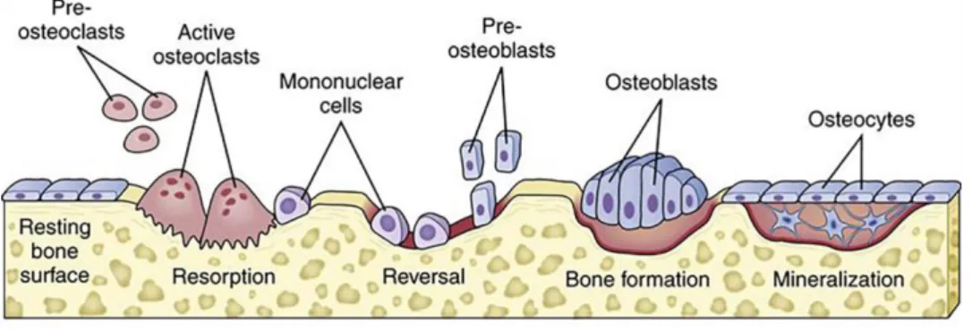
Figure 8. Schematic representation of the different phases and acting cells in the bone remodeling process. Initially a biochemical or biomechanical signal promote pre-osteoclast migration to its resorptive area. In the Resorption phase, mature osteoclasts in the Howship lacunae actively resorb the damaged or old bone. In the Reversal phase pre-osteoblasts are recruited into the area and differentiate into mature osteoblasts. During the Formation phase, osteoblasts actively secrete organic matrix (collagen type I) and initiate its mineralization. Finally in the Formation phase the organic matrix is fully mineralized, osteoblasts either undergo apoptosis or differentiate into osteocytes or bone lining cells (adapted from [38]).
The bone remodeling process comprises four main phases (activation, resorption, reversal and formation stages (ARRF sequence), as shown in Figure 8) and it lasts about 3-6 months before its fully complete in humans [36]. Moreover, bone remodeling is not only required to replace dead or damaged tissue, but also confers bone the capacity to adapt to loading variations or respond to nutritional and metabolic changes.
Activation phase
The first stage of bone remodeling involves the detection of an initiation signal, like mechanical strain, damage or biomolecules released into the bone microenvironment [31, 36]. In response to these stimulus, osteoblasts start recruiting osteoclast precursors to the remodeling site, through the release of a chemoattractant, MCP-1 (monocyte chemoattractant protein) [35]. In case of structural damage, insulin growth factor-I (IGF-I), tumor necrosis factor-α (TNF-α),
11 Hormones may also trigger osteoclast activation and differentiation. PTH acts as an endocrine remodeling signal involved in the maintenance of calcium homeostasis. This hormone is secreted by the parathyroid glands in response to low calcium levels in the blood serum, and can lead both to bone formation, when secreted intermittently, or to bone resorption when secreted continuously [6, 35]. In bone tissue, PTH activates the PTH receptor (a G-protein-coupled receptor found on the surface of osteoblasts) [30], that activates protein kinase A, protein kinase C and intracellular calcium signaling pathways. This osteoblast activation causes a wave of transcriptional responses that modulate the secretion of molecules responsible for the recruitment, differentiation, and activation of osteoclasts, which are one of the main players in bone resorption [18, 35].
Resorption phase
The resorption phase, which lasts on average 30-40 days, consists in the attachment of differentiated osteoclasts to depressions or resorptive bays (known as Howship lacunae) present in the bone matrix [35, 36]. Cell anchorage is performed via αvβ3 integrin molecules, upon
adhesion, osteoclasts suffer cytoskeletal reorganization creating an isolated microenvironment underneath the cell, known as the sealed zone [18]. Subsequently, osteoclasts promote the bone demineralization process, in two steps [36]:
i) acidification of the bone matrix though proton pumps, that leads to a higher concentration of hydrogen ions in the sealed zone [35, 36, 39]
ii) release of lysosomal (e.g. cathepins K) and non-lysosomal (e.g. collagenase) enzymes that are involved in the degradation of the organic component of the bone [18, 35]. This exposes arginine-glycine-aspartic acid (RGD) sequences that enhance osteoclast attachment and consequently increase bone resorption [35]. To avoid excessive bone resorption, osteoclasts suffer apoptosis after this stage.
Reversal phase
Reversal phase marks the transition from osteoclast to osteoblast activity, with osteoblast precursors being recruited and differentiated into their mature state [39, 40]. This phase lasts approximately 9 days, and during this period, bone lining cells (mononuclear cells of osteoblastic origin) prepare the bone surface for the subsequent bone deposition performed by osteoblasts [30, 35, 36]. This stage is also characterized by the presence of activation of factors (IGF-2 and TGF-β) that stimulate osteoblast precursors to proliferate [7, 35, 36].
Formation phase
The formation phase is characterized by the release of a variety of growth factors stored in the bone matrix, such as bone morphogenic protein (BMP), fibroblast growth factor (FGF) and TGF-β that are responsible for the recruitment of osteoblasts into the absorbed area [33, 41]. At this stage, differentiated osteoblasts synthetize new organic matrix (collagen type I) and
release vesicles that contain calcium, phosphate and enzymes that destroy the mineralization inhibitors, such as pyrophosphate or proteoglycans [7, 30, 35, 36].
The remodeling cycle is concluded when all reabsorbed bone is replaced. At the end of the cycle, osteoblasts either suffer apoptosis, revert back to bone lining phenotype, or differentiate into osteocytes that remain within the matrix [30, 41]. Even though the bone remodeling process is one of the most reliable biological process in the body, there are circumstances where it fails. Indeed, bone disorders are almost always related to the deregulation of this cycle, influencing either bone formation or resorption [33, 42]. Such can compromise the architecture, structure and mechanical strength of bone tissue, leading to clinical symptoms such as pain, deformity, fractures and abnormalities of calcium and phosphate metabolism.
1.4. Bone Disorders
Bone defects can be caused by trauma, tumors, infection or bone diseases. Bone diseases comprise abnormal bone growth, that can lead to gigantism or dwarfism; abnormal collagen contents that can lead to osteogenesis imperfecta; Mineral and vitamin deficiencies cause rickets; bacterial infections that can provoke bone destruction, as is the case of osteomyelitis [30, 43]; Osteomalacia and osteoporosis are also responsible for bone damage through decalcification [15]. Figure 9 represents three of the most common disorders associated with bone tissue.
13
1.4.1. Osteoporosis
Osteoporosis is the most common disease affecting bone [44]. A study performed in 2010 estimates that 27.5 million people were affected by the disease only in the European Union [45]. Osteoporosis is characterized by low bone mass and structural deterioration, causing an increased bone fragility and vulnerability to fractures. This disorder is more common in older populations, especially in women, due to menopausal estrogen deficiencies that increase the bone resorption [33]. This disease usually results from an imbalance between bone resorption and bone formation [21, 30, 42, 46]. It is believed that sex steroid hormones, either directly or indirectly, regulate the production of cytokines (e.g. M-CSF) that promote the production of osteoclasts [42, 47].
Nevertheless, excluding the hormonal effects of menopause, this disease has the same rate of progress in both genders [44].
The most common osteoporotic fractures occur in the hip, vertebral column, and forearm. These fractures may result in morbidity or in severe cases can lead to patient death [33]. Unhealthy diet, sedentary lifestyle, nulliparity, aging, smoking, and low body weight are also risk factors that may trigger the development of osteoporosis [21].
1.4.2. Pagets’ Disease
Pagets’ is the second most common bone disease [30]. It is characterized by focal areas of excessive bone resorption, alternated with areas of increased bone formation, leading to the formation of abnormal bone, pain, pathologic fractures, deafness and nerve compression syndromes [30, 33, 46, 48, 49].
Although the causes are not entirely known, it is believed that this disease can be provoked by a viral infection during childhood, and several genetic mutations have also been identified on patients with this disease [39, 44, 49]. Nevertheless, sedentary lifestyle and deficient nutrition are also reported as factors that may be involved in the Pagets’ disease arising.
1.4.3. Osteomyelitis
Osteomyelitis is defined as an inflammation of bone tissue accompanied by its destruction, due to microbial infection [43, 46, 50]. Moreover, it can also be caused by fractures that occur due to trauma or other diseases [46]. Upon fracture, microorganisms (like Staphylococcus aureus) produce a range of extracellular components and cell-associated factors, that facilitate its colonization capacity and virulence [43].
1.5. Bone Grafts
As previously described, there are certain critically sized osseous defects that the organism is unable to heal without medical assistance [51]. In such cases, bone grafts have been used for bone tissue reconstruction. The capacity of bone grafts to regenerate tissue is measured by their osteogenic, osteoconductive and osteoinductive potential [52]. The bone grafts are divided into three main categories: autografts, allografts and xenografts as represented in Figure 10 [52]. Nowadays, autografts are the most used for bone tissue regeneration. However, their use is limited due to the low availability of graft tissue and considerable donor site morbidity, which is proportional to the amount of harvested bone [51, 53, 54]. Furthermore, the harvesting procedure induces bleeding, hematoma, infection and in some cases chronic pain to the patients [52, 55, 56]. In addition, there is no guaranty that the cellular components survive the transplantation [56], and questions have been raised about the osteoinductivity of these grafts, since the uncertainty about the grafts’ growth factor content can lead to insufficient tissue regeneration [57]. To overcome the lack of autografts, allografts have also been used for bone tissue replacement. However, since the grafts are from different donors (of the same species), there is always a risk of disease transmission and of triggering of an immune response from the host [51, 52, 55, 56]. Unfortunately the processing techniques used to decrease the associated risks also reduce the mechanical resistance of the grafts, and usually remove the cellular component from the bone tissue [56].
Xenografts are obtained from different species, which can lead to the rejection of the graft by the host. To overcome this drawback the biological components are removed in order to increase the safety of grafts [55].
Due to all aforementioned limitations, bone graft substitutes have been the focus of intense research in the area of Bone Tissue Engineering [58]. This area intends to develop three-dimensional (3D) structures capable of mimicking the bone structure and microenvironment [52].
15
1.6. Tissue Engineering
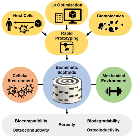
Tissue Engineering (TE) is an interdisciplinary field of research that applies the principles of engineering and life sciences, to develop new therapeutic approaches that allow the restoration, maintenance or improvement of a particular tissue function [2]. In particular, bone tissue engineering aims to produce grafts with the capacity to induce the restoration of bone structure and functions (Figure 11) [14, 20, 58-61].
Figure 11. Experimental approach used to develop biomimetic 3D scaffolds.
Nonetheless, scaffold production is limited by the current manufacturing techniques, and also by the random distribution of cells, matrix and bioactive molecules within the scaffolds’ structure. As such, mimicking the functional and biological complexity of bone tissue is seen as the current challenge to achieve full tissue regeneration [62].
1.6.1. 3D Scaffolds
Scaffolds are porous 3D matrices that act as temporary templates, that allow cell adhesion and proliferation, as well as provide mechanical support until new bone tissue is formed at the defect site.
Scaffolds have been produced using a variety of techniques, such has selective laser sintering (SLS) [63], 3D printing (3DP) [64], stereolithography (SLA) [65] and fused deposition modelling (FDM) [66], using different biomaterials that present certain key properties (biocompatibility, biodegradability, surface properties, porosity and mechanical properties) that are fundamental for scaffolds to promote osteoinductivity, osteoconductivity of bone-producing cells and neovascularization [14, 67-70].
• Biocompatibility
Biocompatibility is considered one of the most important properties that any type of implant or biomedical device must display [17]. Biocompatibility represents the ability of a material to support normal cellular activity including adhesion, proliferation and differentiation without eliciting any local or systemic response from the immune system [14, 58, 67]. Generally, scaffolds should be produced from materials that do not trigger any immunogenic reactions. Furthermore, the degradation products of the scaffolds must also be biocompatible and easily removed from the body [67, 71].
• Biodegradability
A scaffold aimed for bone regeneration may be designed to serve as a temporary matrix, and eventually, be replaced by bone tissue. In addition, the degradation products of the scaffolds must not elicit any adverse effect on the host [14]. The scaffolds’ degradation rates ideally should accompany the rate of regeneration of new bone [58, 60, 71]. As a result, a scaffold
17 the implant at the lesion site and also provide structural integrity to the injury location. Moreover, the clot can serve as a bridge for cells to migrate from the surrounding bone tissue to the scaffolds’ surface [2, 14, 51]. On the other hand, osteoinduction is a process by which the osteoprogenitor cells are stimulated to undergo osteogenic differentiation [67]. Hence, the use of osteoconductive and osteoinductive materials in bone tissue regeneration are required for bone structure and function to be reestablished [2]. The rough and positively charged surfaces promote the osteoconduction, creating a matrix that enhances cell adhesion and proliferation [14, 60, 67, 72]. In addition, the scaffolds may also release osteoinductive signals through the action of growth factors or bioactive molecules that induce differentiation of bone cells [2, 51, 67].
• Mechanical Properties
In order to provide adequate mechanical support until the new bone is fully matured, a bone scaffold must have mechanical properties that fulfill all the requirements of the host bone [14, 58, 60, 67, 68]. Young’s modulus presented by cortical bone is comprised between 15-20GPa, while for cancellous bone it is between 0.1-2GPa [14]. In turn, the compressive strength is comprehended between 100-200MPa for cortical bone, and 2-20MPa for cancellous bone [14]. Since these properties depend on bone type and location, scaffolds must be tailored for a specific application, taking into account the loads that they will have to bear once implanted. Additionally, finding a harmonious balance between scaffolds’ mechanical properties and their degradation kinetics is essential, since this grants integrity to the fracture site while scaffold degrades and new bone is formed [60, 67].
• Porosity
The porosity is defined as the percentage of void spaces available inside of the scaffolds. The scaffolds must possess pores with inter-connectivity among them, to ensure the nutrients and gaseous exchanges within the scaffold, processes that are crucial for maintaining cell viability and promoting bone regeneration [14, 17, 67]. Taking this into consideration, an ideal 3D scaffold should have a suitable interconnected porosity (higher than 90% to replicate trabecular bone) to allow cell penetration, differentiation, and consequently improve new bone tissue formation [54]. Additionally to facilitate bone ingrowth, scaffolds must possess pores with a minimum diameter of 100µm, with an ideal range comprehended between 200-300µm [14]. The pores size and distribution must be tailored so that biological functions and mechanical stability of the scaffolds are preserved. It is known that scaffolds with a high porosity tend to present lower mechanical properties and higher degradation rates [58, 67, 71]. Thus, it is important to
develop a 3D scaffold that provides the best compromise between porosity, mechanical performance and degradation rates.
1.6.2. Biomaterials for scaffold fabrication
Scaffolds composition will be decisive for their performance, since the materials used for their production will ultimately be responsible for the properties displayed by the scaffolds. Several materials (ceramics, metals, polymers and composites) have been employed so far in the production of 3D scaffolds that meet the requirements mentioned in the previous section [53, 60, 73].
1.6.2.1. Ceramics
Ceramic based scaffolds usually present high mechanical stiffness, low elasticity and hard surface [2]. 3D scaffolds are usually produced using calcium rich substances like hydroxyapatite (HA) and tricalcium phosphate (TCP, shown in Figure 12) [2, 19, 53, 73].
Figure 12. Chemical structure of tricalcium phosphate.
19 limitations, like brittleness and shape related issues that decrease their applications in the clinic [73].
1.6.2.2. Metals
Metals are characterized by presenting a great compressive strength and fatigue resistance [75]. Titanium is mostly used for scaffold production, due to its mechanical properties. However, they are not biodegradable and require coatings and surface treatments to allow the immobilization of biologically active molecules on their surface [14, 78].
1.6.2.3. Polymers
Polymers are macromolecules formed by repetition of one or several subunits, that have excellent biological properties such as biodegradability, biocompatibility, flexibility and bioactivity. Such features make them very suitable materials for bone tissue engineering applications [20, 58, 67, 73]. The polymers can be categorized as natural and synthetic, according to their source [79].
• Natural Polymers
Natural polymers are obtained directly from natural sources, such as animals and plants. Up to now there are several natural polymers that have been used in bone tissue engineering like: Alginic Acid ((AA) obtained from brown seaweeds); Cellulose (obtained from plants); Chitosan (obtained from crustaceans); Silk (obtained from plant sources); Collagen, Gelatin and Hyaluronic acid (obtained from animal tissues upon proper treatment) [20, 75, 80].
These materials usually present a high cytocompatibility and low risk of immunogenic response [75, 79, 81]. On the other hand, the limitations of naturally derived polymers include weak mechanical behavior, fast degradation rates and in some cases have associated hard processing and purification steps [74, 75, 81].
Figure 13. Chemical structure of alginic acid.
AA is an example of a natural polymer (depicted in Figure 13) and it is composed by β-d-mannuronate (M) and α-l-guluronate (G) [82]. The physical properties displayed by this material are strongly influenced by the chain length as well as the ratio of G residues [82, 83]. AA with a high content of G residues becomes a stiff and stable material, whereas the polymer with a low content of G residues becomes a more elastic and less mechanically stable material [83, 84]. In addition, this polymer can form stable hydrogels cross-linked with divalent cations. This crosslinking happens between two G blocks of adjacent polymer chains, through the interaction with the carboxylic groups. Hence, the stiffness of the produced gel is directly proportional to the M/G ratio of the polymer and to the amount of free divalent cations available in solution [82-84]. Still the dissolution of ionically cross-linked alginates cannot be perfectly controlled, and it presents slow degradation profiles in vivo.
• Synthetic Polymers
Synthetic polymers are chemically synthetized and are widely used in the bone tissue engineering field, due to their high reproducibility and versality [14, 58, 67, 81]. Generally, they have enhanced mechanical properties although they are less biocompatible, when compared to natural polymers. PCL as a synthetic material, is an aliphatic, linear polyester, which is synthesized through ring-opening polymerization of ɛ-caprolactone [14, 58]. Despite
21 groups on its edges and basal planes, as shown in Figure 14 [87-89]. These oxygen functional groups confer GO a good dispersibility and hydrophilicity.
Figure 14. Molecular representation of a GO flake. Highlighted in green are its groups: epoxy (A); hydroxyl (B) and carboxyl (C).
In recent years it has been demonstrated that by performing GO incorporation in composite materials their mechanical strength could be increased [88, 90]. This property confers GO a good potential for being applied in bone tissue engineering.
Moreover, GOs’ charged chemical groups and surface area have been correlated with its potential to promote cell adhesion, proliferation and differentiation towards the osteogenic lineage, without the need for osteogenic factors [91]. Additionally, GO has also been linked to enhanced osteoinductivity and osteoconductivity by promoting surface adsorption of minerals (calcium and phosphate) that improve cellular attachment and osteointegration of organic/inorganic composite scaffolds aimed for bone tissue regeneration [92-94].
Furthermore, several studies have shown that GO is biocompatible [85, 86] and that it can suffer extracellular biodegradation, making it suitable for bone tissue engineering applications [95].
1.6.2.5. Composites
Composite materials have in their constitution two or more distinct materials, for example ceramics and polymers [14, 19]. These combinations aim to mimic the structure of the native bone (organic and inorganic matrices) and try to overcome the drawbacks of the brittleness of ceramic scaffolds by adding a polymeric compound, that is inherently more flexible [96]. The ceramic/polymer combinations allow the production of structures with an excellent balance between strength, toughness and flexibility, making them more suitable for bone tissue engineering applications [73, 96].
1.6.3. Techniques used in scaffold fabrication
In order to produce scaffolds aimed for bone regenerating applications various techniques can be used [58, 97-99]. Until now, the most used included particulate leaching [100, 101], gas foaming [102, 103], solvent casting [104-106], vapor deposition [107, 108] and freeze drying [109, 110]. These methods allow the reliable production of macroscopic 3D structures. However, they present some disadvantages, such as the use of toxic solvents, the inability to create large structures with suitable mechanical properties or lack of precise microstructure and porosity control [111]. The advances in computer technology allowed the development of rapid prototyping (RP) techniques that can be used to produce 3D scaffolds for tissue engineering applications. Several new methods have been developed for the accurate fabrication of complex 3D structures [97, 98, 111-113], such has selective laser sintering (SLS) [63], 3D printing (3DP) [64], stereolithography (SLA) [65] and fused deposition modelling (FDM) [66]. These techniques enable the manufacturing of highly reproducible 3D scaffolds, with increased complexity and interconnected porosity [14, 97, 99, 112, 114]. Moreover, the model used for scaffold production can be based on medical data, and thus be tailored specifically for a particular damaged tissue with high anatomic accuracy [114-116].
One of the most used RP techniques in the area of tissue engineering is 3D plotting [1, 117]. In 3D plotting a blend is dispensed through a syringe onto a platform. The deposition can be achieved by pneumatic action, screw-driven, or piston action, with the latter providing the best flow control. The robotic deposition has resolutions of the order of 200µm, with high fabrication speeds, and is one of the most promising technologies in the field [62].
In this work, a Fab@home 3D plotter was used for scaffold production which has been previously used for tissue engineering applications [118-120].
1.6.3.1. Scaffold fabrication with a Fab@home 3D Plotter
The Fab@home model has advantages over other equipment, allowing the employment of different samples, like composites and viscous solutions, as hydrogels. However, the printing
23 specific software, such as CAD software (e.g. SolidWorks™) or by using real data from patients (computerized tomography (CT) scans). This strategy provides a precise model of the lesion site, enabling the production of a 3D model with the exact size of the defect [116].
Subsequently, the CAD or the scanned model are converted to Standard Tessellation Language (STL) format. This is then loaded onto the Fab@home software, that slices the model into several layers. With the syringe loaded with the solution and equipment properly configured, the scaffold is produced layer-by-layer onto the plotter platform [120, 121]. This feasible, cheap and reproducible technique allows the control over pore size, pore network orientation and morphology when compared to other methods [79, 122]. Also, the templates fabricated by this method present higher mechanical properties which are crucial to fulfill the demands of bone healing applications [79, 122]
1.7. Aims
The overall aim of the present thesis was to design, fabricate and characterize a new 3D cylindrical composite scaffolds functionalized with Graphene Oxide for bone tissue regeneration applications.
The specific objectives of this study were:
• Design of a new 3D template (cylindrical) of scaffolds to be produced, using a computer assisted design software;
• Optimization of the composition of the blends (TCP, AA and GO) to be used in scaffold production;
• Production of the 3D scaffolds loaded/unloaded with GO;
• Evaluation of the mechanical, physicochemical and biological properties of the 3D scaffolds.
2. Materials and methods
2.1. Materials
Alginic Acid (AA), Alizarin Red S (ARS), Amphotericin B, Ascorbic Acid, Calcein powder (MW= 622.53g/mol), Calcium Chloride (CaCl2), Dulbecco’s modified Eagle medium: nutrient mixture
F12 (DMEM-F12), Gentamicin, Graphite, and Trypsin were bought from Sigma-Aldrich (Sintra, Portugal). 3-(4,5-dimethylthiazol-2-yl)-5-(3-carboxymethoxyphenyl)-2-(4-sulfophenyl)-2H-tetrazolium salt (MTS) was purchased from Promega (Madison, USA). Fetal Bovine Serum (FBS) was acquired from Biotecnómica (São Mamede de Infesta, Portugal). Sodium Hydrogen Carbonate (NaHCO3), Sulfuric Acid (H2SO4) and Tricalcium Phosphate (TCP, MW=310.2g/mol)
were obtained from Panreac (Barcelona, Spain). Phosphoric Acid (H3PO4) was obtained from
VWR (Carnaxide, Portugal). Glutaraldehyde 25% (v/v) and Potassium Permanganate (KMnO4)
were purchased from Acros Organic (Geel, Belgium). Normal Human Osteoblast (hOB), (406-05f) cryopreserved cells were acquired from Cell Applications, Inc. (San Diego, USA). Wheat germ agglutinin conjugate Alexa® 594 (WGA-594) was bought from Invitrogen (USA). Double deionized and filtered water was obtained using a Milli-Q Advantage A10 ultrapure Water Purification System (resistivity = 18.2 MΩ/cm at 25°C).
2.2. Methods
2.2.1. Layer design and assembly of the scaffold 3D model
Scaffolds were designed through computer assisted design software (SolidWorksTM design
software). As represented in Figure 15A, two types of layers were designed, wherein each layer acts as a support for the next layer stacked on top, in order to allow the scaffold to sustain its’ structural integrity, while it is being printed.
27
2.2.2. GO synthesis
To obtain Graphene Oxide (GO), graphite oxide was initially synthesized by using a modified version of the improved Hummer’s method as we previously described [123]. Shortly, a H2SO4/H3PO4 solution (9:1 v/v, 67mL) was slowly added to a KMnO4 (3.10g) and graphite (0.51g)
mixture in an ice bath. The resulting solution was left to react over a period of 4 days at room temperature (RT), under constant stirring.
Afterwards, the mixture was poured into 67mL of frozen water and H2O2 was added until the
solution displayed a yellow coloration. The product was then purified through several centrifugations steps (with HCl (3.7%) and water). The resulting material was dialyzed against water for 5 days. Finally, graphite oxide was submitted to sonication cycles, yielding GO (Vibra-Cell VC600-2, Sonic & Materials, CT, USA).
2.2.3. Production of 3D hybrid scaffolds
Scaffolds were fabricated by employing a RP technique, i.e. a Fab@home 3D printer, as previously described by our group [119]. In scaffolds’ production, different ratios of TCP/AA (w/w) were used: 60/40, 70/30 and 80/20 respectively. The alginic acid solution (15% (w/v)) was prepared by dissolving the polymer in double deionized and filtered water. Subsequently, TCP powder was added and homogenized to obtain mixtures with different TCP/AA ratios. The resulting TCP/AA polymeric mixtures were homogenized for 30min, using an X10/25 Ultra-turrax (Ystral, Germany). Following, a 5% CaCl2 solution was added to the mixtures, at ratio 1:2
of CaCl2:AA, for allowing the partial cross-linking of AA chains (sodium substitution by calcium).
The mixtures of TCP/AA were then loaded into a syringe (10cc Luer Lock) and the scaffolds were then printed.
Thereupon the scaffolds were immersed in a 5% CaCl2 (w/v) solution overnight to achieve full
cross-linking of AA chains in the scaffolds’ exterior and interior, in order to enhance their structural integrity. Lastly, scaffolds were removed from the CaCl2 solution and air-dried at
room temperature (RT), during 48h.
2.2.3.1. Incorporation of GO into the TCP/AA blend
A blend of TCP/AA (60/40 ratio) was chosen as the ideal candidate for GO incorporation, since this is the ratio that better mimics the inorganic/organic ratio found in native bone. Moreover, previous studies demonstrated that 60/40 ratio on composite TCP/AA blends exhibited better mechanical and biological properties [117]. The blend was prepared by adding the TCP powder to the 15% AA solution, followed by the incorporation of GO, until a ratio of 0.5% (w/w), relative to TCP/AA content, was reached. The 60/40_GO blend obtained was then homogenized, loaded
into the Fab@home printer, extruded and crosslinked following the protocol previously described in section 3.2.3.
2.2.4. Scaffolds Physio-Chemical Characterization
2.2.4.1. Attenuated total reflectance—Fourier transform infrared spectroscopy analysis
To characterize the chemical composition of the scaffolds, Attenuated Total Reflectance-Fourier Transform Infrared spectroscopy (ATR-FTIR) was used, following a protocol previously described by our group elsewhere [124]. The acquired spectra represent the average of 128 scans, between 400 and 4000cm-1, with a spectral resolution of 32cm-1. All the samples were
crushed into powder, mounted on a diamond window and spectra were recorded with a Nicolet iS10 FTIR spectrophotometer (Thermo Scientific, Waltham, MA, USA). All the components used for scaffold production were also analyzed in a pure state for comparison purposes.
2.2.4.2. Scanning electron microscopy analysis
Scanning electron microscopy (SEM) analysis of the scaffolds was performed in order to characterize the morphology, porosity and surface of the scaffolds. Samples were mounted onto aluminum stubs with araldite glue and sputter-coated with gold, using a Quorum Q150R ES sputter coater (Quorum Technologies, UK). The SEM images were acquired with different magnifications, at an acceleration voltage of 20kV, using a Hitachi S-3400N scanning electron microscope (Hitachi, Japan).
29
2.2.4.4. Characterization of the mechanical properties of the scaffolds
The mechanical properties of scaffolds were evaluated in dry and wet conditions, to mimic the conditions found in the human body. For that, scaffolds were immersed in Simulated Body Fluid (SBF) for 1h. The SBF solution was prepared in accordance with ionic concentrations found in human blood plasma (142.0mM Na+, 5.0mM K+, 1.5mM Mg2+, 2.5mM Ca2+, 147.8mM Cl-, 4.2mM
HCO3-, 1.0mM HPO42-, and 0.5mM SO42-) [125]. All samples (n=5) were subjected to compressive
assays using Zwick® 1435 Material Prüfung (Ulm, Germany). A crosshead speed of 3mm/min and a load cell of 5kN were used to analyze five specimens of each formulation in each assay. The compressive strength (CS) was determined using Equation 1 [126].
(1)
𝐶
𝑠=
𝐹 𝑤∗𝑙Where F is the load at the time of fracture, w and l represents the width and length of the scaffolds, respectively. Scaffold’s Young Modulus (YM) was determined by the stress-strain relation, using Equation 2 [126].
(2) 𝑌𝑀 = 𝐶𝑠 𝐻𝑑
Where Cs represents the scaffolds compressive strength and Hd stands for the height
deformation at maximum load.
2.2.4.5. Swelling assays
The swelling capacity of the scaffolds was determined following a protocol previously described in the literature [82]. Briefly, the scaffolds were immersed in Tris buffer solution (1M, pH 7.4) at 37°C, using a stirring speed of 60rpm, during 55h (n=5). At predetermined intervals, samples were withdrawn from solution, and the excess Tris was removed using filter paper. The scaffolds were weighted and re-immersed in the solution. Swelling ratio was calculated through Equation 3.
(3) 𝑆𝑤𝑒𝑙𝑙𝑖𝑛𝑔 𝑅𝑎𝑡𝑖𝑜 (%) = (𝑊𝑡−𝑊𝑜 𝑊0 ) ∗ 100
Where Wo and Wt represent the scaffolds’ weight at the beginning and end of the assay,
2.2.4.6. Determination of the water contact angles
The measurement of the water contact angles (WCA) was performed using a OCAH 200 Contact Angle System (DataPhysics Instruments, Germany), operated in a static mode at RT. In this assay, water was used as a reference fluid [119]. For each sample, a water drop (4µL) was placed in different locations on the scaffolds’ surface and the contact angle values were determined. The reported contact angles are the average of five measurements.
2.2.4.7. Evaluation of porosity of the scaffolds
Scaffolds’ total porosity was determined by a liquid displacement method, according to the procedure previously reported [126]. In brief, scaffolds were weighted, immersed in an absolute ethanol solution (EtOH) for 48h, and weighed again. EtOH was chosen due to its capacity to penetrate inside the scaffolds’ structure, without causing neither swelling nor matrix shrinkage as well as avoiding any possible structural changes [127]. The obtained weight values were applied in Equation 4 and scaffolds’ porosity was calculated:
(4) 𝑃𝑜𝑟𝑜𝑠𝑖𝑡𝑦 (%) = 𝑊𝑓−𝑊𝑖
𝑑𝑒𝑡ℎ𝑎𝑛𝑜𝑙∗𝑉𝑠𝑐𝑎𝑓𝑓𝑜𝑙𝑑
Where Wf and Wi represent the final and initial scaffold’s weight respectively, dethanol is the
ethanol solution density and Vscaffold represents the volume of the scaffolds.
2.2.4.8. Characterization of the degradation profile of the scaffolds
Scaffolds’ degradation profile was evaluated using a previously described method [128, 129]. Briefly, scaffolds were immersed in DMEM-F12 medium at 37°C under stirring at 60rpm. At predetermined timepoints, the scaffolds were removed from the solution, freeze-dried for 2h and weighted. The degradation percentage at each point was calculated using Equation 5.
31
2.2.4.9. In vitro biomineralization assay
In order to evaluate the bioactivity of the scaffolds, the 3D structures were immersed in a SBF solution (prepared as described in section 3.2.4.4.), at pH 7.4 and incubated at 37°C during 1, 3, 7, 14 and 21 days [130]. At the predetermined intervals, scaffolds were removed from the solution and rinsed with double deionized water to remove excess soluble inorganic ions. The quantification of the calcium and phosphate ions was achieved through EDS analysis. In turn, the deposition and formation of the apatite layers on the surface of the scaffolds was also evaluated by SEM analysis.
2.2.5. Characterization of the biological properties of the scaffolds
2.2.5.1. Evaluation of cell viability and proliferation in the presence of the scaffolds
Cell viability was assessed by growing hOB cells in DMEM-F12, supplemented with 10% heat inactivated FBS, amphotericin B (100µg/mL) and gentamicin (100µg/mL) in 75cm2 T-flasks. Cells
were incubated at 37°C, in 5% CO2 humidified atmosphere, until cell confluence was attained.
Before seeding, scaffolds were cut into smaller pieces, placed in a 96-well plate and sterilized by UV radiation for 1h. Following, a cell density of 10 × 103 per well was used to assess cell
viability over 1, 3 and 7 days. The culture medium was replaced every 3 days until the end of the assays. At the end of each timepoint, microscopic images were acquired to assess cell morphology and growth.
The scaffolds’ cytotoxic profile was evaluated through a MTS assay, where the cellular metabolism was assessed through metabolic conversion of MTS into water soluble formazan [110]. Shortly, at the predetermined incubation times, medium in each well was replaced by a mixture of 100µL of DMEM-F12 and 20µL of MTS. Then, the plate was incubated for 4h, at 37°C, in 5% CO2 humidified atmosphere. Finally, the absorbance of the samples was read at
492nm, using a microplate reader (Biorad xMark microplate spectrophotometer). Cells incubated without the materials were used as negative control (K−), while cells cultured with
EtOH (70%) were used as a positive control (K+).
2.2.5.2. Characterization of cell adhesion on surface of the scaffolds
In order to evaluate the cellular attachment on the surface of the scaffolds, hOB cells were seeded in contact with scaffold samples an incubated during 1, 3 and 7 days [115]. At the predetermined timepoints, samples were washed and fixed with 2.5% (v/v) glutaraldehyde, for 1h. After, samples were dehydrated with growing concentrations of EtOH (50, 60, 70, 80, 90, and 99.9% w/v), and subsequently they were frozen at -80ºC and freeze-dried for 3h. Then,
samples were analyzed through SEM analysis, according to what was described in section 3.2.4.2.
2.2.5.3. Alizarin Red S staining
To quantify and evaluate the scaffolds’ ability to promote calcium deposition by hOB cells, an Alizarin Red S (ARS) staining method was conducted following a protocol already optimized by our group [117]. Briefly, cells were seeded (10 x 103 cells/well) in contact with scaffolds, in
96-well plates, during 1, 3 and 7 days. After each timepoint, the samples were fixed with 4% (w/v) paraformaldehyde (PFA) solution during 1h. Subsequently, samples were stained with 100µL of ARS (40mM, pH = 4.1-4.3) during 30min, under gentle shaking. Following, the excess of dye was removed from samples by rinsing them 3 times with double deionized and filtered H2O. Then
microscopic images were then acquired to visualize the calcium deposits.
To quantify the calcium deposition, the adsorbed ARS was eluted with 100µL of 10% (v/v) acetic acid solution for 30min, under shaking. Afterwards, samples were vortexed for 30s and the liquid phase was heated to 85°C for 10min. Thereupon, the samples were centrifuged at 14,000g, during 25min, and 100µL of supernatant was transferred to new microtubes. Then the neutralization of the samples was performed by adding 37.5µLof ammonium hydroxide (10% v/v). Finally, the absorbance was read at 405nm using a microplate reader (Biorad xMark microplate spectrophotometer). A calibration curve was performed using solutions with known concentrations of ARS. The experiment was performed for all scaffold formulations, with n = 5.
2.2.5.4. Confocal microscopic analysis
Confocal Laser scanning microscopy (CLSM) was used to characterize cell distribution within scaffolds. In this assay, only the 60/40 and 60/40_GO scaffolds were selected, since these formulations exhibited the better mechanical, physicochemical and biological properties. Here,
![Figure 2. Cross-sectional view of compact bone showing the basic structural unit: the osteon (adapted from [9])](https://thumb-eu.123doks.com/thumbv2/123dok_br/18847506.929245/26.892.179.751.105.500/figure-cross-sectional-compact-showing-structural-osteon-adapted.webp)


![Figure 7. Representation of the types of cells found in bone (adapted from [9]).](https://thumb-eu.123doks.com/thumbv2/123dok_br/18847506.929245/30.892.241.694.247.556/figure-representation-types-cells-bone-adapted.webp)