SPECIAL ISSUE PATTERNS & PHENOTYPES
Comparative Expression of Mouse and Chicken
Shisa Homologues During Early Development
Ma´ rio Filipe,1†Lisa Gonc¸alves,1,2†Margaret Bento,1,2†Ana Cristina Silva,1,2andJose´ Anto´ nio Belo1,2*
During vertebrate embryogenesis, fibroblast growth factor (FGF) and Wnt signaling have been implicated in diverse cellular processes, including cell growth, differentiation, and tissue patterning. The recently identified Xenopus Shisa protein promotes head formation by inhibiting Wnt and FGF signaling through its interaction with the immature forms of Frizzled and FGF receptors in the endoplasmic reticulum, which prevents their posttranslational maturation. Here, we describe the mouse and chicken homologues of Xenopus Shisa. The mouse and chicken Shisa proteins share, respectively, 33.6% and 33.8% identity with the Xenopus homolog. In situ hybridization analysis shows that mouse shisa is expressed throughout embryonic development, predominantly in the anterior visceral endoderm, headfolds, somites, forebrain, optic vesicle, and limb buds. Cross-species comparison shows that the expression pattern of cshisa closely mirrors that of mshisa. Our observations indicate that the Shisa family genes are typically expressed in tissues known to require the modulation of Wnt and FGF signaling. Developmental Dynamics 235:2567–2573, 2006.
©2006 Wiley-Liss, Inc.
Key words: mouse; chicken; shisa; presomitic mesoderm; somite; anterior visceral endoderm; limb buds
Accepted 30 April 2006
INTRODUCTION
The establishment of the anteroposte-rior (AP) axis in vertebrates has been postulated to be under the control of two distinct head and trunk organiz-ing centers (Spemann, 1931; Mangold, 1933). In mammals, the head-induc-ing activity is thought to reside in the anterior visceral endoderm (AVE) and later in the axial mesendoderm, whereas trunk-inducing and pattern-ing activities reside in the more poste-rior primitive streak/node (Belo et al., 1997; Bouwmeester and Leyns, 1997; Beddington and Robertson, 1999).
The AVE is an extraembryonic tis-sue required for early anterior neural
specification in the mouse embryo (Thomas and Beddington, 1996). The AVE is induced at the distal tip of the 5.5 days postcoitum (dpc) embryo and then migrates to the prospective ante-rior side, where it imparts anteante-rior identity upon the underlying epiblast (Rivera-Perez et al., 2003; Yamamoto et al., 2004; Srinivas et al., 2004; Ro-driguez et al., 2005).
Signaling molecules play crucial roles in developmental events, and their actions are highly regulated by endogenous modulators and antago-nists to obtain precisely balanced out-puts. The process of neural AP pat-terning involves the integration of
various signals such as retinoic acid (RA), fibroblast growth factors (FGF), and members of the Wnt family. The combined inhibition of bone morpho-genetic protein-4 (BMP4), Nodal, and Wnt8 signaling has been demon-strated to be necessary for the specifi-cation of anterior neural tissues (Glinka et al., 1997; Piccolo et al., 1999; Silva et al., 2003). Several se-creted antagonists of the BMP, Nodal, and Wnt pathways, such as Cerl, Lefty1, and Dkk-1, are expressed in the mouse AVE underlying the pro-spective anterior neuroectoderm (Belo et al., 1997; Glinka et al., 1998; Oulad-Abdelghani et al., 1998). Likewise, the
1Instituto Gulbenkian de Cieˆncia, Oeiras, Portugal
2Centro de Biomedicina Molecular e Estrutural, Universidade do Algarve, Campus de Gambelas, Faro, Portugal †Drs. Filipe, Gonc¸alves, and Bento contributed equally to this work.
*Correspondence to: Jose´ A. Belo, Centro de Biomedicina Molecular e Estrutural, Universidade do Algarve, Campus de Gambelas, 8005-139 Faro, Portugal. E-mail: [email protected]
DOI 10.1002/dvdy.20862
Published online 13 June 2006 in Wiley InterScience (www.interscience.wiley.com).
acknowledged topological and func-tional equivalent of the AVE in chick, the hypoblast, also expresses Nodal, BMP, and Wnt antagonists, such as Caronte, Dkk-1, and Crescent (Pfeffer et al., 1997; Rodriguez Esteban et al., 1999; Foley et al., 2000).
Development of the vertebrate limb bud involves a series of cell and axis specification and patterning processes directed by specialized structures such as the zone of polarizing activity (ZPA), the apical ectodermal ridge (AER), and the nonridge ectoderm. The organizing and patterning activi-ties of these regions are mediated by specific genes that have been shown to be regulated by a complex network of transforming growth factor-beta (TGF-), BMP, FGF, and Wnt signal-ing pathways (reviewed in Capdevila and Izpisua Belmonte, 2001). FGFs expressed in the AER, such as FGF2, 4, and 8, promote the proliferation of the mesenchymal limb bud cells in the progress zone and are absolutely re-quired for limb outgrowth. Wnt3A, initially expressed in the limb surface ectoderm and subsequently restricted to the AER cells, plays an essential role in controlling the induction of the AER. Another Wnt factor, Wnt7A, is expressed in the dorsal ectoderm and is involved in the specification of dor-sal identities in the limb. FGFs also have been shown to oppose TGF-2– induced chondrogenesis, and this in-hibition is necessary to keep the pro-liferating mesenchymal cells of the progress zone in an undifferentiated state and maintain limb outgrowth. A strong argument can be made, there-fore, for the important role that mod-ulation mechanisms for such signal-ing pathways must play in the positioning and outgrowth of the limbs.
Metameric organization of the ver-tebrate body plan is established by somitogenesis, a process by which the paraxial mesoderm becomes seg-mented into somites, which later will give rise to the vertebrae, skeletal muscles, and part of the dermis (re-viewed in Pourquie, 2001). Wnt and FGF signaling pathways are key ele-ments in almost all steps of this pro-cess. Correct specification of paraxial mesoderm, a prerequisite event for somitogenesis, is dependent on Wnt and FGF patterning signals (Deng et
al., 1994; Yoshikawa et al., 1997; Sun et al., 1999). The precise spatial and temporal formation of somites relies on the concerted action of two major mechanistic components: the segmen-tation clock, a molecular oscillator that drives the cyclic expression of a set of genes, setting the periodicity of somite formation; and the determina-tion front, a dynamic morphogen gra-dient that confers positional respon-siveness of the presomitic mesoderm (PSM) cells to the clock signals, thereby defining the segmentation boundaries (reviewed in Pourquie, 2004; Dubrulle and Pourquie, 2004a; Aulehla and Herrmann, 2004). Pro-gression of the determination front in-volves the establishment of a caudor-ostral gradient of FGF8/Wnt3A activities along the PSM (Dubrulle et al., 2001; Aulehla et al., 2003; Dubrulle and Pourquie, 2004b). Fur-thermore, evidence suggests that the oscillations in notch signaling, which controls the expression of cyclic genes linked to the segmentation clock, are dependent on Wnt3A in the posterior PSM (Aulehla et al., 2003). The formed somites undergo a maturation process in response to signals emerg-ing from surrounding structures, which leads to the differentiation of three compartments: the sclerotome, the myotome, and the dermatome. The sclerotome gives rise to the verte-brae and ribs and forms from a ven-tromedial epithelium that has ac-quired mesenchymal character. The dorsolateral epithelium that remains forms a cap, the dermomyotome, gives rise to the dermatome, from which the dorsal skin dermis originates, and to the myotome, which will form skeletal muscle. Instructive Wnt and FGF sig-nals, among others, are responsible for the specification of the different cell fates in the somite. Particularly, Wnt signaling from the dorsal neural tube and adjacent ectoderm (Stern and Hauschka, 1995; Wagner et al., 2000) and FGFs from the somite itself (Crossley and Martin, 1995; Grass et al., 1996; Pirskanen et al., 2000) have an important role in the specification and maintenance of myogenic fates.
A recently described Xenopus tein termed Shisa, was shown to pro-mote head formation through the in-hibition of both Wnt and FGF signaling pathways by a novel ER
re-tention mechanism (Yamamoto et al., 2005). Secreted antagonists that com-petitively bind to caudalizing/ventral-izing factors (Piccolo et al., 1996, 1999; Zimmerman et al., 1996) or to their receptors preventing ligand binding (Mao et al., 2001) play a major role in the head-inducing activity of the orga-nizer. However, Shisa, which is ex-pressed in the organizer and anterior endomesoderm as well as in the ante-rior neuroectoderm, is able to inhibit Wnt and FGF signals in a cell-auton-omous manner. It does so by physi-cally interacting with the immature forms of the Wnt and FGF receptors within the ER and preventing their posttranslational modification and trafficking to the cell surface (Yamamoto et al., 2005).
Here, we report the identification of the mouse and chicken homologues of Xenopus Shisa. We present a detailed description of the expression patterns of mshisa and cshisa during mouse and chick development and compare them with Xshisa expression in Xeno-pus.
RESULTS AND DISCUSSION
Cloning and Sequence
Analysis of Mouse and
Chicken shisa
To gain further insight into the molec-ular mechanisms involved in the early steps of forebrain specification, we have carried out a screening for differ-entially expressed genes in the mouse AVE (Filipe et al., unpublished re-sults). Briefly, a transgenic mouse line was generated in which enhanced green fluorescent protein (EGFP) is expressed in the AVE, under the con-trol of the promoter region of the Cerl gene (TgN(Cerl-GFP)328Belo; Mes-nard et al., 2004). In this transgenic line, the AP axis reorientation could be followed, by the fluorescently la-beled AVE cells, even before gastrula-tion. Gene expression profiling using GeneChips (Affymetrix) identified several new transcripts expressed in the AVE at the very early stages of AP axis establishment.
One of the novel genes identified in this screening and provisory named MAd2 (Mouse Anterodistally ex-pressed gene 2, probe set ID 1423852_at), was found to display a
particularly interesting dynamic ex-pression pattern that warranted a more detailed analysis. A BLAST search (Altschul et al., 1990) of the Xenopus laevis expressed sequence tag (EST) database using the MAd2 sequence as query, returned a poten-tial homolog, which was reported re-cently by Yamamoto et al. (2005) as shisa. In view of this finding, the MAd2 gene was henceforth desig-nated as mouse shisa (GenBank acces-sion no. DQ342342). The EST clone BC057640, obtained from RZPD (IMAGp998G149268Q3), was se-quenced and found to contain the en-tire putative coding sequence (CDS) of mshisa as well as 5⬘ and 3⬘ untrans-lated regions (UTRs). This putative CDS consists of an 888-bp open read-ing frame (ORF) that encodes a pre-dicted 295-amino acid protein with a calculated molecular weight of 31.6 kDa, whose sequence is identical to that reported by Yamamoto et al. (2005) for the mouse Shisa homolog.
The cDNA sequence of mshisa was then used to Blast the Gallus gallus sequence databases for potential ho-mologs. This search led to the identi-fication of two mRNAs (GenBank ac-cession no. NM_204501, AF257354) and three EST clones (GenBank ac-cession no. DR424805, BU205915, BM488505). An 855-bp ORF from the AF257354 RNA was identified as the putative cshisa CDS, which encodes for a 284-amino acid protein with a predicted molecular weight of 29.9 kDa. The cshisa cDNA sequence was then assembled in silico from the re-trieved sequences and submitted to GenBank with the accession no. DQ342343.
Sequence comparison of the Shisa homologs reveals two highly con-served cysteine-rich domains (CRD; Fig. 1). The three proteins are also relatively well conserved over their entire sequence, with the murine and chicken Shisa showing, respectively, 33.6% and 33.8% overall identity and 49.5% and 49.2% overall similarity to the Xenopus protein. The Shisa pro-teins of the two amniote vertebrates are even more closely related to each other, sharing 81% identity and 85.8% similarity.
The gene structure of the mouse and chicken shisa was deduced from cDNA-genomic alignments and by
us-ing the Genscan gene prediction pro-gram (http://genes.mit.edu/GENSCAN. html; Burge and Karlin, 1997). The mouse shisa gene is composed of two exons, each containing one of the CRDs and separated by a 3,234-bp phase 1 intron, inserted between the first and second base of the codon for the first valine in the conserved se-quence VPIYVPFLIV. An identical two-exon gene structure was reported for two other mammalian homologs, the rat and human shisa (Katoh and Katoh, 2005). Despite the still prelim-inary nature of the first draft of the chicken genome assembly, which did not allow the unequivocal determina-tion of the exon structure of the cshisa gene, it was nevertheless possible to identify a 1,140-bp intron placed at the exactly same position as the mouse shisa intron. Another evidence supporting the homology of the mu-rine and chicken shisa comes from the chromosomal location of these two genes, which map to syntenic regions in the mouse chromosome 14C3 and chicken chromosome 1, as annotated in the Ensembl genome databases (v.37 - Feb2006; http://www.ensembl. org/; Birney et al., 2006).
Expression of mshisa During
Mouse Development
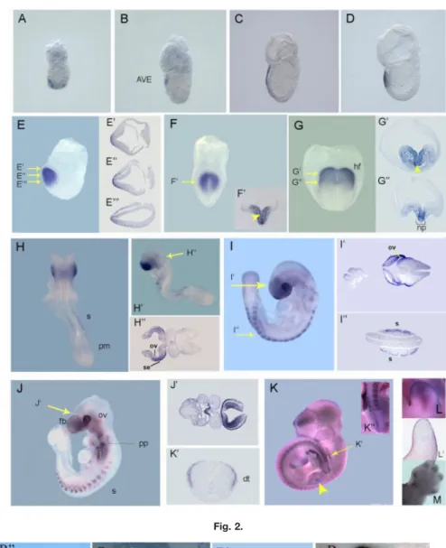
In situ hybridization analysis was used to examine the expression of mshisa transcripts during mouse em-bryogenesis. The expression of mshisa can be seen as early as 5.5 dpc and continues throughout embryonic de-velopment (Fig. 2). At pre- to early streak stages, mshisa is specifically expressed in the AVE as it migrates to the anterior side (Fig. 2A–C). By late streak stage, expression is found in a patch of anterior definitive endoderm (ADE) cells that has replaced the AVE (Fig. 2D).
In early allantoic bud embryos (Fig. 2E), around embryonic day (E) 7.25– E7.5, mshisa transcripts can only be detected in the ADE and subjacent cranial mesoderm (Fig. 2E⬘–E⬘⬙), while by early headfold stage, mshisa is also induced in the anterior neural plate (Fig. 2F⬘). Up to this point, mshisa expression seems to be ex-cluded from the midline axial mesen-doderm (Fig. 2F). As the embryo reaches stage E8.0, mshisa is
ex-pressed in the cephalic mesenchyme and presumptive forebrain neuroecto-derm (Fig. 2G–G⬙). Expression is also present in the endoderm lining the fo-regut pocket and in the rostral end of the notochordal plate (Fig. 2G⬙).
By E8.5, mshisa expression marks the prospective eye and forebrain re-gions (Fig. 2H–H⬙). Expression of mshisa is maintained in the optic ves-icles of E9.0 –E9.5 embryos (Fig. 2I– I⬘,J), and the same is true for the ex-pression in the forebrain, which can be more precisely located to the sur-face ectoderm and neuroepithelium of the prosencephalic vesicle (Fig. 2J⬘). Other expression domains found at this stage include the pharyngeal pouches (Fig. 2J), the lateral region of the invaginating otic pit (Fig. 2H⬙,I⬘), and the ventral endoderm of the fo-regut and immediately adjacent mes-enchyme (Fig. 2J⬘). Later in develop-ment, mshisa expression in the forebrain appears to become progres-sively confined to the dorsal telen-cephalon (Fig. 2K).
With the onset of somitogenesis, mshisa starts to be expressed in the forming somites, but is apparently ab-sent from the presomitic mesoderm (Fig. 2H–K). Somitic expression of mshisa is restricted to the dorsolat-eral part that constitutes the dermo-myotome (Fig. 2I⬙,K⬘). This expression pattern persists through later stages, albeit gradually decreasing to lower levels in older somites (Fig. 2H–K).
Expression of mshisa in the devel-oping limb buds can first be seen in a proximal domain (arrowhead, Fig. 2K) that subsequently shifts toward the distal tip as the bud grows (Fig. 2L). The expression in the limb bud is re-stricted to the ectoderm, as shown in Figure 2L⬘. At E13.5, mshisa expres-sion can still be detected in the tip of the forming digits (Fig. 2M), in the region undergoing chondrogenesis.
Expression of cshisa During
Chick Development
Embryos from prestreak to mid-limb stages of development (Hamburger and Hamilton, 1951) were examined by in situ hybridization (Fig. 3). Our observations reveal that the expres-sion pattern of cshisa is very similar to that of its murine counterpart.
Fig. 1. Sequence alignment of XShisa, cShisa, mShisa, rShisa, hShisa, and zShisa. Predicted amino acid sequences of cysteine-rich domains are underlined in orange. Identical amino acids among all are shaded red, whereas identical amino acids in only two sequences are shaded blue.
Fig. 2.
and Hamilton stage 1, HH1), cshisa transcripts were strongly detected in the hypoblast (Fig. 3A,A⬘). As gastru-lation begins and the primitive streak is formed, cshisa expression becomes restricted to the anterior part of the embryo, more specifically to the endodermal layer (stage HH3⫹; Fig. 3B,B⬘,B⬙). By stage HH5 (Fig. 3C), cshisa is expressed in the prospective neural plate tissue. Transverse sec-tions showed that cshisa transcripts are still present in the endodermal layer and also start to be expressed in mesodermal cells (Fig. 3C⬘,C⬙,C⬘⬙). This expression pattern is consistent with the observation that Xshisa is essential for vertebrate head forma-tion (Yamamoto et al., 2005). At stage HH6, cshisa mRNA is present in high levels in the head folds and neural plate region (Fig. 3D). Transverse sec-tions show that cshisa transcripts are localized to the ectodermal cells (Fig. 3D⬘,D⬙).
With the beginning of somitogen-esis, cshisa starts also to be expressed in the somitic territories. At stage HH7, cshisa can be detected in the first forming somite (not shown). By
stage HH9⫺ cshisa is strongly ex-pressed in the head folds and in the developing somites (Fig. 3E). A dy-namic expression pattern is observed throughout somitogenesis (Fig. 3E–H). cshisa transcripts are absent from the posterior region of the presomitic me-soderm but can be detected at low lev-els at its rostral end. The expression is strongest in the newly formed somites and gradually decreases as the somites mature. A transverse section at the somite level of a stage HH7 embryo shows that cshisa is expressed in the entire somite as well as in the lateral plate mesoderm and notochord (Fig. 3E⬘). Later in development, cshisa transcripts are also present in the pro-spective eye, forebrain, branchial arches, and otic vesicle (Fig. 3F–H). As the optic vesicles evaginate, expression is seen in the lens vesicle and anterior surface ectoderm of the frontonasal mass (Fig 3H–H⬙). At stage HH25, cshisa expression in the somite is re-stricted to the dermomyotome (Fig. 3H⬘,H⬙), resembling that of mshisa (Fig. 2K⬘).
During early limb bud stages, cshisa starts to be detected in the
more proximal region of the limb buds (not shown) and later, as limbs de-velop, the expression shifts toward the distal region (Fig. 3I,J). This expres-sion pattern in the limb buds resem-bles the one observed for MyoD, a marker of differentiating myogenic cells (Gamer et al., 2001; Fig. 3I,J).
As demonstrated above, the murine and chicken shisa are very closely re-lated to each other both in terms of their sequence similarity and the evo-lutionarily conserved expression pat-tern. The early expression of mshisa and cshisa in the AVE/hypoblast and anterior neuroectoderm also recapitu-lates the deep endomesoderm and pro-spective head ectoderm expression previously described for the Xenopus shisa, the founding member of this gene family (Yamamoto et al., 2005). Mouse and chicken shisa, however, additionally are expressed in struc-tures such as the somites, pharyngeal region. and the eye.
Being members of the Xshisa fam-ily, an antagonist of Wnt and FGF signalling (Yamamoto et al., 2005), the conserved expression patterns of both mshisa and cshisa reflect the
im-Fig. 2. Expression pattern of mshisa during mouse development. Analysis performed by in situ hybridization. All sections are 8m thick. The level at
which each section was taken is indicated on panels with yellow arrows, and the sections are shown next to the relevant panels. A–D: mshisa is expressed in the AVE in embryonic day (E) 5.5, E6.5, E6.75, and E7.0 mouse embryos. E: At E7.25 mshisa is detected in the prospective head fold.
Eⴕ,Eⴖ, Eⴕⴖ: Transverse sections of an E7.25 embryo show that mshisa is only expressed in the anterior definitive endoderm and subjacent cranial
mesoderm. F: In an E7.5 embryo, mshisa is expressed in the head folds. F⬘: Transverse section of E7.5 embryo shows that mshisa is also induced in the anterior neural plate. G: In an E8.0 embryo, mshisa is expressed in the head folds. Transverse sections show that mshisa is present in the cephalic mesenchyme, in the presumptive forebrain neuroectoderm, in the endoderm lining the foregut pocket (G⬘) and in the rostral end of the notochordal plate (G⬙). H,Hⴕ: At E8.5 mshisa transcripts are expressed in the prospective eye, forebrain, and somites. H⬙: Transverse section of E8.0 embryo shows that mshisa is expressed in the optic pit and surface ectoderm. I: At E9.0, mshisa is expressed in the eye, somites, and forebrain. Iⴕ,Iⴖ: Transverse sections of an E9.0 embryo show expression of mshisa in the optic vesicle, optic eminence, and somites. J: At E9.5, mshisa is expressed in the eye, forebrain, somites, and pharyngeal pouches. Jⴕ: Transverse section shows that mshisa is present in the surface ectoderm and in the ventral endoderm of the foregut and immediately adjacent mesenchyme. K: At E11.5, transcripts of mshisa are expressed in the dorsal telencephalon, in the eye, in the somites, and limb buds. Kⴕ: Transverse section of the tail at E11.5 shows mshisa in the somite is restricted to the dermomyotome. Kⴖ: Amplification of the tail shows that mshisa is absent from the presomitic mesoderm. L: Amplification of the limb bud at E11.5 shows mshisa expression. Lⴕ: Sagittal section of the limb bud at E11.5 shows that mshisa expression is restricted to the surface ectoderm. K: Amplification of the limb at 13.5 shows mshisa is detected in the tip of the forming digits. AVE, anterior visceral endoderm; dt, dermatome; fb, forebrain; fg, foregut; hf, head fold; np, notochordal plate; ov, optic vesicle; pm, presomitic mesoderm; pp, pharyngeal pouches; s, somite; se, surface ectoderm.
Fig. 3. Localization of cshisa transcripts in developing chicken embryos detected by in situ hybridization. A, C, E and F are ventral views, whereas B
and D are dorsal views of whole-mount embryos. All sections are transverse 16-m cryo-sections. The level at which each section was taken is indicated on panels with yellow arrows, and the sections are shown next to the relevant panels. A: cshisa is expressed in the hypoblast in Hamburger and Hamilton stage (HH) 1 chicken embryo. Anterior is to the top. Aⴕ: Transverse section of a HH1 chicken embryo showing cshisa expression exclusively in the hypoblast. B,Bⴕ: Stage HH3⫹embryo showing expression of cshisa restricted to the endodermal layer. C: At HH5, cshisa transcripts are expressed in the prospective neural plate. Cⴕ,Cⴖ,Cⴕⴖ: Transverse sections of the embryo in C showing cShisa staining in endoderm and ectoderm.
D: At HH6, cshisa expression appears restricted to the neural plate and primitive folds. Dⴕ,Dⴖ: Transverse sections a HH6 embryo show that cshisa
transcripts are located in the ectodermal cells. E: In stage HH9, cshisa is expressed in the head folds and the somites. There is an absence of cshisa transcripts within the presomitic mesoderm of the embryo. Eⴕ: Section taken at the level of the somites shows cshisa expression within the somite and in the lateral plate mesoderm. The notochord is also positive for cshisa expression. F: At HH11, cshisa transcripts are observed in the forming brain, prospective eye, and at the somite level. In the somites, cshisa expression is strongest in the recently formed somites. G: By stage HH18, cshisa can be detected in the forebrain, eye, otic vesicle, pharyngeal pouches, branchial arches, somites, and the developing limb buds. H: cshisa expression in HH25 remains in the otic vesicle, forebrain, branchial arches, eye, somites, and limb buds. Hⴕ,Hⴖ: Transverse sections show that cshisa transcripts in the somite are restricted to the dermatome. I,J: cshisa expression in the early limb buds (J, stage HH22; I, stage HH25) has a very dynamic pattern. cshisa starts to be expressed more posteriorly and then migrates toward more distal region. Forelimbs are shown in the top and hindlimbs in the bottom. ba, branchial arches; dt, dermatome; ey, eye; fb, forebrain; hf, head fold; hyp, hypoblast; lpm, lateral plate mesoderm; ov, otic vesicle; nc, notochord; np, neural plate; pm, presomitic mesoderm; s, somite.
portance they may have during em-bryonic development of the mouse and chick embryos, patterning topologi-cally equivalent regions in these ver-tebrate embryos.
Assuming, based on their homology with the Xenopus protein, that the mouse and chicken Shisa also function as antagonists of Wnt and FGF signal-ling, then their expression in the AVE and hypoblast may seem, at a first glance, hard to conciliate with the role attributed to these tissues in early neural induction. In fact, recent find-ings strongly suggest that, at least in chicken, FGF and Wnt signalling are required for neural induction at a very early stage, even before gastrulation. However, it should be taken into con-sideration that Shisa acts cell-autono-mously; therefore, its expression in the AVE/hypoblast is unlikely to in-hibit FGF signalling in the overlying epiblast. Shisa might instead play an indirect role in promoting neural in-duction by participating in the speci-fication and/or maintenance of the AVE/hypoblast identities, for example through repression of the autocrine action of FGF-8, which is expressed in the AVE (Crossley and Martin, 1995). Later on, Shisa is expressed in the neural plate, and it’s plausible then that, like in Xenopus, it inhibits the caudalizing Wnt and FGF signals in this tissue. A similar reasoning can be applied to the function of Shisa in the developing limb buds, where Wnt and FGF signaling is known to direct out-growth and patterning. shisa is ex-pressed in the ectoderm layer of the limbs, where most of the Wnt and FGF signalling centers are also lo-cated. Again it is conceivable that Shisa is not antagonizing these sig-nalling pathways in the target mesen-chymal cells but is instead acting on some of the signalling centers, per-haps protecting them from their own signals. During somitogenesis Shisa might be involved in the process of somite differentiation and condensa-tion through the inhibicondensa-tion the FGF signalling coming from the posterior presomitic mesoderm. Subsequently expression in the dorsolateral com-partment of the somite suggests that Shisa could be repressing the FGF-and Wnt-mediated myogenic signals in these cells, which as a result will be specified as dermatome. These
consid-erations are purely hypothetical, how-ever, and a more conclusive character-ization of the biological function of the mouse and chicken Shisa in embry-onic development will require further biochemical and genetic analyses.
EXPERIMENTAL
PROCEDURES
Chicken and Mouse Embryo
Collection
Fertilized chicken eggs were pur-chased from local suppliers. Eggs were incubated at 37°C in a humidified in-cubator until the desired developmen-tal stage. Embryos were staged ac-cording to Hamburger and Hamilton (1951).
Mouse embryos were obtained crossing B6SJL/F1 hybrids main-tained on a 19-hour light to 5-hour dark cycle and mated overnight. Noon of the day of vaginal plug detection was designated 0.5 dpc. Embryos were dissected from the uterus in phos-phate buffered saline and further staged by morphological landmarks (Downs and Davies, 1993).
Cloning of mshisa and
cshisa cDNAs
The EST clone BC057640, containing the entire predicted coding sequence of mshisa as well as the 5⬘- and 3⬘-UTRs, was obtained from RZPD (IMAGp998G149268Q3). To isolate a fragment of the cshisa coding se-quence (323– 829), total RNA from stage HH22 chick embryos (Ham-burger and Hamilton, 1951) was iso-lated using Trizol reagent (Invitrogen) according to the manufacturer’s proto-col. Random-primed cDNA synthe-sized from these samples with H mi-nus M-MuLV reverse transcriptase (Fermentas) was subjected to 25 cy-cles of amplification by polymerase chain reaction (PCR), at an annealing temperature of 55°C. The following primers were used: forward, 5⬘- CAT-TGTCGGCTCCGTCTTCGTC-3⬘; re-verse, 5 ⬘-TTCTGCTCTCCGCCTGCA-TG -3⬘. The PCR product was cloned into the pGEM-T Easy vector (Pro-mega). The sequence of PCR-amplified cshisa cDNA was determined on an ABI sequencer. The sequence of chicken shisa cDNA was deposited in
the GenBank database under the ac-cession no. DQ342343.
Whole-Mount In Situ
Hybridization and Histology
Single whole-mount in situ hybridiza-tion and antisense-probe preparahybridiza-tion were performed as previously de-scribed (Belo et al., 1997). Digoxige-nin-labeled mshisa antisense RNA probe was synthesized by linearizing the BC05764 clone with BglII and transcribing with T7 RNA polymer-ase. To generate the digoxigenin-la-beled cshisa antisense RNA probe, the plasmid containing cshisa coding se-quence fragment (pGEM-Teasy.c-shisa) was linearized using SalI and transcribed using T7 RNA polymer-ase.After staining, embryos were re-fixed in 4% paraformaldehyde and photographed by using a Leica DFCM20 digital camera. Some em-bryos were embedded in 15% sucrose, 7.5% gelatin, frozen, and sectioned (16 m) using a Leica CMM0S0 S cryo-stat; others were embedded in paraf-fin and sectioned (8m) using a mic-rotome Leica RM2135. The sections were examined and photographed us-ing a Leica DM LB2 microscope and a Leica DFCM20 digital camera.
ACKNOWLEDGMENTS
We thank Ana T. Tavares and Nuno Afonso for critically reading of this manuscript. M.F., L.G., and A.C.S. are recipients of F.C.T. PhD fellowships. M.B. is recipient of a F.C.T. fellow-ship. This work was supported by re-search grants from F.C.T. and IGC/ Fundac¸a˜o Calouste Gulbenkian to J.A.B., where he is a Principal Inves-tigator.
REFERENCES
Altschul SF, Gish W, Miller W, Myers EW, Lipman DJ. 1990. Basic local alignment search tool. J Mol Biol 215:403–410. Aulehla A, Herrmann BG. 2004.
Segmen-tation in vertebrates: clock and gradient finally joined. Genes Dev 18:2060 –2067. Aulehla A, Wehrle C, Brand-Saberi B, Kemler R, Gossler A, Kanzler B, Herr-mann BG. 2003. Wnt3a plays a major role in the segmentation clock control-ling somitogenesis. Dev Cell 4:395–406. Beddington RS, Robertson EJ. 1999. Axis
development and early asymmetry in mammals. Cell 96:195–209.
Belo JA, Bouwmeester T, Leyns L, Kertesz N, Gallo M, Follettie M, De Robertis EM. 1997. Cereberus-like is a secreted factor with neutralizing activity expressed in the anterior-primitive endoderm of the mouse gastrula. Mech Dev 68:45–57. Birney E, Andrews D, Caccamo M, Chen Y,
Clarke L, Coates G, Cox T, Cunningham F, Curwen V, Cutts T, Down T, Durbin R, Fernandez-Suarez XM, Flicek P, Gra¨f S, Hammond M, Herrero J, Howe K, Iyer V, Jekosch K, Ka¨ha¨ri A, Kasprzyk A, Keefe D, Kokocinski F, Kulesha E, Lon-don D, Longden I, Melsopp C, Meidl P, Overduin B, Parker A, Proctor G, Prlic A, Rae M, Rios D, Redmond S, Schuster M, Sealy I, Searle S, Severin J, Slater G, Smedley D, Smith J, Stabenau A, Stalker J, Trevanion S, Ureta-Vidal A, Vogel J, White S, Woodwark C, Hubbard TJP. 2006. Ensembl 2006. Nucleic Acids Res 34:D556 –D561.
Bouwmeester T, Leyns L. 1997. Vertebrate head induction by anterior primative endoderm. BioEssays 19:855– 863. Burge C, Karlin S. 1997. Prediction of
com-plete gene structures in human genomic DNA. J Mol Biol 268:78.
Capdevila J, Izpisua Belmonte JC. 2001. Patterning mechanisms controlling ver-tebrate limb development. Annu Rev Cell Dev Biol 17:87–132.
Crossley PH, Martin GR. 1995. The mouse Fgf8 gene encodes a family of polypeptides and is expressed in regions that direct out-growth and patterning in the developing embryo. Development 121:439.
Deng CX, Wynshaw-Boris A, Shen MM, Daugherty C, Ornitz DM, Leder P. 1994. Murine FGFR-1 is required for early postimplantation growth and axial orga-nization. Genes Dev 8:3045–3057. Downs KM, Davies T. 1993. Staging of
gas-trulating mouse embryos by morpholog-ical landmarks in the dissecting micro-scope. Development 118:1255–1266. Dubrulle J, Pourquie O. 2004a. Coupling
segmentation to axis formation. Develop-ment 131:5783–5793.
Dubrulle J, Pourquie O. 2004b. fgf8 mRNA decay establishes a gradient that couples axial elongation to patterning in the ver-tebrate embryo. Nature 427:419 –422. Dubrulle J, McGrew MJ, Pourquie O. 2001.
FGF signaling controls somite boundary position and regulates segmentation clock control of spatiotemporal Hox gene activation. Cell 106:219 –232.
Foley AC, Skromne I, Stern CD. 2000. Rec-onciling different models of forebrain in-duction and patterning: a dual role for the hypoblast. Development 127:3839 – 3854.
Gamer LW, Cox KA, Small C, Rosen V. 2001. Gdf11 is a negative regulator of chondrogenesis and myogenesis in the developing chick limb. Dev Biol 229:407– 420.
Glinka A, Wu W, Onichtchouk D, Blumen-stock C, Niehrs C. 1997. Head induction
by simultaneous repression of Bmp and Wnt signalling in Xenopus. Nature 389: 517–519.
Glinka A, Wu W, Delius H, Monaghan AP, Blumenstock C, Niehrs C. 1998. Dick-kopf-1 is a member of a new family of secreted proteins and functions in head induction. Nature 391:357–362. Grass S, Arnold HH, Braun T. 1996.
Alter-ations in somite patterning of Myf-5-de-ficient mice: a possible role for FGF-4 and FGF-6. Development 122:141. Hamburger V, Hamilton HL. 1951. A
se-ries of normal stages in the development of the chick embryo. J Morphol 88:49 – 92.
Katoh Y, Katoh M. 2005. Comparative genomics on Shisa orthologs. Int J Mol Med 16:181.
Mangold O. 1933. U¨ ber die Induktionsfa¨-higkeit der verschiedenen Bezirke der Neurula von Urodelen. Naturwissen-schaften 21:761–766.
Mao B, Wu W, Li Y, Hoppe D, Stannek P, Glinka A, Niehrs C. 2001. LDL-receptor-related protein 6 is a receptor for Dick-kopf proteins. Nature 411:321–325. Mesnard D, Filipe M, Belo JA,
Zernicka-Goetz M. 2004. The anterior-posterior axis emerges respecting the morphology of the mouse embryo that changes and aligns with the uterus before gastrula-tion. Curr Biol 14:184 –196.
Oulad-Abdelghani M, Chazaud C, Bouillet P, Mattei MG, Dolle P, Chambon P. 1998. Stra3/lefty, a retinoic acid-induc-ible novel member of the transforming growth factor-beta superfamily. Int J Dev Biol 42:23–32.
Pfeffer PL, De Robertis EM, Izpisua-Bel-monte JC. 1997. Crescent, a novel chick gene encoding a Frizzled-like cysteine-rich domain, is expressed in anterior re-gions during early embryogenesis. Int J Dev Biol 41:449 –458.
Piccolo S, Sasai Y, Lu B, De Robertis EM. 1996. Dorsoventral patterning in Xeno-pus: inhibition of ventral signals by di-rect binding of chordin to BMP-4. Cell 86:589 –598.
Piccolo S, Agius E, Leyns L, Bhattacharyya S, Grunz H, Bouwmeester T, De Robertis EM. 1999. The head inducer Cereberus is a multifunctional antagonist of Nodal, BMP and Wnt signals. Nature 397:707– 710.
Pirskanen A, Kiefer JC, Hauschka SD. 2000. IGFs, insulin, Shh, bFGF, and TGF-beta1 interact synergistically to promote somite myogenesis in vitro. Dev Biol 224:189.
Pourquie O. 2001. Vertebrate somitogen-esis. Annu Rev Cell Dev Biol 17:311–350. Pourquie O. 2004. The chick embryo: a leading model in somitogenesis studies. Mech Dev 121:1069 –1079.
Rivera-Perez JA, Mager J, Magnuson T. 2003. Dynamic morphogenetic events characterize the mouse visceral endoderm. Dev Biol 261:470 –487.
Rodriguez TA, Srinivas S, Clements MP, Smith JC, Beddington RS. 2005. Induc-tion and migraInduc-tion of the anterior vis-ceral endoderm is regulated by the extra-embryonic ectoderm. Development 132: 2513–2520.
Rodriguez Esteban C, Capdevila J, Econo-mides AN, Pascual J, Ortiz A, Izpisua Belmonte JC. 1999. The novel Cer-like protein Caronte mediates the establish-ment of embryonic left-right asymmetry. Nature 401:243–251.
Silva AC, Filipe M, Kuerner KM, Stein-beisser H, Belo JA. 2003. Endogenous Cereberus activity is required for ante-rior head specification in Xenopus. De-velopment 130:4943–4953.
Spemann H. 1931. U¨ ber den Anteil von Implantat und Wirtskeim an der Orien-tierung und Beschaffenheit der induz-ierten Embryonalanlage. W Roux Arch Entwicklungsmech 123:390 –516. Srinivas S, Rodriguez T, Clements M,
Smith JC, Beddington RS. 2004. Active cell migration drives the unilateral movements of the anterior visceral endoderm. Development 131:1157–1164. Stern HM, Hauschka SD. 1995. Neural tube and notochord promote in vitro myogenesis in single somite explants. Dev Biol 167:87.
Sun X, Meyers EN, Lewandoski M, Martin GR. 1999. Targeted disruption of Fgf8 causes failure of cell migration in the gastrulating mouse embryo. Genes Dev 13:1834 –1846.
Thomas P, Beddington R. 1996. Anterior primitive endoderm may be responsible for patterning the anterior neural plate in the mouse embryo. Curr Biol 6:1487– 1496.
Wagner J, Schmidt C, Nikowits W Jr, Christ B. 2000.Compartmentalization of the somite and myogenesis in chick em-bryos are influenced by wnt expression. Dev Biol 228:86.
Yamamoto M, Saijoh Y, Perea-Gomez A, Shawlot W, Behringer RR, Ang SL, Hamada H, Meno C. 2004. Nodal antag-onists regulate formation of the antero-posterior axis of the mouse embryo. Na-ture 428:387–392.
Yamamoto A, Nagano T, Takehara S, Hibi M, Aizawa S. 2005. Shisa promotes head formation through the inhibition of re-ceptor protein maturation for the caudal-izing factors, Wnt and FGF. Cell 120:223– 235.
Yoshikawa Y, Fujimori T, McMahon AP, Takada S. 1997. Evidence that absence of Wnt-3a signaling promotes neuraliza-tion instead of paraxial mesoderm devel-opment in the mouse. Dev Biol 183:234 – 242.
Zimmerman LB, De Jesus-Escobar JM, Harland RM. 1996. The Spemann orga-nizer signal noggin binds and inactivates bone morphogenetic protein 4. Cell 86: 599 –606.