JOSÉ MOISÉS BATISTA PEREIRA FILHO
Filogenia da tribo Attacobiini Roewer, 1955 (Araneae, Corinnidae, Corinninae)
Belém, 2015
Filogenia da tribo Attacobiini Roewer, 1955 (Araneae, Corinnidae, Corinninae)
Orientador: Dr. Alexandre Bragio Bonaldo
Belém, 2015
Dissertação apresentada ao Programa de Pós-graduação em Zoologia, do convênio da Universidade Federal do Pará e Museu Paraense Emílio Goeldi, como requisito parcial para obtenção do título de Mestre em Zoologia.
Área de concentração: Evolução
Esta dissertação não é válida como publicação, confirme capítulo 3 do CÓDIGO
INTERNACIONAL DE NOMENCLATURA ZOOLÓGICA. Portanto, os novos
nomes e mudanças taxonômicas propostas aqui não têm validade para fins de
nomenclatura ou prioridade.
WARNING
JOSÉ MOISÉS BATISTA PEREIRA FILHO
Filogenia da tribo Attacobiini Roewer, 1955 (Araneae, Corinnidae, Corinninae)
Dissertação apresentada ao Programa de Pós-Graduação em Zoologia, do convênio
da Universidade Federal do Pará e Museu Paraense Emílio Goeldi, como requisito
parcial para obtenção do título de Mestre em Zoologia, sendo a COMISSÃO
JULGADORA composta pelos seguintes membros:
DR. ALEXANDRE BRAGIO BONALDO
INSTITUIÇÃO: MUSEU PARAENSE EMILIO GOELDI (PRESIDENTE)
DR. GUSTAVO RODRIGO SANCHES RUIZ
INSTITUIÇÃO: UNIVERSIDADE FEDERAL DO PARÁ
DRA. REGIANE SATURNINO FERREIRA
INSTITUIÇÃO: MUSEU PARAENSE EMILIO GOELDI
DR. ANTÔNIO DOMINGOS BRESCOVIT
INSTITUIÇÃO: INSTITUTO BUTANTAN
DR. RICARDO OTT
INSTITUIÇÃO: MUSEU DE CIÊNCIAS NATURAIS
DRA. DANIELE POLOTOW GERALDO
INSTITUIÇÃO: UNIVERSIDADE DE SÃO PAULO
Aprovada em: 02 de abril de 2015
Agradeço inicialmente ao meu orientador Dr. Alexandre Bragio Bonaldo pelos ensinamentos, incentivo e paciência que permitiram a finalização deste trabalho. Sua orientação foi de grande importância para o amadurecimento do meu conhecimento científico e desenvolvimento profissional.
Agradeço ao Conselho Nacional de Desenvolvimento Científico e Tecnológico (CNPq) pela concessão da bolsa que deu suporte ao andamento do projeto. Ao Programa de Pós-graduação em Zoologia da Universidade Federal do Pará (UFPA), em convênio com o Museu Paraense Emílio Goeldi (MPEG) por todo apoio e suporte necessário durante os anos de desenvolvimento dessa dissertação.
A todos os curadores e assistentes de curadoria, no Brasil e no exterior, que enviaram material ou fotografias: Antônio Brescovit, Márcio Luiz de Oliveira, Ricardo Ott, Luis Alberto Pereira, Ricardo Pinto-da-Rocha, Adalberto dos Santos, José Roberto Pujol Luz.
Aos amigos e colegas de laboratório: Regiane Saturnino, Bruno Rodrigues, Manoel Barros, Emanuel Cafofo, Laura Miglio, Daniella Moss, Lúciu Rezende, Níthomas Feitosa e Yulie Shimano, pelas discussões de cunho científico que em muito contribuíram para minha dissertação, bem como pelos momentos de confraternização, descontração e diversão que também contribuíram bastante, pois me ajudavam a diminuir a tensão além de tornarem meus dias no laboratório mais agradáveis. Aos meus amigos, os de infância, de Soure e os do Colégio Sophos (GDQ) por todo momento de diversão e descontração nesse período.
Aos meus pais (in memorian) que mesmo não estando presentes sempre foram fonte de inspiração, tanto como pessoas quanto como profissionais. O que me incentiva cada vez mais a estudar e buscar meus objetivos sempre com muita fé.
Agradeço especialmente a minha esposa Thalita, por também ser exemplo de profissional dedicada e comprometida. E por todos os momentos felizes que me proporcionou durante esse período, por ter compreendido os momentos em que tive de abrir mão de sua companhia para estar trabalhando no laboratório, e, principalmente, gostaria de agradece-la por estar carregando o maior presente que ela poderia me dar, a nossa filha Laura.
1.Introdução Geral ... 1
2.Referências bibliográficas ... 23
3.Artigo: A Cladistic Analysis of the Tribe Attacobiini Roewer, 1955 (Araneae, Corinnidae, Corinninae) ... 25
Abstract ... 26
Key words... 26
Introduction ... 26
Material and Methods ... 27
Result and Discursion ... 30
Character coding and optimization ... 30
Recognized clades ... 48
Character systems ... 53
Acknowledgements ... 55
Legends ... 57
Figures ... 59
References ... 69
Appendix 1 ... 71
As aranhas aqui tratadas são pertencentes à família Corinnidae, que somente se estabeleceu como tal a partir das evidencias do trabalho de Lehtinen (1967) sobre a artificialidade de Clubionidae, família que incluía Corinninae desde o trabalho de Simon (1897). Recentemente a composição da família sofreu alterações, passando a ser composta apenas por duas subfamílias, Corinninae e Castianeirinae, além de um agrupamento de gêneros considerados como Incertae sedis (Ramirez, 2014), passando a a ser composta por 659 espécies distribuídas em 58 gêneros (WSC, 2015). As subfamílias de Corinnidae são facilmente reconhecidas pelas morfologias do palpo do macho. Castianeirinae apresenta o bulbo do palpo em forma de pêra, enquanto Corinninae apresenta duto espermático com percurso helicoidal. Ambas as subfamílias compartilham uma placa epiginal única nas fêmeas; os palpos dos machos apresentam um alargamento no setor distal do ducto espermático; normalmente são desprovidos de apófise média, exceto em Attacobiini (tribo de Corinninae), nos gêneros neotropicais Ianduba Bonaldo e Olbus Simon e nos gêneros africanos Mandaneta Strand, Procopius Thorell e Pseudocorinna Simon (Corinnidae insertae sedis). Além disso, o corpo fortemente esclerotinizado, principalmente a carapaça (exceto em algumas espécies de Olbus e em alguns gêneros de Corinninae, incluindo os pertencentes à Attacobiini) e os espiráculos pulmonares largos, com bordas esclerotinizadas, são também características informativas para o reconhecimento da família.
nos novos formigueiros.
As primeiras contribuições para a taxonomia destas aranhas foram confusas. A subfamília Myrmecobiinae foi proposta por Mello-Leitão (1923), para agrupar uma única espécie, Myrmecobius luederwaldti. Na ocasião o autor defendeu a proposição da subfamília devido a estas aranhas possuírem características tão singulares que não se enquadrariam em quaisquer das subfamílias até então propostas em Clubionidae. Posteriormente foi proposto Attacobius como um novo nome para o gênero, pois Myrmecobius estava pré-ocupado por um marsupial (Mello-Leitão, 1925). Mello-Leitão (1947) sinonimizou Myrmeques Roewer (1935) com Attacobius, sinonimizando a espécie-tipo, Myrmeques attarum Roewer, 1935, com Attacobius luederwaldti. Roewer (1955) alocou o nome do grupo tribal Attacobieae, na subfamília Liocraninae, Clubionidae, grupo que posteriormente foi elevado a família. Platnick & Baptista (1995) transferiram a tribo Attacobiini de Liocranidae para Corinninae (Corinnidae), tornando-a a única tribo aceita até o momento na subfamília, além de Corinnini, a tribo-tipo. Além disso, estes autores removeram Attacobius attarum da sinonímia de A. luederwaldti, trazendo à luz dois sinônimos adicionais do gênero, Morenilia e Achalaicola, propostos por Mello-Leitão (1942; 1943) em Gnaphosidae e Prodidomidae, respectivamente. Uma vez que Morenilia e Achalaicola são gêneros monotípicos e dado que que suas espécies-tipo foram sinonimizadas por Platnick & Baptista (1995), apenas uma espécie foi acrescida ao gênero na revisão de Platnick & Baptista, A. nigripes (Mello-Leitão, 1942). Uma nova espécie de Attacobius foi descoberta somente 56 anos após a última contribuição de Mello-Leitão, com a descrição de A. verhaagui por Bonaldo & Brescovit (1998). Contudo, a real diversidade de Attacobius foi reconhecida apenas após o trabalho de Bonaldo & Brescovit (2005) que acrescentaram seis espécies ao gênero, quatro das quais conhecidas por machos. Esta contribuição permitiu pela primeira vez uma apreciação da variação dos caracteres morfológicos do palpo do macho, uma vez que a única forma masculina conhecida até aquele momento era a de A. attarum.
sinapomorfia de Attacobiini, mas do gênero. Eles também propuseram duas sinapomorfias putativas adicionais para a tribo: labium muito mais largo do que longo (característica já observada por Mello-Leitão em 1923 em Attacobius luederwaldti) e palpo do macho com um processo retrolateral longo e fino, medialmente embutido no tegulum, denominado como processo tegular Attacobiini, que mais tarde foi considerado homólogo à apófise média por Bonaldo & Brescovit (2005). Ecitocobius distingue-se de Attacobius pela ausência dos olhos posteriores e laterais anteriores e pela presença de um espinho curvo não pareado na região ventral apical dos metatarsos de todas as pernas. Outras características adicionais são a ausência de qualquer padrão distintivo nos olhos medianos anteriores e tarsos das pernas não modificados (Bonaldo & Brescovit, 1998).
Atualmente a tribo Attacobiini é composta por dois gêneros, Attacobius e Ecitocobius. O gênero Attacobius compreende 10 espécies válidas. As aranhas deste gênero ocorrem no Brasil e na Argentina. O gênero Ecitocobius, é representado por uma única espécie, conhecida apenas por um indivíduo macho coletado na Reserva Florestal Adolpho Ducke, Manaus, Amazonas, Brasil.
QUADRO 1. Lista de espécies válidas da tribo Attacobiini, espécies tipos dos gêneros marcadas com (*).
Espécie Autor Sexo conhecido
Ecitocobius comissator* Bonaldo & Brescovit, 1998 Macho Attacobius luederwaldti* (Mello-Leitão 1923) Fêmea
Attacobius attarum (Roewer 1935) Ambos
Attacobius nigripes (Mello-Leitão 1942) Fêmea Attacobius verhaaghi Bonaldo & Brescovit, 1998 Fêmea Attacobius tucurui Bonaldo & Brescovit, 2005 Macho Attacobius lamellatus Bonaldo & Brescovit, 2005 Ambos Attacobius uiriri Bonaldo & Brescovit, 2005 Macho Attacobius blakei Bonaldo & Brescovit, 2005 Macho Attacobius carranca Bonaldo & Brescovit, 2005 Ambos Attacobius kitae Bonaldo & Brescovit, 2005 Fêmea
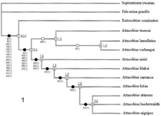
primeiro composto por A. tucurui, A. lamellatus e A. verhaaghi, sendo estes dois últimos mais intimamente relacionados. O segundo clado é composto pelos demais representantes do gênero, sendo que um clado formado por três espécies, A. attarum, A. luederwaldti e A. nigripes, é o melhor suportado dentro do gênero com 4 sinapomorfias e SB = 3.0.
FIGURA 1: Filogenia de Attacobiini proposta por Bonaldo & Brescovit. Percebe-se que o suporte dos grupos de espécies de Attacobius foi mais fraco do que os suportes obtidos para a Tribo e para o gênero Attacobius. FONTE: Bonaldo e Brescovit (2005).
No intuito de obter uma análise mais completa e com maiores resolução e suporte, o presente trabalho realizou uma nova filogenia utilizando, além de todos os terminais usados por Bonaldo e Brescovit (2005), novos terminais (espécies descritas após o trabalho de Bonaldo e Brescovit, 2005), complementando a matriz com informação adicional sobre terminais incluídos originalmente (sexos não conhecidos naquela ocasião). Assim, apresenta-se a seguir a análise de uma matriz mais completa, incorporando as informações taxonômicas disponibilizadas por Pereira-Filho & Bonaldo (em preparação), que propuseram duas novas espécies de Attacobius (Attacobius sp. n. PAR e Attacobius sp. n. TOC) bem como apresentaram pela primeira vez as descrições do macho de A. verhaaghi e das fêmeas de A. uiriri e A. blakei.
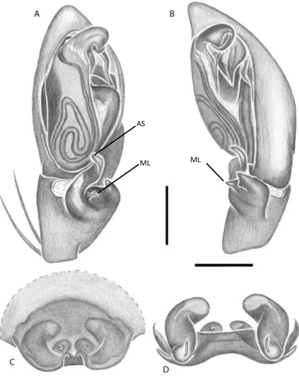
Xeropigo cotijuba De Souza & Bonaldo (Figura 2):
FIGURA 2: A-D Xeropigo cotijuba, Palpo do macho: A – posição ventral; B – posição retrolateral; Epígino: C – posição ventral; D - posição dorsal. Abreviatura: ML, Median Lobe of RTA; AS, apical spur. Barra de escala, 0,50 mm. FONTE: Modificado de De Souza & Bonaldo (2007).
ML ML
Septentrinna yucatan Bonaldo (Figura 3):
Falconina gracilis (Keyserling) (Figura 4):
FIGURA 4: A-D Falconina gracilis, Palpo do macho: A – posição ventral; B – Tíbia retrolateral; Epígino: C – posição ventral; D - posição dorsal. Abreviaturas: PVTK, prolateral ventral tegular keel; AS, apical spur; ML, median lobe. Barra de escala, 0,25 mm. FONTE: Modificado de Bonaldo (2000).
ML
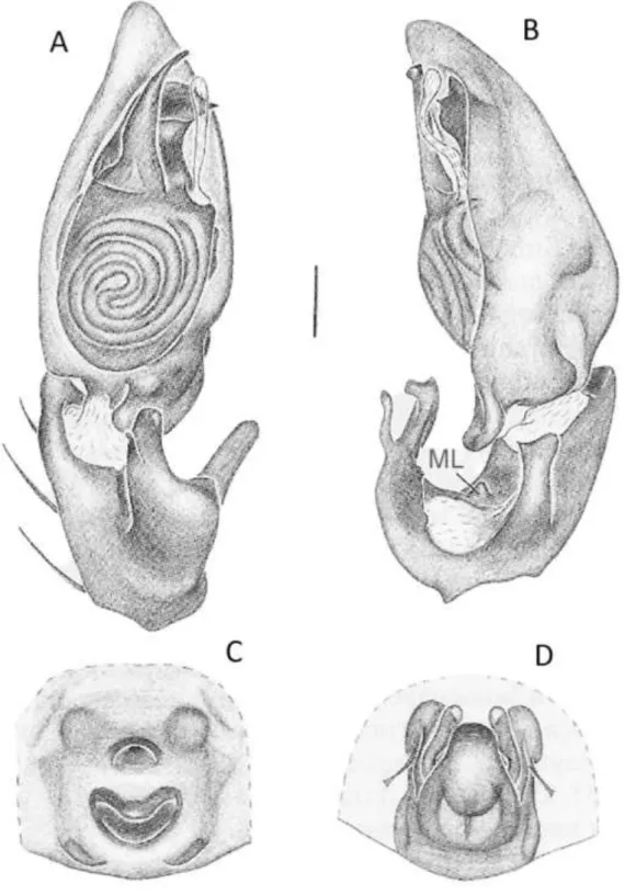
Ecitocobius comissator Bonaldo & Brescovit (Figura 5):
FIGURA 5: A-C Ecitocobius comissator, A – cefalotórax e abdome, vista dorsal; Palpo do macho: B – posição ventral; C – posição retrolateral. Abreviaturas: CT, cymbial tubercle; VTK, ventral tegular keel; AS, apical spur; dMA, distal end of median apophysis; pMA, proximal end of median apophysis. Barra de escala, 0,25 mm. FONTE: Modificado de Bonaldo e Brescovit (1998).
VTK
AS
dMA
Attacobius tucurui Bonaldo & Brescovit (Figura 6):
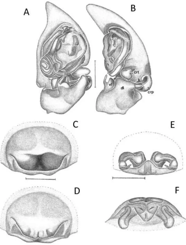
FIGURA 6: A-B Attacobius tucurui, Palpo do macho: A – posição ventral; B – posição retrolateral. Abreviaturas: AS, apical spur of VL; CRP, cymbial retrodorsal process; CRT, cymbial retrolateral tubercle; DL, dorsal lobe of RTA; VL, ventral lobe of RTA; dMA, distal end of median apophysis; mMA, median extension of median apophysis; pMA, proximal end of median apophysis; PVTK, prolateral ventral tegular keel; VTK, ventral tegular keel. Barra de escala, 0,25 mm. FONTE: Modificado de Bonaldo e Brescovit (2005).
PVTK
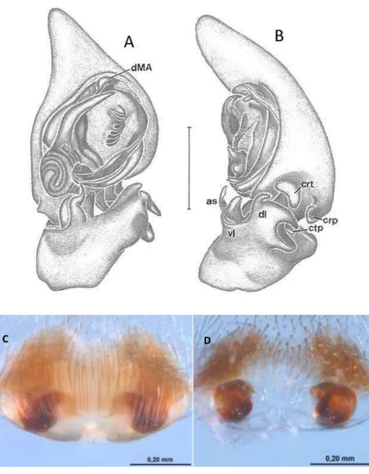
Attacobius uiriri Bonaldo & Brescovit (Figura 7):
FIGURA 7: A-D Attacobius uiriri, Palpo do macho: A – posição ventral; B – posição retrolateral. Barra de escala, 0,25 mm. FONTE: Modificado de Bonaldo & Brescovit (2005). Epígino da fêmea: C – posição ventral; D – posição dorsal. FONTE: Pereira-Filho & Bonaldo (em preparação). Abreviaturas: AS, apical spur of VL; CRP, cymbial retrodorsal process; CRT, cymbial retrolateral tubercle; DL, dorsal lobe of RTA; VL, ventral lobe of RTA; dMA, distal end of median apophysis; mMA, median extension of median apophysis; pMA, proximal end of median apophysis; PVTK, prolateral ventral tegular keel; VTK, ventral tegular keel.
C D
PVTK
Attacobius blakei Bonaldo & Brescovit (Figura 8):
FIGURA 8: A-D Attacobius blakei, Palpo do macho: A – posição ventral; B – posição retrolateral. Barra de escala, 0,25 mm. FONTE: Bonaldo e Brescovit (2005). Epígino da fêmea: C – posição ventral; D – posição dorsal. FONTE: Pereira-Filho & Bonaldo (em preparação). Abreviaturas: AS, apical spur of VL; CRP, cymbial retrodorsal process; CRT, cymbial retrolateral tubercle; DL, dorsal lobe of RTA; VL, ventral lobe of RTA; dMA, distal end of median apophysis.
Attacobius attarum (Roewer) (Figura 9):
FIGURA 9: A-E Attacobius attarum, Epígino: A – posição ventral; B – posição posterior; C – posição dorsal. FONTE: Modificado de Platnick & Baptista (1995). Palpo do macho: D – posição ventral; E – posição retrolateral. FONTE e de Bonaldo e Brescovit (1998). Abreviaturas: C, condutor; CP, processo cimbial; CT, tubérculo cimbial; dMA, distal end of median apophysis; pMA, proximal end of median apophysis; VTF, ventral tegular flange; AS, apical spur; ML, median lobe. Barra de escala, 0,25 mm para os palpos; epíginos sem escala.
VTF
AS
ML pMA
Attacobius carranca Bonaldo & Brescovit (Figura 10):
Attacobius lamellatus Bonaldo & Brescovit (Figura 11):
Attacobius luederwaldti (Mello-Leitão) (Figura 12):
Attacobius nigripes (Mello-Leitão) (Figura 13):
Attacobius verhaaghi Bonaldo & Brescovit (Figura 14):
FIGURA 14: A-F Attacobius verhaaghi, A – Carapaça, posição dorsal; Epígino: B – posição ventral; C – posição dorsal; D - posição posterior. Barra de escala, 0,25 mm. FONTE: Modificado de Bonaldo e Brescovit (1998). Palpo do macho: E – posição ventral; F – posição retrolateral. FONTE: Pereira-Filho & Bonaldo (em preparação).
Attacobius kitae Bonaldo & Brescovit (Figura 15):
Attacobius PAR Pereira-Filho & Bonaldo (em preparação) (Figura 16):
FIGURA 16: A-F Attacobius PAR. Palpo do macho: A – posição ventral; B – posição retrolateral. FONTE: Pereira-Filho & Bonaldo (em preparação).
Attacobius TOC Pereira-Filho & Bonaldo (em preparação) (Figura 17):
REFERÊNCIAS BIBLIOGRÁFICAS
BONALDO, A.B. & BRESCOVIT, A.D. (1998): On Ecitocobius , a new genus from Central Amazonia with comments on the tribe Attacobiini (Arachnida, Araneae, Corinnidae, Corinninae). Spixiana 21/2: 165-172.
BONALDO, A.B. & BRESCOVIT, A.D. (2005): On new species of the Neotropical spider genus Attacobius Mello-Leitão, 1923 (Araneae, Corinnidae, Corinninae), with a cladistics analysis of the tribe Attacobiini. Insect Syst.Evol. 36: 35-56.
DA SILVA CAMARGO, R., FORTI, L. C., DE MATOS, C. A. O., & BRESCOVIT, A. D. (2015). Phoretic behaviour of Attacobius attarum (Roewer, 1935) (Araneae: Corinnidae: Corinninae) dispersion not associated with predation?. Journal of Natural History, 1-6.
ERTHAL JR, M., & TONHASCA JR, A. (2001). Attacobius attarum Spiders (Corinnidae): Myrmecophilous Predators of Immature Forms of the Leaf-Cutting Ant Atta sexdens (Formicidae) 1. Biotropica, 33(2), 374-376.
ICHINOSE, K., RINALDI, I. & FORTI, L.C. (2004): Winged leaf-cutting ants on nuptial flights used as transport by Attacobius spiders for dispersal. Ecological Entomology, 29, 628-631.
LEHTINEN, P.T. 1967. Classification of the cribellate spiders and some allied families, with notes on the evolution of Araneomorphae. Annales Zoologici Fennici, Helsinki, 4(3): 1-199.
MELLO-LEITÃO, C.F. de (1923) Sobre uma aranha parasita de saúva. Revista do Museu Paulista, São Paulo, 13, 523–525
MELLO-LEITÃO, C.F. de (1925) Pequenas notas arachnologicas. Boletim do Museu Nacional do Rio de Janeiro 1: 455-463.
MELLO-LEITÃO, C.F. de (1943) Arañas nuevas de Mendoza, La Rioja y Córdoba colectadas por el professor Max Birabén. Ibid. 3: 101-121.
MELLO-LEITÃO, C.F. de (1947) Aranhas do Paraná e Santa Catarina, das coleções do Museu Paranaense. Arch. Mus. Paranaense 6: 231-304.
PEREIRA-FILHO, J. M. B. & BONALDO, A. B. On two new species of Attacobius (Araneae, Dionycha, Corinnidae, Corinninae), with descriptions of the females of A.blakei and A. uiriri and the male of A. verhaaghi (EM PREPARAÇÃO)
PLATNICK, N. I.; BAPTISTA, R. L.C. (1995) On the spider genus Attacobius (Araneae, Dionycha). American Museum Novitates; no. 3120.
RAMÍREZ, M. J. (2014). The morphology and phylogeny of dionychan spiders (Araneae: Araneomorphae). Bulletin of the American Museum of Natural History 390: 1-374.
ROEWER, C.F. (1935) Zwei myrmecophile Spinnen-Arten Brasiliens. Veröff. deuts. Kolon-. Ubersee-Mus. Bremen 1: 193-197.
ROEWER, C.F. (1955). Katalog der Araneen von 1758 bis 1940, bzw. 1954. Bruxelles, 2: 1-1751.
SIMON, E. 1897. Histoire naturelle des araignées. Paris, 1: 1-192.
A cladistic analysis of the tribe Attacobiini Roewer, 1955 (Araneae, Corinnidae, Corinninae)
PEREIRA-FILHO, J.M.B. & BONALDO, A.B. Museu Paraense Emílio Goeldi, Departamento de Zoologia, Laboratório de
Aracnologia. Av. Perimetral, nº 1901, CEP 66077-830, Belém, Pará, Brazil:
[email protected]; [email protected]
Table of contents
Abstract ... 26 Key words ... 26 Introduction ... 26 Material and Methods ... 27 Result and Discursion ... 30 Character coding and optimization ... 30
Abstract
A cladistic analysis of tribe Attacobiini (Corinnidae, Corinninae) with 17 taxa and 109 characters is presented. The outgroup is composed by four species (Castianeira aff rubicunda ACR; Xeropigo cotijuba; Septentrinna yucatan and Falconina gracilis) all of them represented by males and females. The ingroup is represented by 13 Attacobiini species, seven of which represented by both sexes (Attacobius TOC; A. attarum; A. verhaaghi; A. lamellatus; A. uiriri; A. blakei and A. carranca), three represented only by females (A. luederwaldti; A. nigripes and A. kitae) and three represented only by males (Ecitocobius comissator; Attacobius PAR and A. tucurui). Regarding to terminals, the present data matrix represent an increase of two species in relation to a previous analysis of the Tribe. The availability of data was improved by adding character states on genitalic features for three terminals, of which the counterpart sex was unknown at the time in which that previous analysis was made (males of A. verhaagui and females of A. blakei and A. uiriri). Furthermore, several characters used in the previous analysis were re-interpreted and some new characters were proposed. A single tree was obtained under equal weights. Attacobinni and Attacobius were retrieved as monophyletic groups but the groups of species of Attacobius depicted here are considerably different from those recognized in the previous analysis. The exact solution under equal weights and all characters running unordered resulted in a fully resolved, single most parsimonious tree. As in the previous analysis, the better-supported clades were Attacobiini and Attacobius. However, the groups of Attacobius species recovered here are considerably different from the ones recovered previously, with the exception of an apical clade composed by A. nigripes, A. kitae, A. attarum and A. luederwaldti, which was recognized in both analyses as the best supported group within the genus.
Key words: Corinnidae; Corinninae; Attacobiini; Ecitocobius; Attacobius; Phylogeny Introduction
were recognized as valid in the first modern revision of the genus by Platnick & Baptista (1995), who presented a synonymic list of five generic names, most of then originally assigned to different family groups. At that time, a total of only 13 adults were reported in the primary taxonomical literature, among them only one male. The rarity of these spiders in collections was probably the major difficulty in accessing the relationships of these enigmatic animals, but the single male known in the group (A. attarum) provided grounds for Platnick & Baptista (1995) to correct the family assignment of Attacobiini, transferring it from Liocranidae to Corinninae, Corinnidae, since the male palp of that species present the coiled sperm duct hypothesized as a synapomorphy for Corinninae. Bonaldo & Brescovit (1998) proposed a second genus of the tribe, the monotypic Ecitocobius, providing some insights on putative synapomorphies that could support Attacobiini and both Attacobius and Ecitocobius as well. Bonaldo & Brescovit (2005) made the first attempt to access the species relationships on Attacobiini, presenting a cladogram that included E. comissator and 10 species of Attacobius, six of which represented also by males. In their analysis, Attacobiini was supported by 18 unambiguous synapomorphies while Attacobius was supported by seven unambiguous synapomorphies. By contrast, the support of Attacobius species clades retrieved at that time was low. From the 11 ingroup terminals (E. comissator and 10 species of Attacobius), only three were known by both sexes. This situation generated several missing entries in the data matrix used in that analysis, since nearly 40% of all characters used were genitalic.
In this paper, a novel cladistic analysis of the Tribe Attacobiini is presented, adding taxonomic information recently made available by Pereira-Filho & Bonaldo (in press), thus bringing an increase of two species of Attacobius in relation to the previous analysis. The outgrup was increased to represent a more comprehensive sample of Corinnidae. We also present character states on genitalic features for three terminals for which the counterpart sex was unknown at the time in which the previous analysis was made. Furthermore, several characters used in that analysis were re-interpreted and some new characters were proposed.
Material and Methods
For the present analysis, 49 specimens from the following institutions were examined: Instituto Butantan, São Paulo (IBSP, A. D. Brescovit); Instituto Nacional de Pesquisas da Amazônia, Manaus (INPA, M. L. Oliveira); Museu de Ciências Naturais (MCN, R. Ott); Museu de La Plata, La Plata (MLP, L. A. Pereira); Museu de Zoologia da Universidade de São Paulo, São Paulo (MZSP, R. Pinto-da-Rocha); Museu Paraense Emílio Goeldi, Belém (MPEG, A. B. Bonaldo); Universidade Federal de Minas Gerais, Belo Horizonte (UFMG, A. J. Santos); Universidade Nacional de Brasília, Brasília (UNB, J. R. P. Luz). We were able to examine 16 types (holotypes or paratypes) of eleven of the thirteen species presently known in the tribe, including those of the two new species described by Pereira-Filho & Bonaldo (in prep). Ordinary specimens of Attacobius attarum were identified through Platnick & Baptista (1995) and Bonaldo & Brescovit (1998). Specimen of Attacobius nigripes were not available for examination. However, photographs of the types of this species and of its junior synonym Achalaicola vestita were made available by the MLP curator, thus allowing the codding of most characters in the data matrix for this terminal. A complete list of the vouchers used here is presented on Appendix 1.
Abbreviations in character descriptions and illustrations
AME, anterior median eyes; ALE, anterior lateral eyes; AS, apical spur of VL; BVS, basal ventral sclerotization; C, conductor; CO, copulatory openings; CRP, cymbial retrodorsal process; CRT, cymbial retrolateral tubercle; CTP, cymbial transverse process; DL, dorsal lobe of RTA; dMA, distal end of median apophysis; DP, dorsal process of DL; dTP, dorsal tegular process; E, embolus; EC, epiginal complex; EVP, epiginal ventral plate; EVS, epiginal ventral surface; ML, median lobe of RTA; mMA, median extension of median apophysis; PLE, posterior lateral eyes; pMA proximal end of median apophysis; PME, posterior median eyes; PVTK, prolateral ventral tegular keel; R, reservoir; RTA, retrolateral tibial apophysis; SD, spermatic duct; Sp, spermatecae; T, tegulum; VL, ventral lobe of RTA; VTK, ventral tegular keel.
Laboratory procedures
genitalia, the epigynum was detached from the body, cleaned mechanically with pins and chemically with pancreatine solution, and immersed in clove oil. Male palps (preferably the left one) were detached from the body and examined immersed in alcohol or, for visualization of internal details, in clove oil. For scanning electron microscopy (SEM), structures were excised, air dried, mounted on stubs with double-face adhesive tape, sprayed coated with gold and examined under a Zeiss LEO 1450 VP from Institutional Laboratory of Electron Microscopy of Scan of Museu Paraense Emilio Goeldi (MPEG).
Cladistic Analyses
The ingroup included all valid species of the tribe Attacobiini known for both males and females or for only one sex (13 taxa). According to Bonaldo (2000) the genera Attacobius and Ecitocobius are composed of highly specialized species, making it difficult to establish hypotheses about its relationship with other members of Corinninae, hindering the choice of external groups. Bonaldo & Brescovit (2005) used only two species as outgroup, Septentrinna yucatan Bonaldo and Falconina gracilis (Keyserling). This choice was made on grounds of three characters shared with Attacobiini and these two genera: labium wider than long, a median epigynal plate produced by a fold in the epiginal ventral surface, and a keel in the male palpal tegulum. Since the informativeness of these characters was not yet tested in a comprehensive analysis of Corinninae, the outgroup representation is here increased to include a putatively basal Corinninae genus (Xeropigo) and a representative of Castianeirinae (Castianeira aff. rubicunda ACR), the only other subfamily of Corinnidae presently recognized. This increased outgroup representation allowed to test the support of the tribe found by Bonaldo & Brescovit (2005), totaling four species in the outgroup and 17 taxa in total.
The most parsimonious tree was searched by submitting implicit enumeration (command "ienum"). The character optimization and tree’s edition was implemented in Winclada 1.00.08 (Nixon, 2002). Ambiguous characters were optimized directly in the obtained trees under either ACCTRAN or DELTRAN regimes, as noted. An asterisk indicates when ACCTRAN and DELTRAN produce the same results (unambiguous). Branch support was estimated by the Bremer Support (BS) (Bremer, 1994), a statistic based on the number of extra steps needed on a tree in order to collapse a given branch. The BS was calculated in TNT.
Results and Discussion
The exact solution under equal weights and all characters running unordered resulted in a fully resolved, single most parsimonious tree (L = 204, CI = 63, RI = 74) (Fig. 1).
Character coding and optimization
Carapace:
Character 1. Relative height of carapace regions (L=2, Ci=100, Ri=100): (0) Cephalic region higher than thoracic region (Bonaldo, 2000: figs. 312, 318); (1) Thoracic region higher than cephalic region (Fig. 9); (2) Domed carapace (Bonaldo, 2000: fig. 295; Bonaldo & Brescovit, 1998: fig. 8). Codified under ACCTRAN optimization. (See Bonaldo & Brescovit, 2005: character 1). State 0 occur only in the
external group, state 1 is a synapomorphy of Attacobiini and state 2 is an
autapomorphy of E. comissator.
Character 2. Carapace shape (L=2, Ci=50, Ri=50): (0) longer than wide (Bonaldo, 2000: fig. 215); (1) approximatelyas long as wide. (Fig. 4; Platnick & Baptista, 1995: fig. 7; Bonaldo & Brescovit, 1998: figs. 7, 14; Bonaldo & Brescovit, 2005: fig. 13; [Introdução Geral: fig. 5A]). Codified under ACCTRAN optimization. State 1 is a synapomorphy of Corinninae, reversed in node 3
diminution on carapace sclerotization and, in this dataset, is a synapomorphy of Attacobiini, independently appeared in S. yucatan.
Character 4. Posterior invagination of carapace edge (L=2, Ci=50, Ri=50): (0) Present (Ramirez, 2014: Fig. 1B); (1) Absent (Fig. 4; Ramirez, 2014: Fig. 1D). Codified under ACCTRAN optimization. In this dataset, the absence of this invagination appears as a synapomorphy of Corinninae, reversed in outgroup node 3. Character 5. Thoracic fovea* (L=2, Ci=50, Ri=0): (0) Superficial (Bonaldo, 2000:
fig. 215; Ramirez, 2014: Fig. 2F); (1) Deep (Figs. 2, 4; Ramirez, 2014: Fig. 2B, 2C). In this dataset, state 1 is a synapomorphy of Corinninae, reversed in F. gracilis. A wider representation of both Castianeirines and Corinnines may refute this statement. Character 6. Stiff feathery setae on carapace, legs and abdomen* (L=1, Ci=100,
Ri=100): (0) Absent; (1) Present. (See Bonaldo & Brescovit, 2005: character 10). The presence of this kind of modified seta is unique to Attacobiini spiders.
Eyes:
Character 7. PME* (L=1): (0) Present (Figs. 4, 10; Platnick & Baptista, 1995: figs. 7, 8; Bonaldo & Brescovit, 1998: fig. 14; Bonaldo & Brescovit, 2005: fig. 13; [Introdução geral: fig. 14A]); (1) Absent (Bonaldo & Brescovit, 1998: figs. 7, 8; [Introdução geral: fig. 5A]). The absence of PME is an autapomorphy of E. comissator.
Character 8. ALE* (L=1): (0) Present (Figs. 4, 10; Platnick & Baptista, 1995: figs. 7, 8; Bonaldo & Brescovit, 1998: fig. 14; Bonaldo & Brescovit, 2005: fig. 13; [Introdução geral: fig. 14A]); (1) Absent (Bonaldo & Brescovit, 1998: figs. 7, 8; [Introdução geral: fig. 5A]). The absence of ALE is an autapomorphy of E. comissator.
[Introdução geral: fig. 5A]). The absence of PLE is an autapomorphy of E. comissator.
Character 10. AME demarcation* (L=1, Ci=100, Ri=100): (0) Absent (Bonaldo & Brescovit, 1998: fig.7; [Introdução Geral: fig. 5A]); (1) Present (Fig. 10; Platnick & Baptista, 1995: figs. 7, 8; Bonaldo & Brescovit, 1998: fig. 14; [Introdução Geral: fig. 14A]). See Bonaldo & Brescovit, 2005: character 4. State 1 is a unambiguous synapomorphy of Attacobius
Character 11. Black spot on AME area (L=4, Ci=25, Ri=0): (0) Absent (Fig. 10; Platnick & Baptista, 1995: figs. 7, 8; Bonaldo & Brescovit, 1998: fig.7; [Introdução Geral: fig. 5A]); (1) Present (Bonaldo & Brescovit, 1998: fig. 14; [Introdução Geral: fig. 14A]). Codified under ACCTRAN optimization. (See Bonaldo & Brescovit, 2005: character 5). The presence of a black spot around AMEs was independently acquired three times: in node 7 with a subsequent loss in Attacobius PAR (polymorphic in A. uiriri); in A. verhaaghi and in A. kitae.
Character 12. Size of AME in relation to the size of PME (L=2, Ci=50, Ri=50): (0) AME larger than PME (Platnick & Baptista, 1995: figs. 7, 8); (1) AME and PME similarly sized (Fig. 10; Bonaldo & Brescovit, 1998: fig. 14; [Introdução Geral: fig. 14A]). Codified under ACCTRAN optimization. (See Bonaldo & Brescovit, 2005: character 6; Bosselaers & Jocqué, 2002: character 96). State 1 is a synapomorphy of node 10, reversed in the node 13. It is inapplicable in E. comissator and polymorphic in Attacobius TOC. In the male of this species AME and PME are similar in size while the female presents AME larger than PME.
Character 13. Anterior lateral and posterior eyes (L=1, Ci=100, Ri=100): (0) Domed-shaped; (1) Flattened (Figs. 4, 9, 10). (See Bonaldo & Brescovit, 2005: character 7; Ramirez, 2014: character 19). Codified under DELTRAN optimization. Inapplicable in E. comissator. State 1 is a synapomorphy of Attacobius.
This character is only applicable to Attacobius species. States 0 was independently acquired in A. tucurui, A. kitae and A. luederwaldti.
Character 15. Flattened PLE shape* (L=4, Ci=50, Ri=0): (0) Circular; (1) Elliptical; (2) Irregular (Fig. 10). This character is only applicable to Attacobius species. State 2 is a synapomorphy of node 10, independently acquired in A. blakei; A. uiriri is coded polymorphic since states 1 (in male) and 2 (in female); state 0 was independently acquired in A. tucurui and A. kitae.
Character 16. PME-AME lenses distance* (L=1): (0) Separated (Fig. 10); (1) Contiguous. Uninformative in this dataset. State 1 is an autapomorphy of A. verhaaghi. Maintained here for documentation purposes.
Character 17. ALE-PLE lenses distance (L=2, Ci=50, Ri=80): (0) Separated; (1) Juxtaposed (Fig. 10). (See Ramirez, 2014: character 8). Codified under DELTRAN optimization. The juxtaposed ALE and PLE occurs in all Attacobius species but those of node 15, in which it reverse to state 0.
Character 18. ALE-AME lenses distance (L=3, Ci=33, Ri=66): (0) Separated; (1) Contiguous (Fig. 10). Codified under DELTRAN optimization. The contiguous ALE and AME is a synapomorphy of Attacobius reversed independently in node 15 and in Attacobius PAR.
Character 19. ALE-PLE tubercle* (L=1, Ci=100, Ri=100): (0) Absent (Fig. 4, 10); (1) Present. (See Ramirez, 2014: character 6). The presence of this tubercle is a synapomorphy of node 2, supporting in this dataset a group composed of Corinninae other than Attacobiini.
Character 20. Anterior eye row (L=1, Ci=100, Ri=100): (0) Procurved; (1) Recurved (Figs. 4, 10). (See Bonaldo & Brescovit, 2005: character 3; Ramirez, 2014: character 9). Codified under DELTRAN optimization. Attacobius is unique in Corinninae for having a recurved anterior eye row.
Character 21. Cheliceral geniculum* (L=5, Ci=20, Ri=42): (0) Absent (Fig. 5); (1) Present (Bonaldo, 2000: Figs. 144, 163, 173, 215). In this dataset State 1 appears as a synapomorphy of Corinninae; The cheliceral geniculum is independently lost in A. tucurui, A. blakei and node 10 (with a single reappearance in A. nigripes).
Character 22. Chelicerae Texture* (L=1, Ci=100, Ri=100): (0) Smooth (Fig. 5); (1) Reticulate (Bonaldo, 2000: Fig. 15; De Souza & Bonaldo, 2007: Figs. 7, 8). State 1 is a synapomorphy of node 2.
Mouthparts:
Character 23. Labium* (L=1, Ci=100, Ri=100): (0) Sub-squared; (1) Trapezoid (Figs. 3, 11). (See Bonaldo & Brescovit, 2005: character 9). A trapezoid labium is a synapomorphy of Attacobiini.
Character 24. Serrula* (L=1, Ci=100, Ri=100): (0) Present; (1) Absent (Figs. 3, 11). (See Bonaldo & Brescovit, 2005: character 8; Ramirez, 2014: character 74). The loss of the serrula is a synapomorphy of Attacobiini.
Character 25. Endites* (L=5, Ci=20, Ri=33): (0) Parallel (Fig. 11); (1) Convergent (Fig. 3; Bonaldo & Brescovit, 1998: fig. 9). Convergent endites appears in the present dataset as a synapomorphy of Corinninae. The parallel condition is a synapomorphy of node 11 reversed in A. kitae, and polymorphic in A. attarum (in female is convergent, however in male is parallel). State 0 also appears independently in Attacobius TOC and A. uiriri.
Legs:
Character 26. Femur I dorsal spines* (L=2): (0) Three spines; (1) Two spines; (2) one spine. (See Bonaldo & Brescovit, 2005: character 11). Uninformative. The lost of two spines in this femoral surface is an autapomorphy of E. comissator. Polymorfic in X. cotijuba, where the same specimen (female) have lost one spine in one leg (right) and two in the other (left).
synapomorphy of node 13, also occurring independently in E. comissator. Polymorphic in Attacobius TOC, present in female but present in male.
Character 28. Femur II dorsal spines* (L=2): (0) Three spines; (1) Two spine; (2) One spines. (See Bonaldo & Brescovit, 2005: character 13). The presence of one spine is an autapomorphy of E. comissator; polymorphic in A. tucurui, where the same specimen have one spine in the right leg but two in the left.
Character 29. Femur II prolateral spine* (L=1, Ci=100, Ri=100): (0) Present; (1) Absent. (See Bonaldo & Brescovit, 2005: character 14). The loss of this spine is a synapomorphy of Attacobiini.
Character 30. Femur III dorsal spines (L=2, Ci=100, Ri=100): (0) Three spines; (1) Two spines; (2) One spine. Codified under ACCTRAN optimization. (See Bonaldo & Brescovit, 2005: character 15). The presence of three spines in this femoral surface is universal in the outgroup. The loss of one spine is a synapomorphy of Attacobiini, while the loss of two spines is an autapomorphy of E. comissator.
Character 31. Femur III prolateral spines (L=3, Ci=66, Ri=50): (0) Three spines; (1) Two spines; (2) None. Codified under DELTRAN optimization. (See Bonaldo & Brescovit, 2005: character 16). The absence of spines in the femur III prolateral surface is a synapomorphy of Attacobiini, as found by Bonaldo & Brescovit, 2005. Character 32. Femur III retrolateral spine* (L=1, Ci=100, Ri=100): (0) Present; (1)
Absent. (See Bonaldo & Brescovit, 2005: character 17). The unarmed retrolateral surface of femur III is a synapomorphy of Attacobiini.
Character 34. Femur IV prolateral spines* (L=1, Ci=100, Ri=100): (0) Two spines; (1) None. (See Bonaldo & Brescovit, 2005: character 19). The loss of these two spines is a synapomorphy of Attacobiini.
Character 35. Femur IV retrolateral spine* (L=3, Ci=33, Ri=66): (0) Present; (1) Absent. (See Bonaldo & Brescovit, 2005: character 20). The femur IV retrolateral spine was independently lost three times in the ingroup: in E. comissator, clade 9 and clade 13. Bonaldo & Brescovit (2005) stated wrongly that the absence of this spine was a synapomorphy of Attacobiini.
Character 36. Tibia I ventral prolateral spines* (L=2, Ci=100, Ri=100): (0) Four or more spines; (1) Three spines; (2) Two spines. (See Bonaldo & Brescovit, 2005: character 21). The presence of three spines is nearly universal across the terminals used here. The Corinninae outgroup acquired a fourth spine and the presence of only one spine is an autapomorphy from A. luederwaldti. Polymorphic in Attacobius TOC (female with 3 spines but male with two).
Character 37. Tibia I ventral retrolateral spines* (L=2, Ci=100, Ri=100): (0) Four or more spines; (1) Three spines; (2) Two spines. (See Bonaldo & Brescovit, 2005: character 22). Nearly the same as previous character but E. comissator presents only two spines in this surface and it is polymorphic in A. luederwaldti instead, where the same specimen have three spines in the right leg and two in the left.
Character 38. Tibia I ventral basal spine (L=2, Ci=50, Ri=0): (0) Absent; (1) Present (Fig. 12). Codified under ACCTRAN optimization. In this dataset the presence of this spine is a synapomorphy of Corinninae, autapomorphically lost in E. comissator. The condition in A. nigripes is unknown.
Character 40. Tibia II ventral retrolateral spines (L=4, Ci=50, Ri=33): (0) Three or more spines; (1) Two spines; (2) One spine. Codified under ACCTRAN optimization. (See Bonaldo & Brescovit, 2005: character 24). Most Corinnines present three or more spines in this surface. State 2 appeared independently in E. comissator and A. luederwaldti, but it is polymorphic in the last (voucher with two spines in right leg and one the left). State 1 appeared independently in Castianeira and node 13, reversed in A. kitae, and is also polymorphic (0,1) in A. uiriri and A. lamellatus (in both species, males with two spines, females with three).
Character 41. Tibia II ventral basal spine* (L=2, Ci=50, Ri=50): (0) Absent; (1) Present. The presence of this spine occurs independently in outgroup node 3 and in A. attarum. The condition in A. nigripes is unknown.
Character 42. Tibia III prolateral spines (L=2, Ci=100, Ri=100): (0) Two spines; (1) One spine; (2) None. Codified under ACCTRAN optimization. (See Bonaldo & Brescovit, 2005: character 25). The presence of two spines is the universal condition in the outgroup. State 1 is a synapomorphy of Attacobiini, while state 2 is a synapomorphy of Attacobius.
Character 43. Tibia III retrolateral spines (L=2, Ci=100, Ri=100): (0) Two spines; (1) One spine; (2) None. Codified under ACCTRAN optimization. (See Bonaldo & Brescovit, 2005: character 26) As in previous character.
Character 44. Tibia III ventral prolateral spines* (L=1, Ci=100, Ri=100): (0) Three spines; (1) None. (See Bonaldo & Brescovit, 2005: character 27). The Corinninae outgroup presents three spines, while the lost of these spines (state 1) is a synapomorphy of Attacobiini.
Character 45. Tibia III ventral retrolateral spines* (L=1, Ci=100, Ri=100): (0) Three spines; (1) None. (See Bonaldo & Brescovit, 2005: character 28). As in previous character.
Character 47. Tibia IV retrolateral spines (L=2, Ci=100, Ri=100): (0) Two or three spines; (1) One spine; (2) None. Codified under ACCTRAN optimization. (See Bonaldo & Brescovit, 2005: character 30). As in character 42.
Character 48. Tibia IV ventral prolateral spines* (L=1, Ci=100, Ri=100): (0) Three spines; (1) None. (See Bonaldo & Brescovit, 2005: character 31). The loss of three spines in this surface is a synapomorphy for Attacobiini.
Character 49. Tibia IV ventral retrolateral spines* (L=1, Ci=100, Ri=100): (0) Three spines; (1) None. (See Bonaldo & Brescovit, 2005: character 32). Same as in previous character.
Character 50. Metatarsus I ventral apical median spine* (L=1): (0) Absent; (1) Present, curved. (See Bonaldo & Brescovit, 2005: character 33). Uninformative. Autapomorphy of E. comissator.
Character 51. Metatarsus II ventral prolateral spines* (L=1): (0) Two spines, (1) One spine. Uninformative. Throughout the data set, the only terminal that have lost a spine in this surface is A. luederwaldti. Maintained here because the condition in A. nigripes is unknown.
Character 52. Metatarsus II ventral apical median spine* (L=1): (0) Absent; (1) Present, Curved. (See Bonaldo & Brescovit, 2005: character 35). Uninformative. The single curved median apical spine in metatarsus II is an autapomorphy of E. comissator.
Character 54. Metatarsus III retrolateral spines* (L=1, Ci=100, Ri=100): (0) Three spines; (1) None. (See Bonaldo & Brescovit, 2005: character 37). The absence of spines on metatarsus III retrolateral surface is a synapomorphy for Attacobiini. Character 55. Metatarsus III ventral prolateral spines (L=3, Ci=66, Ri=75): (0)
Two or three spines; (1) One spine; (2) None. Codified under ACCTRAN optimization. (See Bonaldo & Brescovit, 2005: character 38). The presence of just one spine is a synapomorphy for Attacobiini. The unarmed metatarsus III ventral prolateral surface is a synapomorphy for Attacobius, but was reversed to state 0 in A. attarum, which presents two spines in this surface.
Character 56. Metatarsus III ventral retrolateral spines (L=4, Ci=50, Ri=60): (0) Two or three spines; (1) One spine; (2) None. Codified under ACCTRAN optimization. (See Bonaldo & Brescovit, 2005: character 39). The presence of just one spine is a synapomorphy for Attacobiini. Unarmed metatarsus III ventral retrolateral surface is a synapomorphy for Attacobius, reversed to state 1 in A. nigripes and to state 0 in A. attarum.
Character 57. Metatarsus III ventral apical median spine* (L=1, Ci=100, Ri=100): (0) Absent; (1) Present. (See Bonaldo & Brescovit, 2005: character 40). The majority of Corinnines present a ventral apical, medially inserted spine in metatarsus III. The loss of this spine is a synapomorphy of Attacobius.
Character 58. Metatarsus III ventral apical median spine shape (L=1): (0) Unmodified; (1) Curved. Codified under DELTRAN optimization. (See Bonaldo & Brescovit, 2005: character 40). Uninformative. State 1 is an autapomorphy of E. comissator. Under ACCTRAN optimization the presence of this modified spine would be regarded as a synapomorphy of the tribe, with all Attacobius species codding as ambiguous, since they do not present such spine.
only two spines on this surface). The loss of these spines is a synapomorphy of Attacobiini.
Character 60. Metatarsus IV retrolateral spines* (L=1, Ci=100, Ri=100): (0) Three spines; (1) Two spines; (2) None. (See Bonaldo & Brescovit, 2005: character 42). Same as previous character.
Character 61. Metatarsus IV ventral prolateral spines* (L=1, Ci=100, Ri=100): (0) Two spines; (1) None. (See Bonaldo & Brescovit, 2005: character 43). The absence of these spines is a synapomorphy of Attacobius.
Character 62. Metatarsus IV ventral retrolateral spines* (L=1, Ci=100, Ri=100): (0) Two spines; (1) None. (See Bonaldo & Brescovit, 2005: character 44). Same as previous character.
Character 63. Metatarsus IV ventral apical median spine* (L=1, Ci=100, Ri=100): (0) Absent; (1) Present. (See Bonaldo & Brescovit, 2005: character 45). As in character 57.
Character 64. Metatarsus IV ventral apical median spine shape (L=1): (0) Unmodified; (1) Curved. Codified under DELTRAN optimization. (See Bonaldo & Brescovit, 2005: character 45). As in character 58.
Character 65. Coloration of tarsus of legs I-IV* (L=1, Ci=100, Ri=100): (0) Same colored or slightly darker than other articles (Fig. 13); (1) Distinctly black (Fig. 14). The contrasting tarsal coloration is a synapomorphy of node 13.
Male palp (unknown for A. nigripes, A. kitae and A. luederwaldti).
Castianeira aff rubicunda ACR present a single lobed RTA (state 0). State 1 is a synapomorphy of Corinninae and state 2 arises independently twice, as synapomorphies of node 2 and 11. F. gracilis has a third, median RTA lobe (contra Bonaldo & Brescovit, 2005), but other species of this genus present only two (Bonaldo, 2000). The median lobe of F. gracilis is multi-tuberculate (Bonaldo, 2000: fig. 223). X. cotijuba also have a third, median lobe (= Pvd, Processo ventral do lobo dorsal in De Souza & Bonaldo, 2007: figs. 51, 52).
Character 67. AS insertion*(L=2): (0) Apical (De Souza & Bonaldo, 2007: fig. 52; Bonaldo, 2000: fig. 158; [Introdução geral: fig. 2A]); (1) Sub-apical (Figs. 6, 7, 20; Bonaldo, 2000: figs. 216, 243; Bonaldo & Brescovit, 2005: figs. 3, 12; [Introdução geral: figs. 3A, 3B, 4A, 6B, 8B, 9D, 9E, 10A, 10B, 11A, 11B, 14A, 14B, 16A, 16B, 17A, 17B]); (2) Basal; (Fig. 16; Bonaldo & Brescovit, 1998: fig. 10; [Introdução geral: fig. 5B]). Uninformative. The Attacobiini apical spur is homologous to the “processo ventral do lobo ventral da ATR (PV)” in Bonaldo (2000) and De Souza & Bonaldo (2007). Not applicable to Castianeira. State 1 is almost universal in this dataset. State 0 appear only in X. cotijuba, and state 2 is an autapomorphy of E. comissator.
Character 68. AS shape* (L=2, Ci=50, Ri=50): (0) Spiniform, with narrow base (Fig. 16; Bonaldo & Brescovit, 1998: fig. 10; [Introdução geral: figs. 2A, 2B, 3A, 3B, 5B]); (1) Lameliform, with broad base (Figs. 6, 7, 20, 21; Bonaldo & Brescovit, 2005: figs. 2, 4, 11, 14; [Introdução geral: figs. , 4A, 6A, 6B, 7A, 7B, 8A, 8B, 9D, 9E, 10A, 10B, 11A, 11B, 14A, 14B, 16A, 16B, 17A, 17B]). Not applicable to Castianeira. State 1 is a synapomorphy of Attacobius and appears independently in F. gracilis.
Character 70. ML of RTA (L=2, Ci=100, Ri=100): (0) Represented by a small sclerotized process (=Pvd in De Souza & Bonaldo, 2007: fig. 52; =PMS in Bonaldo, 2000: figs. 220, 223, 243; [Introdução Geral: figs. 3A, 3B, 4B, 5C]); (1) Represented by two unsclerotized lobes (Fig. 18); (2) Represented by a lameliform or finger-like process (Fig. 6, 7, 8; Bonaldo & Brescovit, 2005: figs. 4, 5; [Introdução Geral: figs. 12A, 12B]). Codified under DELTRAN optimization. Applicable only in the Corinninae outgroup (state 0) and three species of Attacobius. State 1 is an autapomorphy of A. attarum and state 2 is a synapomorphy of node 12.
Character 71. DL shape* (L=3, Ci=66, Ri=75): (0) Small, truncated (DL=LD in Bonaldo, 2000: fig. 223; DL=Pdd in De Souza & Bonaldo, 2007: fig. 52; [Introdução geral: fig. 2B, 4B]); (1) Long, finger-shaped (Figs. 8, 18; Bonaldo & Brescovit, 2005: figs. 5; [Introdução geral: fig. 11B]); (2) Long, truncated (Fig. 17; Bonaldo & Brescovit, 2005: figs. 3, 10, 12; [Introdução geral: fig. 6B, 7B, 8B]). Not applicable to Castianeira. State 2 is a synapomorphy of Attacobiini, state 1 is a synapomorphy of node 11 and appear independently in S. yucatan.
Character 72. DL Long, finger-shaped (L=2, Ci=50, Ri=0): (0) With prolateral flattening (Bonaldo, 2000: figs. 242, 243; Bonaldo & Brescovit, 1998: figs. 10, 11; [Introdução geral: figs. 3A, 3B, 5B, 5C]) (1) Cylindrical (Figs. 8, 18; Bonaldo & Brescovit, 2005: fig. 5 [Introdução geral: figs. 11B]). Codified under DELTRAN optimization. Not applicable to the majority of terminals in the present data matrix. State 1 appears independently twice, in A. verhaagui and in A. attarum.
Character 73. DL long, truncated (L=3, Ci=33, Ri=0): (0) Entire (Fig. 17; Bonaldo & Brescovit, 2005: fig. 15 [Introdução geral: fig. 10B]); (1) Subdivided (Bonaldo & Brescovit, 2005: fig. 12 [Introdução geral: fig. 8B]). Codified under DELTRAN optimization. Not applicable to the majority of terminals in the present data matrix. State 1 appears independently three times, in A. tucurui, in Attacobius PAR and in A. carranca.
10B]). The presence of a dorsal accessory process on the ATR dorsal lobe is a synapomorphy of Attacobiini, lost independently twice, in node 11 and in A. tucurui. Character 75. Subapical process on the ventral margin of ATR’s dorsal lobe (L=1):
(0) Absent (Fig. 17); (1) Present (Bonaldo & Brescovit, 2005: fig. 12 [Introdução geral: fig. 8B]). Codified under DELTRAN optimization. Uninformative. State 0 is an autapomorphy of A. carranca.
Character 76. Prolateral tubercle on ATR’s dorsal lobe* (L=2, Ci=50, Ri=66): (0) Absent; (1) Present (Fig. 24). The presence of this structure is a synapomorphy of node 8, independently acquired by A. carranca.
Character 77. ATR’s basal ventral sclerotization - BVS* (L=1, Ci=100, Ri=100):
(0) Absent (Figs. 15, 16); (1) Present (Figs. 20, 21, 22, 23). The presence of this sclerotized area is a synapomorphy of Attacobius.
Character 78. Cymbial Retrolateral Tubercle - CRT* (L=1): (0) Absent (Fig. 15); (1) Present (Figs. 17, 18, 19, 20, 21, 22, 24). (See Bonaldo & Brescovit, 2005: character 48). This structure is present in all Corinninae represented in the dataset. Character 79. CRT shape (L=3, Ci=33, Ri=50): (0) Blunt (Figs. 17, 18, 19, 20, 21);
(1) Laminar (Fig. 22, 23, 24). Codified under ACCTRAN optimization. (See Bonaldo & Brescovit, 2005: character 48). State 1 is a synapomorphy in node 7, reversed in A. uiriri and appears independently in node 12.
Character 80. CRT size* (L=3, Ci=33, Ri=33): (0) Massive (Figs. 17, 18, 19 20, 21; Bonaldo & Brescovit, 2005: figs. 3, 10; Bonaldo & Brescovit, 1998: fig. 13 [Introdução geral: fig. 6B, 7B, 9E]); (1) Small (Fig. 22, 24; Bonaldo & Brescovit, 2005: fig. 5 [Introdução geral: fig. 11B]). State 1 appeared independently three times: in X. cotijuba, node 9 and A. lamellatus.
Character 82. CRP shape* (L=1, Ci=100, Ri=100): (0) Spoon-shaped; (1) Ventral surface concave (Figs. 8, 17, 18, 20). (See Bonaldo & Brescovit, 2005: character 50). State 0 is a synapomorphy of node 8.
Character 83. Apical projection on CRP (L=2, Ci=50, Ri=0): (0) Absent (Fig. 8); (1) Present (Figs. 17, 18). Codified under ACCTRAN optimization. (See Bonaldo & Brescovit, 2005: character 51). The presence of an apical projection on the cymbial retrodorsal process is a synapomorphy in node 10, reversed in node 12.
Character 84. Basal projection on CRP* (L=1): (0) Absent; (1) Present (Fig. 17). Uninformative. State 1 is an autapomorphy of A. carranca.
Character 85. Cymbial transverse process - CTP* (L=1, Ci=100, Ri=100): (0) Absent (Figs. 15, 16); (1) Present (Figs. 6, 7, 8, 22, 24). (See Bonaldo & Brescovit, 2005: character 50). The presence of this structure is a synapomorphy of Attacobius. Character 86. Direction of the apical cymbial apices* (L=2, Ci=50, Ri=0): (0) Apical (Fig. 15; Bonaldo, 2000: fig. 242; [Introdução geral: fig. 3A]); (1) Prolateral (Figs. 16, 19, 20, 21, 22, 23; Bonaldo & Brescovit, 1998: figs. 10, 12; Bonaldo & Brescovit, 2005: figs. 2, 4, 9, 11, 14; [Introdução geral: figs. 5B, 6A, 7A, 8A, 9D, 10A, 11A, 14E, 16A, 17A]). In the present dataset, the curved cymbial apices is a synapomorphy of Corinnidae reversed in S. yucatan.
Character 87. Ventral Tegular flange* (L=1, Ci=100, Ri=100): (0) Absent (Figs. 15, 16, 19, 21, 22; [Introdução geral: figs. 16A, 17A]; (1) Present (Figs. 18, 20, 23; Bonaldo & Brescovit, 1998: fig. 12; [Introdução geral: fig. 9D]). The presence of this structure is a synapomorphy of node 11. This structure went unnoticed in previous studies, likely because it is conspicuous only in A. verhaaghi, which male was only recently discovered.
also in E. comissator. However, the recognition of a second tegular keel (which occurs concomitantly with the tegular keel in five terminals), led to the reinterpretation of this character. Thus, character 88 codes a tegular keel inserted near the embolus, in a prolatero-ventral position, while the next character (89) codes a tegular keel secluded from embolar insertion, in a more ventral position.
Character 89. Ventral tegular keel - VTK* (L=3, Ci=33, Ri=66): (0) Absent (Figs. 15, 17, 18, 19, 20, 21); (1) Present (Figs. 6, 16, 22, 23). (See Bonaldo & Brescovit, 2005: character 54). The presence of this structure appeared three times in the ingroup, in E. comissator and in Attacobius nodes 7 and 12.
Character 90. Tegular retrolateral groove* (L=1, Ci=100, Ri=100): (0) Absent (Figs. 15, 16); (1) Present (Figs. 19, 20, 21, 22, 23, 24). (See Bonaldo & Brescovit, 2005: character 55). State 1 is a synapomorphy of Attacobius.
Character 91. Spermatic duct course * (L=1): (0) Folded (Fig. 15); (1) Coiled (Figs. 16, 19, 20, 21, 22, 23, 24; Bonaldo, 2000: figs. 216, 242; De Souza & Bonaldo, 2007: fig. 51; Bonaldo & Brescovit, 1998: figs. 10, 12; Bonaldo & Brescovit, 2005: figs. 2, 4, 9, 11, 14; [Introdução geral: figs. 2A, 3A, 4A, 5B, 6A, 7A, 8A, 9D, 10A, 11A]). The coiled spermatic duct is a synapomorphy of Corinninae (node 1).
Character 92. Coiled spermatic duct* (L=1, Ci=100, Ri=100): (0) Ventrally coiled (Fig. 16); (1) Prolaterally coiled (Figs. 19, 20, 21, 22, 23, 24). (See Bonaldo & Brescovit, 2005: character 57). Not applicable to Castianeira. Attacobius species are unique in Corinninae by presenting spermatic duct coilings shifted prolaterally on the tegulum.
Character 93. Central spermatic duct loop orientation (L=3, Ci=33, Ri=0): (0) Retrolateral (Figs. 20, 21); (1) Proximal (Figs. 16, 19, 22, 23, 24). Codified under DELTRAN optimization. Not applicable to Castianeira. State 0 appear three times in this dataset, in F. gracilis, Attacobius TOC and in A. attarum.
Ramirez, 2014: character 356). In this dataset the presence of median apophysis is a synapomorphy of Corinninae, reversed in F. gracilis.
Character 95. Median apophysis* (L=1, Ci=100, Ri=100): (0) Massive (Bonaldo, 2000: figs. 242; De Souza & Bonaldo, 2007: fig. 51; [Introdução geral: figs. 2A, 3A]); (1) Composed of 3 parts (Figs. 16, 19, 20, 21, 22, 23; Bonaldo & Brescovit, 1998: figs. 10, 12; Bonaldo & Brescovit, 2005: figs. 2, 4, 9, 11, 14; [Introdução geral: figs. 5B, 6A, 7A, 8A, 9D, 10A, 11A, 14E, 16A, 17A]). Not applicable to Castianeira. State 1 is a synapomorphy of Attacobiini.
Character 96. Proximal end of median apophysis* (L=2, Ci=50, Ri=50): (0) Unfolded (Figs. 16, 21); (1) Folded over itself (Figs. 6, 19, 20, 22, 23, 24). (See Bonaldo & Brescovit, 2005: character 59). Inapplicable in the outgroup. State 1 appears independently as synapomorphies of nodes 8 and 10.
Character 97. Median extension of median apophysis* (L=1): (0) Directed retrolaterally (Fig. 16); (1) Folded ventrally (Figs. 6, 17, 18, 19, 20, 21, 22, 23). (See Bonaldo & Brescovit, 2005: character 61). Uninformative. Inapplicable in the outgroup. State 1 is a synapomorphy of Attacobius.
Character 98. Median extension of median apophysis* (L=3, Ci=66, Ri=75): (0) Entire, smooth (Figs. 6, 16, 20, 23); (1) With two pieces, smooth (Fig. 21); (2) With two pieces, the proximal one sculptured (Fig. 17, 19, 22). (See Bonaldo & Brescovit, 2005: character 62). Inapplicable in the outgroup. State 1 is a synapomorphy of node 6, changing to state 2 in node 9. A. carranca also has state 2.
Character 99. Distal end of median apophysis - dMA* (L=1): (0) Directed retrolaterally (Fig. 16); (1) Folded ventrally (Figs. 6, 19, 20, 21, 22, 23). (See Bonaldo & Brescovit, 2005: character 63). Same as character 97.
Female genitalia: (Unknown for E. comissator, A. tucurui and Attacobius PAR).
Brescovit, 2005: character 65). An epiginal ventral plate appeared independently twice as synapomorphies of outgroup node 3 and ingroup node 10.
Character 101. Epiginal ventral surface* (L=1, Ci=100, Ri=100): (0) Flattened (Figs. 27, 28, 29, 30, 31, 32, 33, 34); (1) Medially projected (Fig. 26, 35, 36, 37, 38, 39, 40). (See Bonaldo & Brescovit, 2005: character 68). A medially projected epigynal surface is a synapomorphy of node 13.
Character 102. Secondary spermathecae* (L=3, Ci=33, Ri=50): (0) Small; (1) Large (Fig. 25). (See Bonaldo & Brescovit, 2005: character 70). The Large secondary spermathecae appears independently three times in this dataset, twice in the outgroup (Castianeira and X. cotijuba) and as a synapomorphy of ingroup node 14.
Character 103. Primary and secondary spermathecae* (L=1): (0) Fused (Fig. 25); (1) Separate. The separated primary and secondary spermathecae appears in this data set as a synapomorphy of Corinninae, a result that may change if more outgroup terminals are added.
Character 104. Number of Copulatory openings * (L=3, Ci=33, Ri=33): (0) Two (Figs. 26, 27, 28, 30, 32, 34, 36, 38, 40, 45; De Souza & Bonaldo, 2007: fig. 53; [Introdução geral: figs. 2C; 7C; 8C; 17C]); (1) One (Figs. 41, 43; Bonaldo, 2000: figs. 221, 244; [Introdução geral: figs. 3C, 4C]). The fusion of the copulatory openings into a single aperture occurred three times in the dataset: as autapomorphies of A. carranca and A. verhaaghi and as a synapomorphy of outgroup node 3.
Character 105. Slit-shaped copulatory openings (L=3, Ci=33, Ri=0): (0) Absent (Figs. 27, 28, 30, 32, 34, 40; Bonaldo, 2000: figs. 221, 244; De Souza & Bonaldo, 2007: fig. 53; [Introdução geral: figs. 2C, 3C, 4C]); (1) Present (Fig. 26, 36, 38, 45). Codified under ACCTRAN optimization. State 1 is a synapomorphy of node 14, reversed in A. luederwaldti, and also appeared independently in A. lamellatus. Character 106. copulatory openings position * (L=2, Ci=50, Ri=66): (0) Lateral
[Introdução geral: figs. 2C, 3C, 4C, 7C, 8C, 17C]). State 1 is a synapomorphy of Corinninae, reversed in node 14.
Character 107. Copulatory openings position * (L=1, Ci=100, Ri=100): (0) Ventral (Figs. 27, 28, 30, 32, 34; Bonaldo, 2000: figs. 221, 244; De Souza & Bonaldo, 2007: fig. 53; [Introdução geral: figs. 2C, 3C, 4C, 7C, 8C, 17C]); (1) Posterior (Fig. 26, 36, 38, 40, 41, 43, 45). The shift of the copulatory openings from a ventral to a posterior position is a synapomorphy of clade 10.
Character 108. Epiginal complex (L=2, Ci=50, Ri=83): (0) Developed anteriorly (Fig. 25; Platnick & Baptista, 1995: figs. 11, 20; Bonaldo, 2000: figs. 222, 245; Bonaldo & Brescovit, 2005: fig. 22; De Souza & Bonaldo, 2007: fig. 54; [Introdução geral: figs. 2D, 3D, 4D, 9C, 12C, 15B]); (1) Restricted to the posterior region of epigynum (Platnick & Baptista, 1995: fig. 14; Bonaldo & Brescovit, 1998: fig. 16; Bonaldo & Brescovit, 2005: figs. 7, 19; [Introdução geral: figs. 7D, 8D, 10E, 11D, 13C, 14C, 17D]). Codified under ACCTRAN optimization. Compact epiginal complex is a synapomorphy of Attacobiini reversed in node 14. We called of epiginal complex the set of all structures which comprise the female epigynum in dorsal view.
Character 109. Spermatecae* (L=4, Ci=25, Ri=40): (0) Attached or contiguous to each other (Figs. 25, 29, 31, 33, 41, 43, 45; Platnick & Baptista, 1995: fig. 11; Bonaldo & Brescovit, 1998: fig. 16; Bonaldo, 2000: figs. 222, 245; Bonaldo & Brescovit, 2005: figs. 7, 19, 22; [Introdução geral: figs. 3D, 4D, 10E, 11D, 12C, 14C, 15B]); (1) Separated (Figs. 28, 30, 32, 34, 42, 44, 46; Platnick & Baptista, 1995: figs. 14, 20; De Souza & Bonaldo, 2007: fig. 54; [Introdução geral: figs. 2D, 7D, 8D, 9C, 13C, 17D]). State 1 is a synapomorphy of node 6, and appears independently in A. nigripes and A. attarum.
Recognized clades
rather weakly supported clades (Fig. 1). Furthermore, the node 13, which includes (A. nigripes (A. kitae (A. attarum; A. luederwaldti))) was also the one with better support in the Attacobius clade with BS=4. In both analyses, the group recovered in clade 13 was the most derivative.
The tribe Attacobiini was once more recovered as a monophyletic group (clade 4, SB=15), and is supported by 16 unambiguous synapomorphies, 15 of then non-homoplastic: similar sclerotization between carapace and legs (Char. 3:1) (the only homoplastic synapomorphy in the clade, appear independently in S. yucatan); presence of stiff feathery setae on carapace, legs and abdomen (Char. 6:1); trapezoid labium (Char. 23:1); absence of serrula (Char. 24:1); absence of prolateral spine in femur II (Char. 29:1); absence of retrolateral spine in femur III (Char. 32:1); absence of prolateral spine in femur IV (Char. 34:1); absence of prolateral spine in tibia II (Char.44:1); absence of ventral retrolateral spine in tibia III (Char. 45:1); absence of prolateral spine in tibia IV (Char. 46:1); the unarmed ventral surface of tibia IV (Chars. 48:1 and 49:1); absence of prolateral spine in metatarsus III (Char. 53:3); absence of retrolateral spine in metatarsus III (Char. 54:1); absence of prolateral spine in metatarsus IV (Char. 59:2); absence of retrolateral spine in metatarsus IV (Char. 60:2). This results corroborate some hypotheses of synapomorphies of the tribe proposed by Bonaldo (2000), Bonaldo & Brescovit (1998) and Bonaldo & Brescovit (2005): presence of stiff feathery setae on carapace, legs and abdomen; labium much wider than long; absence of serrula and reduced leg spination.