Phakic posterior chamber intraocular lenses
Lentes intraoculares fácicas de câmara posterior
Mara Barreto Theiss
1, Marcony R. Santhiago
1,2,3,41Sector of Ophthalmology, Hospital Federal de Bonsucesso, Rio de Janeiro (RJ), Brazil. 2Department of Ophthalmology, Universidade de São Paulo, São Paulo, SP, Brazil.
3Department of Ophthalmology, Universidade Federal do Rio de Janeiro, Rio de Janeiro (RJ), Brazil. 4Department of Ophthalmology, University of Southern California, California, CA, USA.
R
ESUMOO objetivo deste artigo foi reunir estudos que reportam resultados disponíveis na literatura cientíica, considerando a previsibi-lidade, segurança, eicácia, e estabilidade das lentes intraoculares fácicas de câmara posterior. E relatar as complicações documentadas para estas lentes. A revisão criteriosa dos estudos publicados na literatura ate o momento revelam resultado satisfatórios quanto à eicácia, elevada previsibilidade, estabilidade e segurança do implante de lente intraocular de câmara posterior, para correção das miopia, hipermetropia e astigmatismo.
Descritores: Lentes intraoculares; Lentes intraoculares fácicas; Erros de refração; Cristalino: Procedimentos cirúrgicos refra-tivos; Complicações pós operatórias
A
BSTRACTThe objective of this article was to gather studies that report results available in the scientific literature, considering the predictability, safety, efficacy, and stability of posterior chamber phakic intraocular lenses. And report the documented complications for these lenses.
Keywords: Intraocular lenses; Phakic intraocular lenses; Refractive errors; Crystalline; Refractive surgical procedures; Posto-perative complications
Received for publication: 19/02/2017 - Accepted for publication: 07/15/2017.
102
I
NTRODUCTIONP
hakic posterior chamber lenses are indicated for patients with moderate and high ametropias, patients with contrain-dications for photoablative surgeries, and those who do not have ideal optical correction with glasses and contact lenses.(1)Implantation of these lenses allows the maintenance of the crys-talline function until the replacement is indicated, with the phakic posterior chamber lens being easily removed in these situations.
Advantages attributed to the implant of these lenses are: a larger amplitude of correctable ametropia, stable refraction, minimally invasive surgery, stability in visual quality, high efi -ciency, rapid visual recovery, preservation of accommodation and reversibility.(2)
The treatment of high ametropias with photoablative pro-cedures (excimer laser) requires the removal of large amount of corneal tissue, increasing the risk of ectasis(3-6), changes the
corneal asphericity, and introduces reduced predictability and stability due to intense changes of the corneal biomechanics and induction of aberrations.(7)
As the implant of phakic lenses or the exchange of crystalli-ne for refractive purposes are techniques requiring the opening of the ocular globe, the present risks inherent to such procedure, such as: retinal detachment, cystoid macular edema, glaucoma and endophthalmitis.(8)
The Visible Implantable Collamer Lens (ICL; STAAR SURGICAL) is currently the only phakic posterior chamber lens approved by the FDA(2) and available in Brazil for the treatment
of myopia, astigmatism(1) and hypermetropia(9).
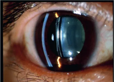
The ICL is a foldable posterior chamber lens made of a biocompatible material called collamer, composed of hydrophilic collagen, a material that does not generate inl ammatory response. (Figure 1) And it has ultraviolet protection. This lens is positioned behind the iris, in front of the anterior capsule of the lens, and with the haptics resting on the ciliary sulcus.(10)
Recently, a new type of Visian ICL was developed: the Vi-sian ICL V4c with central l ow technology. A central peritoneum called KS-AquaPORT has been added to the ICL optical center to improve the circulation of the aqueous humor in the eye and reduce the risk of cataract formation. This new construction eliminates the need for peripheral preoperative iridotomy or even intraoperative peripheral iridectomy, which simplii es the surgical procedure and signii cantly reduces the complications associated with iridotomy, such as hyphema, inl ammation and vitreous detachment or retina regmatogenic.(11-12)
There are some published studies that have evaluated the distance between the ICL / crystalline using ultrasound biomi-croscopy(13), equipment with Scheimpl ug technology(14) and
op-tical coherence tomography (OCT). (15) The new Spectralis OCT
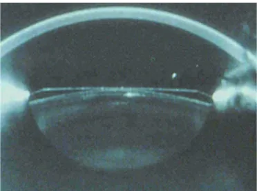
(Heidelberg Engineering, Heidelberg, Germany) with an anterior segment module provides anterior chamber image acquisition and provides high-resolution measurements of the distance between the posterior ICL surface and the anterior surface of the crystalli-ne. This distance known as the vault is an important point, and is related to some complications induced by the ICL, as pupillary block(16) and cataract(17). In the post-operative the vault of ICL
must be between 250 and 750 µm (Figure 2).
There are continuing concerns about the risk of late-onset cataract formation, probably resulting from direct physical con-tact between the ICL and the crystalline, and interruptions in the aqueous l ow that interfere with lens nutrition causing metabolic disturbances in the crystalline.(18) Visian ICL V4c with central l ow
was developed to soften these disadvantages.
Phakic posterior chamber lenses have the additional ad-vantage over anterior chamber lenses of a reduced chance of endothelial touch, as well as not causing pupil ovalization and requiring a smaller incision, which reduces the risk of iatrogenic astigmatism.
Effi cacy
Salera et al. in their study to correct hypermetropia con-cluded that the efi cacy of the procedure can be verii ed by the observation that 61.3% of the eyes presented visual acuity without postoperative correction of 20/40 or better (before the surgery this value was 12, 8%), whereas before surgery 87.1% had this same visual acuity, but with correction. There was a statistically signi-i cant dsigni-ifference signi-in the vsigni-isual acusigni-ity wsigni-ithout correctsigni-ion (p<0.01) before and after surgery. It was concluded that the phakic poste-rior chamber lens corrected hyperopia in this group studied.(19) Rosen et al. found that 14 (56%) of the 25 eyes operated in
Figure 1 - New generation lens model ICL V4c: This new ICL model for myopia and myopic astigmatism (V4c) was developed to minimize complications of increased intraocular pressure by incorporating a 0.36mm artii cial orii ce in the center (KS-Aquaport / CentraFlow), potentially improving aqueous humor circulation and eliminating the need for peripheral laser iridotomy or intraoperative iridectomy
the hypermetropic group presented visual acuity without postope-rative correction better than the best corrected visual acuity in the preoperative period. And in 12 (48%), the best corrected visual acuity of the preoperative period was equal to the uncorrected visual acuity in the postoperative period.(20)
Guimarães et al. showed in their study of myopia correction that the eficacy of the procedure can be easily veriied by the observation that 70% of the eyes presented postoperative AVSC of 20/40 or better, whereas before surgery only 78% presented the same visual acuity, but with correction. This study shows that 11% of eyes presented preoperative visual acuity of 20/20 or better, 56% of eyes achieved the same visual acuity with correction after surgery, and 22% without correction. In this study, 68.8% (64 eyes) of eyes achieved a spherical equivalent within ± 1.00 R of emmetropia, and 41.9% (39 eyes) within ± 0.50 R of emmetropia in the last exam.(21)
Sanders et al. in their study of the treatment of myopia de-termined that the postoperative AVSC for the entire population studied was 20/20 or better in 59.3% of eyes and 20/40 or better in 94.7%; in the preoperative these values were 40.8% and 81.3%.(2)
Alfonso et al.(1) and Pesando et al.(6) showed 100% and 96%
of the eyes respectively with ± 1.0 R of the desired correction. Fernández et al. (20) showed 22.2% of eyes with ± 0,5R of
the desired correction and 61.1% with ± 1.0R of the desired correction.
Safety
The analysis of visual acuity by loss and gain of sight lines is a good parameter to verify the safety of the procedure. In the study of Rosen et al.(20) no eye lost more than one line of best
corrected visual acuity; 2 eyes (8%) lost one line of sight, 8 eyes (32%) gained 1 line of sight, 3 eyes (12%) gained 2 lines of sight, and 12 (48%) remained unchanged.
Guimarães et al.(21) compare in their study the pre- and
post-operative corrected visual acuity in the last exam, having: 2 eyes (2.15%) lost two lines of sight, 2 eyes (2.15%) lost one line of sight, 18.28% of eyes (17 eyes) maintained preoperative visual acuity, whereas 33.3% (31 eyes) gained one line of sight, 27.96% (26 eyes) gained two lines, 11.83% (11 eyes) gained three lines, and 5.38% (5 eyes) gained more than three lines of sight.
Alfonso et al.(1) veriied the safety rate (1.07 in 12 months);
with no eye missing 1 or more lines of sight.
Pesando et al.(9) found that the best corrected visual acuity
remained unchanged in 64.4% of eyes, improved one line of sight n 15.2%, improved 2 lines of sight in 8.3%, improved 3 lines of sight in 8.3%, and reduced 1 line of sight in 8.3%.
The work of Salera et al.(19) showed that: when compared
to the pre- and postoperative visual acuity without correction, there was no loss of lines of sight in any of the cases, and 20 eyes (64.5%) gained more than three lines of sight. When compared to the visual acuity with pre- and postoperative correction, three eyes (9.7%) lost one line of sight, 19 eyes (61.3%) had the same visual acuity, six eyes (19.3%) gained one line of sight, and in three eyes (9.7%) there was gain of two lines of sight.
Fernández et al.(22) determined that 7 eyes (38.8%) gained
1 or more lines of sight, 55.5% kept the same visual acuity, and 1 eye (5.5%) lost more than 2 lines of sight. A recent systematic review showed that implanting phakic LIOs may be as safe as laser excimer ablations.(23)
Tychsen et al.(24) showed that the phakic posterior chamber
LIO is also an option with satisfactory results in children with high myopia.
Stability
It is the ability to maintain a constant, stable, solid result. In all the aforementioned studies there is stability during the follow-up period.
We can mention: Rosen et al.(20) with six months of
pos-toperative follow-up, Guimarães RQ et al.(2) with nine months
of follow-up, Sanders DR et al.(3) with 3 years of postoperative
follow-up.
Pesando et al.(6) found a good stability of refraction in 10
years of follow-up after surgery; this was the study with the longest follow-up among those reported.
As it does not depend on the cicatricial process of the eye for the refractive result, there are no signiicant variations of the result over time.
Complications
The most commonly reported complications for these lenses are lens opacities, IOP increase, pupillary block, loss of endothelial cells and pigment deposits on the anterior surface of the lens. Most
104
of the ICL-associated cataracts were reported as being anterior subcapsular. Phakic lens implants have a potential risk of intrao-cular complications such as endophthalmitis (0.0167%) and retinal detachment (3%), usually related to the axial length 30mm.(25)
The endothelial loss observed in the irst year after ICL is 4.7% to 8.4%, and it continues with a rate of 2% to 3% per year in the irst 3 years due to cellular remodeling; after that, it occurs due to natural loss.(24)
Acute pupillary block(2) and subsequent iridocorneal angle
closure are considered primary causes of IOP elevation, often associated with inadequate preoperative iridotomy or excessive vault.(26)
Less than 260 µm Vault (reduced Vault - Figure 2/Figure 3) may induce more cataracts due to contact and mechanical trauma to the anterior capsule, as well as lead to aqueous low disturbances (poor circulation) by interfering with the nutrition of the crystalline and causing metabolic disorders.(8,15)
The central or peripheral contact of the ICL with the crys-talline may be responsible for the development of an anterior subcapsular cataract; eyes with insuficient vault (distance between the posterior surface of the lens and the anterior surface of the crystalline) are more predisposed to the secondary formation of cataract.(8,11,12,24)
The development of cataracts is more common in older patients and in patients with greater myopia; in addition, the incidence increases with the duration of the follow-up. (8,26,27)
A study carried out in Spain at Instituto Oftalmológico Férnadez-Vega showed the development of anterior and poste-rior subcapsular cataracts in 3 eyes, 1 eye developed anteposte-rior and nuclear subcapsular cataracts, and 17 eyes developed anterior subcapsular cataracts. In the eyes that developed cataract, the majority occurred due to peripheral contact in eyes with high myopia. The mean vault of the eyes that developed cataract was 103 ± 69µm (ranged from 40 to 270µm). In 15 eyes the vault was less than 100µm, and in 6 eyes the vault was between 100 and 270µm. And most eyes developed cataracts between the third and fourth year after LIO implant.(27)
Schmidinger et al.(11) reported a signiicant and continuous
reduction of the central vault over the 10-year follow-up of patients with the ICMV4 model who developed cataract in the middle periphery due to the contact of the same with the anterior surface of the crystalline.
In Rosen et al.(20) 1 patient developed pupillary block and
secondary glaucoma. Guimarães et al.(21) showed that 2 patients
developed signiicant corneal edema in one of the eyes operated during the postoperative period, but the edema reverted quickly. However, signiicant endothelial loss was observed in both eyes (approximately 40%). In 2 eyes (2.15%) there was pupillary block on the irst postoperative day, but it was reversed immediately upon diagnosis. Anterior subcapsular opaciication was observed in 11 eyes (11.82%). In 5 of these eyes (5.3%) the opaciications were peripheral and asymptomatic. About 20% of the eyes had deposits of ine pigments in the lens without any subjective com-plaint of degradation of image quality.
United States Food and Drug Administration clinical trial(2)
showed that the incidence of anterior subcapsular opacities was 2.1% within 1 year and 2.7% within 3 years after lens implant. They reported 2 retinal detachments, 5 eyes (0.9%) developed nuclear opacity, and of these 2 also developed posterior subcap-sular opacity.
Alfonso et al.(1) in their study did not verify a chronic
increase in the IOP or anterior subcapsular cataract during a
12-month follow-up.
Pesando et al.(9) reported 1 patient who developed pupillary
block, 1 patient in which ICL was inadvertently placed upside down, but removed 1 day later and replaced in the correct position, 1 patient developed non-progressive paracentral subcapsular opa-city, 1 patient developed anterior subcapsular cataract, 2 patients complained of halos and glare.
In Salera et al.(19), the most common complication was the
presence of deposit of ine pigments on the anterior surface of the lens, found in 13 eyes (41.9%). But this inding was not associated to any subjective complaint of worsening of sight. The second most common complication was glare, reported by 3 patients (18.7%), without any biomicroscopic alterations justifying such a complaint. In one eye (3.2%), it was observed that the lens was partially captured by the iris, and its repositioning was indicated. In 1 eye (3.2%) the presence of spontaneous seidel was detected in the immediate postoperative period, where suture was performed. No lens opaciication was seen.
Fernández et al.(22) demonstrated 2 eyes (11.1%) that
de-veloped pupillary block, 4 eyes (22.2%) presented deposits of pigments on the anterior surface of the lens, 1 eye (5.5%) deve-loped anterior subcapsular opaciication, 5 eyes (28%) devedeve-loped transient ocular hypertension secondary to the use of corticoid eyedrops.
C
ONCLUSIONThe careful review of the literature published so far has shown satisfactory results regarding the eficacy, high predictability, stabi-lity and safety of posterior chamber intraocular lens implantation for the correction of myopia, hyperopia and astigmatism.
R
EFERENCES1. Alfonso JF, Baamonde B, Madrid-Costa DR, Fernandes P, Jorge J, Montés-Micó R. Collagen copolymer toric posterior chambre phakic intraocular lenses to correct high myopic astigmatismo. J Cataract Refract Surg. 2010;36(8):1349-57.
2. Sanders DR, Doney K, Poco M. United States Food and Drug Ad-ministration clinical trial of the implantable collamer lens (ICL) for moderate to high myopia: three-year follow-up. Ophthalmology. 2004; 111(9):1683-92.
3. Santhiago MR, Smadja D, Gomes BF, Mello GR, Monteiro ML, Wilson SE, et al. Association between the percent tissue altered and post-laser in situ keratomileusis ectasia in eyes with normal preoperative topography. Am J Ophthalmol. 2014;158(1):87-95. 4. Santhiago MR, Wilson SE, Hallahan KM, Smadja D, Lin M, Ambrosio
R Jr, et al. Changes in custom biomechanical variables after femto-second laser in situ keratomileusis and photorefractive keratectomy for myopia. J Cataract Refract Surg. 2014;40(6):918-28.
5. Santhiago MR, Smadja D, Wilson SE, Randleman JB. Relative contribution of lap thickness and ablation depth to the percent tissue altered (PTA) in post-LASIK ectasia. J Cataract Refract Surg. 2015;41(11):2493-500.
6. Santhiago MR, Smadja D, Wilson SE, Krueger RR, Monteiro ML, Randleman JB. Role of percent tissue altered on ectasia after LASIK in eyes with suspicious topography. J Refract Surg. 2015;31(4):258-65. 7. Dada T, Sudan R, Sinha R, Ray M, Sethi H, Vajpayee RB. Results of laser in situ keratomileusis for myopia of -10 to -19 diopters with a Technolas 217 laser. J Refract Surg. 2003;19(1):44-7.
8. Rezende F, Bisol RR, Bisol T. Troca do cristalino com inalidade refrativa (TCR). Rev Bras Oftalmol. 2009; 68(3):180-7.
9. Pesando PM, Ghiringhello MP, Di Meglio G, Fanton G. Posterior chambre phakic intra-ocular lens (ICL) for hyperopia: ten year follow-up. J Cataract Refract Surg. 2007; 33(9):1579-84.
10. Lovisolo CF, Reinstein DZ. Phakic intraocular lenses. Surv Ophthal-mol. 2005; 50(6):549-87.
11. Schmidinger G, Lackner B, Pieh S, Skorpik C. Long-term changes in posterior chamber phakic intraocular collamer lens vaulting in myopic patients. Ophthalmology. 2010; 117(8):1506-11.
12. Kumar N, Feyi-Waboso A. Intractable secondary glaucoma from hyphema following YAG iridotomy. Can J Ophthalmol. 2005;40(1):85-6.
13. Trindade F, Pereira F, Cronemberger S. Ultrasound biomicroscopic imaging of posterior chamber phakic intraocular lens. J Refract Surg. 1998;14(5):497-503
14. Lindland A, Heger H, Kugelberg M, Zetterstrom C. Vaulting of myopic and toric Implantable Collamer Lenses during accom-modation measured with Visante optical coherence tomography. Ophthalmology. 2010;117(6):1245-50.
15. Alfonso JF, Lisa C, Palacios A, Fernandes P, Gonzelez-Meijome JM, Montes-Mico R. Objective vs subjective vault measurement after myopic implantable collamer lens implantation. Am J Ophthalmol. 2009;147(6):978-83.
16. Bylsma SS, Zalta AH, Foley E, Osher RH. Phakic posterior chamber intraocular lens pupillary block. J Cataract Refract Surg. 2002;28(12):2222-8.
17. Sanders DR. Anterior subcapsular opacities and cataracts 5 years after surgery in the Visian implantable collamer lens FDA trial. J Refract Surg. 2008;24(6):566-70.
18. Khalifa YM, Moshirfar M, Miflin MD, Kamae K, Mamalis N, Wer-ner L. Cataract development associated with collagen copolymer posterior chamber phakic intraocular lenses: clinicopathological correlation. J Cataract Refract Surg. 2010; 36(10):1768-74.
19. Salera CM, Servian EE, Guimarães MR, Castro RD, Guimarães RQ. Implante de lentes intra-oculares de câmara posterior em olhos fácicos para correção de hipermetropia. Arq Bras Oftalmol. 2003;66(6):823-9.
20. Rosen E, Gore C. Staar Collamer posterior chamber phakic intrao-cular lens to correct myopia and hyperopia. J Cataract Refract Surg. 1998;24(5):596-606.
21. Guimarães RQ, Castro R, Navarro MP, Guimarães MR. Lente fácica de câmara posterior para correção da miopia. Arq Bras Oftalmol. 2001;64(1):21-6.
22. Fernández AP, Jaramillo J, Vargas J, Jaramillo M, Jaramillo J, Galín-dez A. Phakic posterior chambre intraocular lens for high myopia. J Cataract Refract Surg. 2004; 30(11):2277-83.
23. Barsam A, Allan BD. Excimer laser refractive surgery versus phakic intraocular lenses for the correction of moderate to high myopia. Cochrane Database Syst Rev. 2014 Jun 17;(6):CD007679
24. Tychsen L, Faron N, Hoekel J. Phakic intraocular collamer lens (Vi-sian ICL) implantation for correction of myopia in spectacle-aversive special needs children. Am J Ophthalmol. 2007; 175(1):77-86. 25. Fernandes P, Gonzáles-Méijome JM, Madrid-Costa D, Ferrer-Blasco
T, Jorge J, Montés-Micó R. Implantable collamer posterior chambre intraocular lenses: a review of potencial complications. J Refract Surg. 2011; 27(10):765-76.
26. Alfonso JF, Lisa C, Abdelhamid A, Fernandes P, Jorge J, Montés--Micó R. Three-year follow-up of subjective vault following myopic implantable collamer lens implantation. Graefes Arch Clin Exp Ophthalmol. 2010; 248(12):1827-35.
27. Alfonso JF, Lisa C, Fernández-Vega L, Almanzar D, Pérez-Vives C, Montés-Micó R. et al. Prevalence of cataract after collagen copoly-mer phakic intraocular lens implantation for myopia, hyperopia, and astigmatismo. J Cataract Refract Surg. 2015;41(4):800-5.
Corresponding author:
Marcony R. Santhiago