Revista Científica da Ordem dos Médicos www.actamedicaportuguesa.com 97
P-Cadherin as Prognostic Factor for
Loco-Regional Relapse in Breast Cancer
Caderina-P: Valor Prognóstico na Recidiva Loco-regional do Cancro da Mama
G.F., I.A.: Faculdade de Medicina da Universidade do Porto / Centro Hospitalar S. João. Porto. Portugal. M.J.C.: Faculdade de Medicina da Universidade do Porto. Porto. Portugal.
D.M., F.S.: IPATIMUP - Instituto de Patologia e Imunologia Molecular da Universidade do Porto. Porto. Portugal. H.B.: Centro Hospitalar São João. Porto. Portugal.
Recebido: 19 de Novembro de 2011 - Aceite: 29 de Maio de 2012 | Copyright © Ordem dos Médicos 2012
Gil FARIA, Maria João CARDOSO, Diana MARTINS, Herberto BETTENCOURT, Isabel AMENDOEIRA, Fernando SCHIMITT
Acta Med Port 2012 Mar-Apr;25(2):97-105
RESUMO
Introdução: O cancro da mama é o tumor maligno mais frequente e a principal causa de morte nas mulheres em Portugal. Devido à
sua relação com a metastização à distância e morte subsequente, a recidiva loco-regional é uma das maiores preocupações no segui-mento destas doentes. São conhecidos diversos factores clássicos de prognóstico para recidiva local, tais como o tamanho do tumor, o estádio tumoral, grau histológico, positividade HER2 e a expressão de receptores hormonais. Contudo, existe heterogeneidade no prognóstico e no comportamento do tumor em doentes com estadiamento semelhante e com a mesma expressão de marcadores mo-leculares de prognóstico. Daí advém a necessidade de descobrir novos factores prognósticos. Uma das possibilidades é a P-caderina, previamente descrita como um possível marcador independente de prognóstico no cancro da mama. O objective deste trabalho foi estudar a correlação da expressão de P-caderina com a recorrência loco-regional do cancro da mama.
Material e métodos: Analisámos os registos clínicos de 1432 doentes consecutivos com cancro da mama e tratados na nossa
insti-tuição durante um período de 10 anos. Os doentes com recorrência loco-regional (n=101) sem evidência ou história de metastização à distância foram selecionados como casos. O grupo de controlo consistiu em doentes com mais de 10 anos de seguimento, sem progressão da doença oncológica. Em ambos os grupos foram analisadas variáveis demográficas, clínicas, patológicas e molecula-res. Para estudo da expressão da P-caderina, foram construídos Tissue Micro-Arrays a partir de 86 tumores com blocos de parafina disponíveis.
Resultados: O tempo médio livre de doença foi de 41 meses e a sobrevida media após a recorrência foi de 33 meses. A taxa de
sobre-vivência aos 5 anos foi de 55%. O tamanho do tumor, estadiamento ganglionar e grau histológico foram identificados como marcadores independentes de prognóstico. A P-caderina associou-se com graus histológicos mais altos e tumores sem expressão de receptores hormonais. A P-caderina foi identificada como uma marcador independente de prognóstico para a recidiva livre de doença, mas não para a sobrevivência global.
Conclusão: A P-caderina surgiu associada a outros factores já conhecidos de pior prognóstico e a uma relação independente com
a sobrevivência livre de doença. A P-caderina pode vir a constituir um alvo terapêutic a explorar, mas o seu real valor biológico ainda não está determinado. Subsiste a dúvida sobre se a P-caderina é um marcador independente de prognósico ou apenas um marcador de um conjunto de características clínico-patológicas relacionadas com pior prognóstico.
ABStRACt
Background: Breast cancer is the most frequent malignant tumor and the leading cause of cancer death in women in Portugal. Due
to its relation to an increase in distant metastasis and subsequent death, loco-regional relapse is one major concern in breast cancer women. Several classic prognostic factors as tumour size, nodal stage, histological grade, HER2 status and hormonal receptors have been identified as the most important factors for determining loco-regional relapse, disease free and overall survival. However, there is heterogeneity in prognosis and tumor behaviour in patients with identical disease staging and a similar pattern of expression of known molecular markers, hence the need to discover new prognostic factors. One of the possibilities is P-cadherin, already described by researchers as a possible independent marker of prognosis in breast cancer. The aim of this work was to study in a retrospective series of patients the correlation of P-cadherin expression with loco-regional recurrence in breast cancer women.
Material and methods: We analyzed the clinical records of 1432 consecutive patients with breast cancer and treated in a University
Hospital over a 10 year period. Patients with loco-regional relapse (n=101) without prior or simultaneous distant disease were selected as case group. Control group consisted of patients with more than 10 years follow-up and without disease progression. For both groups demographic, clinical, pathological and molecular markers were analyzed. Tissue micro-arrays were constructed to study P-cadherin expression from 86 tumors with available paraffin embedded blocks.
Results: Mean time to recurrence was 41 months and mean survival time after recurrence was 33 months, with a 5-year survival rate
of 55%. Tumour size, nodal status and histological grade were identified as independent markers of prognosis. P-cadherin was associ-ated with higher histological grades and hormone negative tumours. P-cadherin was identified as an independent prognostic marker for disease free survival, but not for overall survival.
Conclusion: P-cadherin was related to other known factors of worse prognosis and had an independent relation to disease-free
sur-vival. P-cadherin might constitute a novel therapeutic target, but its real biological value is yet to be determined. Doubt persists whether it is an independent marker of tumour behaviour or only a surrogate marker of a set of clinical and molecular features related with worse prognosis.
AR
INtRODUCtION
Breast cancer is the most frequent malignant tumour and the leading cause of cancer related death in woman, with one million cases and half a million deaths each year worldwide.1 Cumulative individual risk of breast cancer is
estimated in 12% (approximately 1 in each 8 women) and the risk of death might be up to 5% (approximately 1 in each 20 women).2
Loco-regional relapse of breast cancer is a frequent concern in the treatment of this disease as it has been es-tablished as an independent prognostic factor for distant metastasis and subsequent death.3-6 However, whether it
constitutes a cause for distant metastasis or only a marker of an existing risk remains a matter of debate.5,7,8
Several clinical and pathological parameters have been used to determine not only prognosis but also the need of adjuvant systemic therapies. The most common of these are: age, size, nodal staging, histological grade, hormonal receptors and HER2 positive.9-12
With the development of new microarrays techniques, it became possible to simultaneously analyze thousands of genes and classify tumours according to their profile of genetic expression. As such, a new classification of breast cancer was developed, based on profiles of genetic ex-pression. Five different groups with prognostic differences were identified: luminal A, luminal B, basal, normal-like and
HER2.13
Although this new classification was based on the hi-erarchical cluster analysis of genetic expression, some currently available imunohistochemistry markers allow the translation of this classification to the routine pathology practice. Specifically, based on 3 markers (ER, PR and HER2), groups can be divided into luminal type (positive for ER or PR), HER2 positive or triple negative (ER, PR and HER2 negative).14 The prognostic evaluation of patients in
the triple-negative group revealed at least 2 groups of tu-mors: one of them expressing markers of basal differentia-tion (CK5, EGFR, P-cadherin) and another without expres-sion of these markers considered unclassified.13
Loco-regional relapse is an early and important marker of disease progression. However, regarding patients with identical staging and a similar pattern of expression of mo-lecular markers there is a significant discrepancy in disease progression and prognosis, hence the need to further dis-cover new prognostic factors and stratify risk for disease progression.12
One of the molecules used to classify the tumour as basal-like, is P-cadherin, which is associated with increased proliferation and undifferentiated phenotype.15
Unlike epithelial cadherin (E-cadherin), P-cadherin expression is usually related to tumorogenic properties, allowing for cellular invasiveness and tumoral
aggressive-AR tIGO ORIGINAL
Global
Study population
Exclusion criteria
• Male gender • Loss of follow-up• Lack of pathological material
TMA’s
1432
patients
Cases
101
Controls
92
70
52
53
33
Revista Científica da Ordem dos Médicos www.actamedicaportuguesa.com 99
table 1 - Sources and dilutions of primary antibodies used in this immnohistochemistry
Antibody Clone Manufacturer Incubation time (m) Dilution Antigen retrieval (m)
ER SP1 Neomarkers 30 1:150 30
PR 1A6 Neomarkers 30 1:40 30
HER-2 SP3 Neomarkers 30 1:80 30
P-cad 56 Transduction 60 1:50 30
CK5 XM26 Neomarkers 60 1:50 30
ness, translating into a worst prognosis in breast cancer patients.16 Its expression is usually associated with other
known factors of worse prognosis (high histological grade, high proliferative rate and lack of estrogen receptors).17,18
Our objective, in this retrospective series of patients, was to evaluate the correlation of P-cadherin expression in breast cancer loco-regional relapse, disease-free and over-all survival.
MAtERIAL AND MEtHODS
We performed a nested case-control study and ana-lyzed the clinical records of 1432 consecutive patients treated and followed at Hospital de São João (University Hospital of Porto Faculty of Medicine) during a 10-year pe-riod (January 1st 1997 to December 31st 2006). The case group consisted of all the patients (n=101; 7%) with loco-regional relapse without previous or concurrent systemic progression.
Loco-regional relapse after breast cancer surgery was defined as the onset of histologically confirmed carcinoma at least in one of the following locations: remaining breast tissue; skin, subcutaneous tissue or muscle of ipsilateral thoracic wall; axillary, supraclavicular or internal mammary lymphnodes.11
As the majority of loco-regional relapses occurs before 10 years after the initial diagnosis,19 for control group we
se-lected patients with more than 10 years of follow-up without disease progression: 92 patients surgically treated between January 1st 1997 and June 30th 1998.
Male patients, patients lost to follow-up and those whithout material available for pathological re-evaluation were excluded from the study. Final case group consisted of 70 patients (69.3% of the initial sample) with loco-regional relapse (cases) and 52 patients (56.5% of the initial sample) without disease progression (control group).
Classical clinical and pathological parameters were evaluated in all patients (age, size, type of surgical treat-ment, TNM staging, histological type, histological grade, presence of associated DCIS, size, lymphatic and venous invasion, Nottingham Prognostic Index [NPI](20) and es-trogen receptors. Molecular classification (Luminal, HER2 [HER2(+)/RE(-)/RP(-)], triple negative) and P-cadherin ex-pression were studied using immunohistochemistry in Tis-sue MicroArrays (TMA’s).
In our series, only 53 cases and 33 controls had paraf-fin-embedded blocks available for the construction of TMA’s
(Fig.1). New sections of the tumour stained with hematoxy-lin-eosin were undertaken in those blocks. Representative areas were selected and marked for TMA construction.11
Representative areas of invasive breast carcinoma were carefully selected on haematoxylin and eosin stained sections and marked on the correspondent individual paraf-fin blocks. Two tissue cores (2mm in diameter) were ob-tained from each selected specimen (donor block) and de-posited into a paraffin block (receptor block) using a TMA workstation (TMA builder ab1802, Abcam, Cambridge, UK). Twenty-two TMA blocks were constructed, each containing 24 tissue cores (4x6). In each TMA block, non-neoplastic breast and liver tissue cores were also included as controls and TMA guide, respectively. After construction, two 2μm tissue sections were cut and adhered to Superfrost Plus glass slides. An HE stained section from each block was reviewed to confirm the presence of morphological repre-sentative areas of the original lesions. Sections were immu-nostained with primary monoclonal antibodies against ER, PR, 2, and P-cad. Immunostaining for ER and HER-2 were performed using the streptavidin-biotin-peroxidase technique (LabVision, Fremont, CA, USA), whereas PR and P-cad immunostaining used the HRP labeled polymer (Da-koCytomation, Carpinteria, CA, USA).
Antigen unmasking for ER, PR and HER-2 was carried out using a dilution of 1:100 from a commercially avail-able solution of citrate buffer, pH=6.0 (Vector Laboratories, Burlingame, CA, USA) at 98ºC, whereas a dilution of 1:10 from tris-ethylenediaminetetraacetic (EDTA) solution with pH=9.0 (DakoCytomation) was used for P-cad.
Antigen retrieval time, antibodies, dilutions and suppli-ers are listed in Table 1. After antigen retrieval procedure, slides were washed in a phosphate buffer solution (PBS), and submitted to blockage of the endogenous peroxidase activity by incubation of the slides in a 3% hydrogen per-oxide (Panreac, Spain) in methanol (Sigma-Aldrich). Slides were further incubated with a blocking serum (LabVision Corporation kit) for 15 min and then incubated with the primary antibodies. After washing, slides were incubated with biotinylated secondary antibody, followed by streptav-idin-conjugated peroxidase (LabVision). Diaminobenzidine (DAB) was used as a chromogen (DakoCytomation). For PR and P-cad staining, secondary antibody was associated with HRP labelled polymer (DakoCytomation) and immedi-ately revealed with DAB. Tissues were then counterstaining with Mayer`s haematoxylin, dehydrated and coverslipped
AR
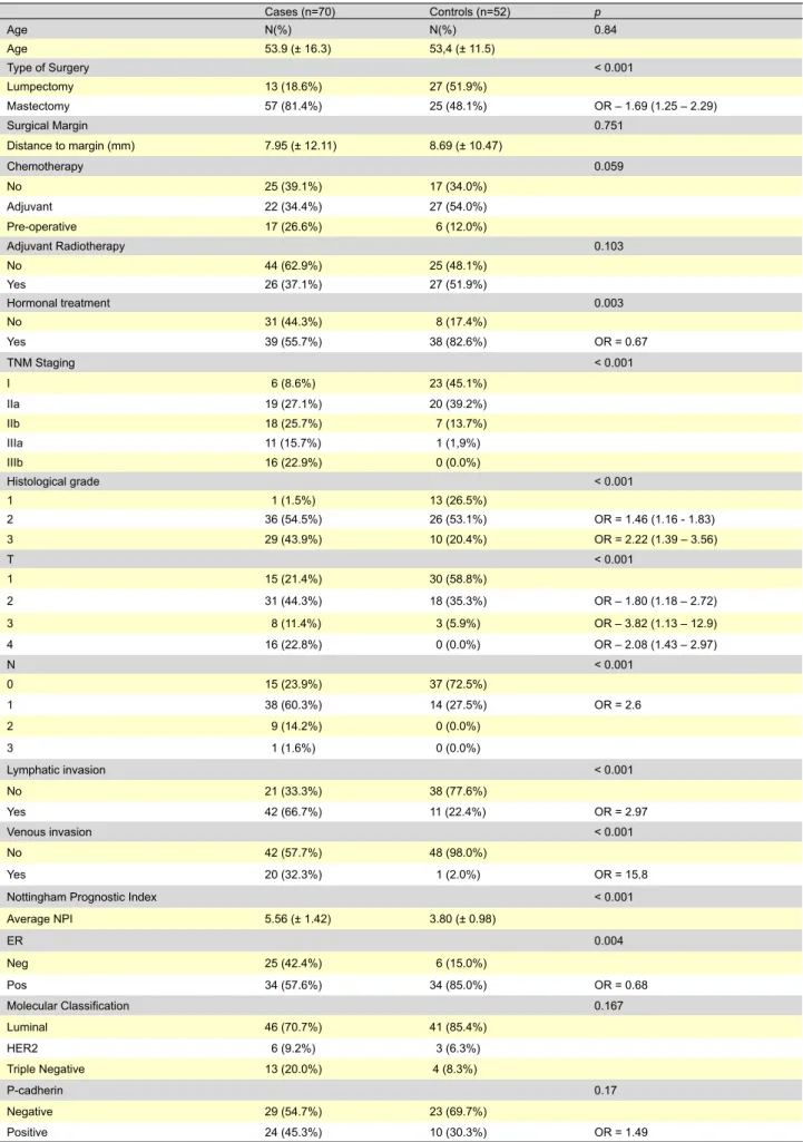
Table 2 - Characterization of the clinical and pathological pattern according to group. Cases (n=70) Controls (n=52) p Age N(%) N(%) 0.84 Age 53.9 (± 16.3) 53,4 (± 11.5) Type of Surgery < 0.001 Lumpectomy 13 (18.6%) 27 (51.9%) Mastectomy 57 (81.4%) 25 (48.1%) OR – 1.69 (1.25 – 2.29) Surgical Margin 0.751 Distance to margin (mm) 7.95 (± 12.11) 8.69 (± 10.47) Chemotherapy 0.059 No 25 (39.1%) 17 (34.0%) Adjuvant 22 (34.4%) 27 (54.0%) Pre-operative 17 (26.6%) 6 (12.0%) Adjuvant Radiotherapy 0.103 No 44 (62.9%) 25 (48.1%) Yes 26 (37.1%) 27 (51.9%) Hormonal treatment 0.003 No 31 (44.3%) 8 (17.4%) Yes 39 (55.7%) 38 (82.6%) OR = 0.67 TNM Staging < 0.001 I 6 (8.6%) 23 (45.1%) IIa 19 (27.1%) 20 (39.2%) IIb 18 (25.7%) 7 (13.7%) IIIa 11 (15.7%) 1 (1,9%) IIIb 16 (22.9%) 0 (0.0%) Histological grade < 0.001 1 1 (1.5%) 13 (26.5%) 2 36 (54.5%) 26 (53.1%) OR = 1.46 (1.16 - 1.83) 3 29 (43.9%) 10 (20.4%) OR = 2.22 (1.39 – 3.56) T < 0.001 1 15 (21.4%) 30 (58.8%) 2 31 (44.3%) 18 (35.3%) OR – 1.80 (1.18 – 2.72) 3 8 (11.4%) 3 (5.9%) OR – 3.82 (1.13 – 12.9) 4 16 (22.8%) 0 (0.0%) OR – 2.08 (1.43 – 2.97) N < 0.001 0 15 (23.9%) 37 (72.5%) 1 38 (60.3%) 14 (27.5%) OR = 2.6 2 9 (14.2%) 0 (0.0%) 3 1 (1.6%) 0 (0.0%) Lymphatic invasion < 0.001 No 21 (33.3%) 38 (77.6%) Yes 42 (66.7%) 11 (22.4%) OR = 2.97 Venous invasion < 0.001 No 42 (57.7%) 48 (98.0%) Yes 20 (32.3%) 1 (2.0%) OR = 15.8
Nottingham Prognostic Index < 0.001
Average NPI 5.56 (± 1.42) 3.80 (± 0.98) ER 0.004 Neg 25 (42.4%) 6 (15.0%) Pos 34 (57.6%) 34 (85.0%) OR = 0.68 Molecular Classification 0.167 Luminal 46 (70.7%) 41 (85.4%) HER2 6 (9.2%) 3 (6.3%) Triple Negative 13 (20.0%) 4 (8.3%) P-cadherin 0.17 Negative 29 (54.7%) 23 (69.7%) AR tIGO ORIGINAL
Revista Científica da Ordem dos Médicos www.actamedicaportuguesa.com 101
using a permanent mounting solution (Zymed, San Fran-cisco, CA, USA).
Positive controls were included in each run, to guar-antee assay reliability. All cases showing an unequivocal nuclear staining for ER and PR in at least 10% of the neo-plastic cells were considered positive. We also considered positive cases with membranous staining for P-cad and in at least 10% of the neoplastic cells. HER2 expression was evaluated according to the DakoCytomation HercepTest scoring system. Cases were considered positive (overex-pression) for HER2 when immunostaining was classified as 3+. All the samples were blinded and reviewed by the same experienced pathologist.
Statistical analysis was done using SPSS 15.0 (SPSS
Inc. Chicago, Illinois, USA). The chi-square contingency test was used for categorical variables and the t-student was used for continuous variables. A p value of less than 0.05 was considered to reflect a significant association. The multivariate analysis was performed with a model of binary logistic regression. The time-dependent variables were an-alyzed with the Cox regression model and the Kaplan-Meier curves were based on life tables. For the multivariate re-gression models, we selected the variables with significant association with the outcome on univariate analysis and in the Cox regression model, we also included the type of sys-temic treatment to check for potential confounding on the effect of P-cadherin.
AR
tIGO ORIGINAL
Table 3 - Logistic regression for loco-regional relapse
p OR Type of surgery 0.241 0.307 Histological grade 0.049 3.802 t 0.034 4.672 N 0.014 8.849 Lymphatic invasion 0.251 3.215 Venous invasion 0.495 2.857 ER 0.530 0.464
Table 4 - P-cadherin expression according to other markers of prognosis
P-cadherin Positive (n=34) Negative (n=52) p
Molecular sub-type 0.002 Luminal 19 (55.9%) 46 (88.5%) HER2 7 (20.6%) 2 (3.8%) Triple negative 8 (23.5%) 4 (7.7%) MIB-1 0.003 Positive 16 (52.9%) 9 (82.7%) Negative 18 (47.1%) 43 (17.3%) tNM stage 0.807 I 7 (20.6%) 8 (15.4%) IIA 11 (36.4%) 15 (28.8%) IIB 5 (14.7%) 13 (25.0%) IIIA 4 (11.8%) 7 (13.5%) IIIB 7 (20.6%) 9 (17.3%) Histological grade 0.008 1 4 (12.1%) 5 (10.0%) 2 10 (30.3%) 32 (64.0%) 3 19 (57.6%) 13 (26.0%) t 0.325 1 12 (35.3%) 15 (28.8%) 2 14 (41.2%) 20 (38.5%) 3 1 (2.9%) 8 (15.4%) 4 7 (20.6%) 9 (17.3%) N 0.779 0 13 (39.4%) 17 (36.2%) 1 16 (48.5%) 25 (53.2%) 2 4 (12.1%) 4 (8.5%) 3 0 (0.0%) 1 (2.1%)
Table 5 - Cox regression – Overall survival and disease free survival
Disease-free survival Overall survival
p HR [95%CI] p HR [95%CI] P-cadherin 0.047 2.108 [1.009; 4.402] 0.129 2.087 [0.807; 5.395] T 0.004 1.822 [1.217; 2.729] 0.003 2.317 [1.325; 4.053] N < 0.001 2.780 [1.609; 4.802] 0.061 1.957 [0.969; 3.954] Grade 0.001 3.326 [1.666; 6.643] < 0.001 8.541 [3.188; 22.883] Molecular class 0.336 0.870 [0.643; 1.178] 0.093 0.688 [0.445; 1.064] Chemotherapy 0.632 0.891 [0.556; 1.427] 0.270 0.702 [0.374; 1.316] Hormone therapy 0.234 0.668 [0.344; 1.298] 0.831 0.909 [0.378; 2.188] Anti-HER2 therapy 0.903 1.057 [0.431; 2.591] 0.980 1.017 [0.275; 3.764] AR tIGO ORIGINAL RESULtS
Mean age at diagnosis was 53.7 years. Mastectomy was the type of surgery performed on the majority of pa-tients (67.2%) and 77% of the papa-tients were classified as stage I or II, according to TNM classification. Predominant histological type was invasive ductal carcinoma (87%) and half the patients (50.8%) had grade II carcinomas (Notting-ham grading system). Forty-seven percent of patients had lymphatic invasion and only 19% had venous invasion. Ax-illary staging was negative (N0) in 45.6%. ER expression was positive in 68.7% of the patients and PR in 52.3% of the patients. The majority of local relapses occurred in the remaining breast tissue or in the thoracic wall (73%) and 56% of the cases were re-excised. After 93 months of mean follow-up, 69% of the patients are alive.
The expression of classical prognostic factors is listed in Table 2. Mastectomy was associated with higher rates of loco-regional relapse but also with the expression of several markers of worse prognosis (larger tumours [70% T1/T2 vs 92.5% for breast conserving surgery; p=.02]; nodal metas-tasis [67.5% vs 27%; p<.001], lymphatic [60.5% vs 19.4%;
p<.001] and venous [24.0% vs 8.3%;p=0.05] invasion and
higher TNM stages [29.6% stage III/IV vs 10.0%; p=.001]). The specimen’s surgical margins were not different be-tween the groups and post-operative radiotherapy was not associated with a decrease in local relapse risk.
Staging was directly associated with relapse risk, as well as histological grade and NPI index. The presence of lymphatic and venous invasion was also strongly associ-ated with loco-regional relapse. Expression of ER was iden-tified as a marker of better prognosis.
Molecular classification was achieved by the use of rou-tine immunohistochemistry and tumours were divided into 3 categories (ER or PR positive, HER2 overexpressing or triple-negative). The majority of patients expressed luminal type markers in both groups (70.7% of the cases vs. 85.4% of the controls). Triple-negative tumours were more frequent in patients with loco-regional relapse (20% vs. 8%) although this value did not reach statistical significance (Table 2).
The logistic regression model (Table 3) identified his-tological grade, size and nodal invasion as independent markers of prognosis for loco-regional relapse. Once cor-rected for other prognostic factors, the type of surgery was no longer related with loco-regional relapse.
P-cadherin was positive in 45.3% of cases and 30.3% of controls (p=0.17), OR=1.49. There was a positive relation of P-cadherin expression with the non-luminal molecular types and with higher proliferative index (p=0.003) as measured by MIB-1. There were no significant relations between P-cadherin expression and other prognostic markers, with the exception of higher histological grade. (Table 4)
P-cadherin expression was related to a significant de-crease (p=0.017) in disease-free survival, from 90.5 months to 55.2 months (Fig.2). However, these earlier recurrences were not related with a decrease in overall survival (135.5 months vs 136.2 months – Fig.3), despite the differences observed in the 5-year survival rate (82.7% vs 58.3%).
Multivariate analysis of prognostic factors for disease-free survival (Table 5) identified P-cadherin expression as an independent factor of prognosis, (HR=2.1) together with the known classical factors of prognosis: tumor size, nodal staging and histological grade. For overall survival the only identified independent factors were tumor size and histo-logical grade.
DISCUSSION
The research around new molecular markers has rised tremendously not only because they have the capacity to add some information and enhance discriminant power to scores already available12 with classical markers but also
because they can bring some new understanding over the oncological biology or arise as new putative therapeutic tar-gets.21
The major limitation of this study is the shortness of the sample, as we could only retrieve 86 tumors for TMA con-struction. Additionally, this is a retrospective study with a 10-year span and during this period the treatment of breast cancer suffered significant variations.
Revista Científica da Ordem dos Médicos www.actamedicaportuguesa.com 103
AR
tIGO ORIGINAL
Fig. 2 - Kaplan-Meier curves for disease free survival according to
P-cadherin expression. FP-cadherin expression.ig. 3 - Kaplan-Meier curves for overall survival according to Several studies have reported the risk of local
recur-rence after breast cancer treatment as being 5-40%.11,22
De-spite therapeutic improvements in the last decade, 40% of the women with local recurrence will have disease progres-sion and eventually death. In our series, local recurrence rate was 7% (101/432) and 54% of these women died from breast cancer, as in most clinical reported studies.22,23
Several studies have reported either a similar24 or
in-creased survival25 with breast conserving surgery when
compared to mastectomy. In our series, breast conserving surgery has a longer median survival (132 vs. 64.5 months for mastectomy). Mastectomy is also related to an increased risk of local relapse (OR = 1.69). However, these results maybe the consequence of a a selection bias, as tumors of patients who had mastectomy, in our series, presented with features of worse prognosis (size, nodal metastasis, histological grade, TNM staging and NPI). Once corrected for these factors, the benefit of conservative surgery is no longer detectable.
Tumor size (p=0.002) and nodal staging (p<0.001) were two important factors of prognosis for local recurrence, which confirms the data of several other studies,22,26,27 and
patients with tumors larger than 5cm had a 4-fold increase in local recurrence as compared with tumors smaller than 2cm (OR=3.82).4 Also as described in the literature,28,29
patients with axillary invasion had an almost 3-fold inrease in local recurrence as compared to patients with node-free disease (OR=2.6). According to some authors, axillary inva-sion might be not just an event related to tumor progresinva-sion, but a biological marker of tumor aggressiveness27
indepen-dently of tumor size, recurrence type or time-to-recurrence. Also according to several studies,5,11 there was a significant
relation between high histological grade and local recur-rence (OR=1.46 for Grade 2 and OR=2.22 for Grade 3;
p<0.001). Regarding all well-known factors our results were
identical to others of similar series.
In one of the first studies about P-cadherin expression
in breast cancer, the molecule was only identified in 4% of invasive breast cancers. In the following studies, its expres-sion was observed in approximately 20% of tumors and inversely related to E-cadherin expression and directly to higher histological grades.17 With the development of
anti-P-cadherin monoclonal antibodies its expression was regis-tered in 30% to 50% of all the invasive ductal cancers.15,30-32
In our series, P-cadherin was expressed in 39.5% of all cases. P-cadherin was more often positive in patients with local recurrence (OR=1.49; p=0.1), although without a sta-tistically significant difference.
Several studies reported that the P-cadherin expres-sion in cancer cells was directly related to other known fac-tors of worse prognosis, such as: tumor size;30 histological
grade;17,30,32 ER negativity17,30,32 and nodal metastization.30 In
multivariate analysis only relation with nodal metastization and histological grade has kept significance.30 Other reports
found no association between P-cadherin expression and tumor size or axillary invasion.32 These conflicting reports
and differing association with known prognostic factors, suggests that P-cadherin might be related to oncological progression of breast cancer, but its real biological behavior is not yet determined.30 In our series, P-cadherin expression
was directly related only with histological grade and ER sta-tus.
Several other reports have shown a direct relation of P-cadherin with other known factors of worse prognosis, such as triple-negative type33-35 and proliferative index.32,36 In this
study, we also confirm these findings of a direct relation between P-cadherin expression and triple-negative tumors (p<0.001) and higher proliferative index (p=0.003), as mea-sured by MIB1.
Several reports observed an inverse relation between P-cadherin expression and hormonal receptors. Most of the P-cadherin expressing tumors lack hormonal receptors ex-pression17,31,32,37,38 and are positive for HER2, EGFR, higher
asso-CONCLUSION
Breast cancer is one of the most prevalent diseases worldwide, being the leading cause of death for cancer in women.1 In the last few years, the mortality due to breast
cancer has been following a downward trend, due to better screening programs and most effective medical care.39
Lo-cal recurrence has been described as a marker of disease progression and an important risk factor for death.3 As a
consequence, several studies have tried to identify risk fac-tors for local recurrence.11
One of the most promising markers for loco-regional dis-ease progression seems to be P-cadherin and in the future, it might even constitute a novel therapeutic target.
P-cadherin, in our study was related to other known fac-tors of worse prognosis, was more frequent in non-luminal type tumors and had an independent relation to disease-free survival. Although it did not affect overall survival or relapse rate, it seemed to be associated with earlier relapse and mortality.
The real biological value of P-cadherin is still undeter-mined raising the question to whether it has an independent relation to tumor behavior or if it constitutes just an indirect marker of a group of clinical and molecular characteristics related to worse prognosis.
CONFLICtS OF INtERESt
The authors declare there are no conflicts of interest.
FUNDING SOURCES
None stated.
AR
tIGO ORIGINAL
ciated with worse prognosis.17,30-32 These authors suggest
that the hormonal negative state is a requirement to the ex-pression of P-cadherin, probably through the differentiation of luminal type cells into myoepithelial cells where P-cad-herin is usually expressed.32 It has been suggested by some
authors that P-cadherin expression in breast cancer cells might represent the differentiation in an embrionary pheno-type, similar to the ductal-extremity cells, which are highly proliferative, negative for ER and positive for P-cadherin.17
Our results, as other before,30 support this hypothesis as
P-cadherin expression was found more often in high histologi-cal grade and ER negative cancers.
Although some studies described impairment in sur-vival for patients with P-cadherin expression, in multivari-ate analysis,30,31,37 our results only confirm a reduction in
disease-free survival (Cox regression; p=0.047), without differences for overall survival (p=0.129). Nevertheless, the Kaplan-Meyer survival curves suggest that there is an effect of P-cadherin on survival, visible at 5-years follow-up and fading progressively, nearly unnoticed at 10 years. Similar data were reported in other studies,30-32 suggesting
this fade-out of effect in long-term follow-up, which explains the lack of association with overall survival but the signifi-cant differences of survival at 5 years (82.7% vs 58.3%). More studies directed to the underlying pathophysiology of P-cadherin will be necessary, in order to unravel this effect and to understand the molecular mechanisms and signaling involved in this process.
REFERENCES
1. Bastos J, Barros H, Lunet N. Evolução da mortalidade por Cancro da Mama em Portugal (1955-2002). Acta Med Port. 2007;20(2):139-144. 2. Soerjomataram I, Louwman M, Ribot J, Roukema J, Coebergh J. An
overview of prognostic factors for long-term survivors of breast cancer. Breast Cancer Res Treat. 2008;107(3):309-330.
3. Barry Feig DB, George Fuhrman. The M.D. Anderson Surgical Oncology Handbook. 3rd ed.
4. Wapnir IL, Anderson SJ, Mamounas EP, Geyer CE Jr, Jeong JH, Tan-Chiu E, et al. Prognosis After Ipsilateral Breast Tumor Recurrence and Locoregional Recurrences in Five National Surgical Adjuvant Breast and Bowel Project Node-Positive Adjuvant Breast Cancer Trials. J Clin On-col. 2006;24(13):2028-2037.
5. Whelan T, Clark R, Roberts R, Levine M, Foster G. Ipsilateral breast tumor recurrence postlumpectomy is predictive of subsequent mortality: results from a randomized trial. Investigators of the Ontario Clinical On-cology Group. Int J Radiat Oncol Biol Phys. 1994;30(1):11-16. 6. Touboul E, Buffat L, Belkacemi Y, Lefranc JP, Uzan S, Lhuillier P, et al.
Local recurrences and distant metastases after breast-conserving sur-gery and radiation therapy for early breast cancer. Int J Radiat Oncol Biol Phys. 1999;43(1):25-38.
7. Schmoor C, Sauerbrei W, Bastert G, Schumacher M. Role of isolated locoregional recurrence of breast cancer: results of four prospective studies. J Clin Oncol. 2000;18(8):1696-1708.
8. Veronesi U, Marubini E, Del Vecchio M, Manzari A, Andreola S, Greco M, et al. Local recurrences and distant metastases after conservative breast cancer treatments: partly independent events. J Natl Cancer Inst. 1995;87(1):19-27.
9. Parikh R, Housman D, Yang Q, Toppmeyer D, Wilson L, Haffty BG. Prog-nostic Value of Triple-Negative Phenotype at the Time of Locally Recur-rent, Conservatively Treated Breast Cancer. Int J Radiat Oncol Biol Phys 2008;72(4):1056-1063.
10. Crowe JP Jr., Gordon NH, Antunez AR, Shenk RR, Hubay CA, Shuck JM. Local-regional breast cancer recurrence following mastectomy. Arch
Surg 1991;126(4):429-432.
11. Carreño G, Casar J, Corte M, González L, Bongera M, Merino A, et al. Local recurrence after mastectomy for breast cancer: analysis of clini-copathological, biological and prognostic characteristics. Breast Cancer Res Treat. 2007;102(1):61-73.
12. Haffty BG, Buchholz TA. Molecular predictors of locoregional recurrence in breast cancer: ready for prime time? J Clin Oncol 2010;;28(10):1627-1629.
13. Perou CM, Sørlie T, Eisen MB, van de Rijn M, Jeffrey SS, Rees CA, et al. Molecular portraits of human breast tumours. Nature 2000;406(6797):747-752.
14. Haffty BG, Yang Q, Reiss M, Kearney T, Higgins SA, Weidhaas J, et al. Locoregional relapse and distant metastasis in conservatively managed triple negative early-stage breast cancer. J Clin Oncol 2006;24(36):5652-5657.
15. Paredes J, Correia AL, Ribeiro AS, Albergaria A, Milanezi F, Schmitt FC. P-cadherin expression in breast cancer: a review. Breast Cancer Res 2007;9(5):214.
16. Paredes J, Correia AL, Ribeiro AS, Milanezi F, Cameselle-Teijeiro J, Schmitt FC. Breast carcinomas that co-express E- and P-cadherin are associated with p120-catenin cytoplasmic localisation and poor patient survival. J Clin Pathol 2008;61(7):856-862.
17. Palacios J, Benito N, Pizarro A, Suarez A, Espada J, Cano A, et al. Anomalous expression of P-cadherin in breast carcinoma. Correlation with E-cadherin expression and pathological features. Am J Pathol 1995;146(3):605-612.
18. Sarrió D, Palacios J, Hergueta-Redondo M, Gómez-López G, Cano A, Moreno-Bueno G. Functional characterization of E- and P-cadherin in invasive breast cancer cells. BMC Cancer. 2009;9:74.
19. Fisher B, Anderson S, Bryant J, Margolese RG, Deutsch M, Fisher ER, et al. Twenty-year follow-up of a randomized trial comparing total mas-tectomy, lumpectomy, and lumpectomy plus irradiation for the treatment of invasive breast cancer. N Engl J Med 2002;347(16):1233-1241.
Revista Científica da Ordem dos Médicos www.actamedicaportuguesa.com 105
AR
tIGO ORIGINAL
20. Galea MH, Blamey RW, Elston CE, Ellis IO. The Nottingham Prognostic Index in primary breast cancer. Breast Cancer Res Treat 1992;22(3):207-219.
21. Ioannidis J. Is Molecular Profiling Ready for Use in Clinical Decision Making? Oncologist 2007;12(3):301-311.
22. Cèfaro G, Genovesi D, Marchese R, Ursini L, Cianchetti E, Ballone E, et al. Predictors of local recurrence after conservative surgery and whole-breast irradiation. Breast Cancer Res Treat 2006;98(3):329-335. 23. Vicini FA, Kestin L, Huang R, Martinez A. Does local recurrence affect
the rate of distant metastases and survival in patients with early-stage breast carcinoma treated with breast-conserving therapy? Cancer 2003;97(4):910-919.
24. van Tienhoven G, Voogd AC, Peterse JL, Nielsen M, Andersen KW, Mi-gnolet F, et al. Prognosis after treatment for loco-regional recurrence after mastectomy or breast conserving therapy in two randomised trials (EORTC 10801 and DBCG-82TM). EORTC Breast Cancer Cooperative Group and the Danish Breast Cancer Cooperative Group. Eur J Cancer 1999;35(1):32-38.
25. Fodor J, Major T, Polgár C, Orosz Z, Sulyok Z, Kásler M. Prognosis of patients with local recurrence after mastectomy or conservative surgery for early-stage invasive breast cancer. Breast 2008;17(3):302-308. 26. Carter CL, Allen C, Henson DE. Relation of tumor size, lymph node
sta-tus, and survival in 24,740 breast cancer cases. Cancer 1989;63(1):181-187.
27. Rack B, Janni W, Gerber B, Strobl B, Schindlbeck C, Klanner E, et al. Patients with recurrent breast cancer: does the primary axillary lymph node status predict more aggressive tumor progression? Breast Cancer Res Treat 2003;82(2):83-92.
28. Mirza NQ, Vlastos G, Meric F, Buchholz TA, Esnaola N, Singletary SE, et al. Predictors of locoregional recurrence among patients with early-stage breast cancer treated with breast-conserving therapy. Ann Surg Oncol 2002;9(3):256-265.
29. Broet P, de la Rochefordiere A, Scholl SM, Fourquet A, De Rycke Y, Pouillart P, et al. Analyzing prognostic factors in breast cancer using a multistate model. Breast Cancer Res Treat 1999;54(1):83-89. 30. Gamallo C, Moreno-Bueno G, Sarrio D, Calero F, Hardisson D, Palacios
J. The prognostic significance of P-cadherin in infiltrating ductal breast carcinoma. Mod Pathol 2001;14(7):650-654.
31. Peralta Soler A, Knudsen KA, Salazar H, Han AC, Keshgegian AA. P-cadherin expression in breast carcinoma indicates poor survival. Cancer 1999;86(7):1263-1272.
32. Paredes J, Albergaria A, Oliveira JT, Jerónimo C, Milanezi F, Schmitt FC. P-Cadherin Overexpression Is an Indicator of Clinical Outcome in Invasive Breast Carcinomas and Is Associated with CDH3 Promoter Hy-pomethylation. Clin Cancer Res 2005;11(16):5869-5877.
33. McSherry E, Donatello S, Hopkins A, McDonnell S. Molecular basis of invasion in breast cancer. Cell Mol Life Sci 2007;64(24):3201-3218. 34. Paredes J, Lopes N, Milanezi F, Schmitt FC. P-cadherin and cytokeratin
5: useful adjunct markers to distinguish basal-like ductal carcinomas in situ. Virchows Arch 2007;450(1):73-80.
35. Rakha E, El-Sayed M, Green A, Lee A, Robertson J, Ellis I. Prognostic markers in triple-negative breast cancer. Cancer 2007;109(1):25-32. 36. Matos I, Dufloth R, Alvarenga M, Zeferino L, Schmitt F. p63, cytokeratin
5, and P-cadherin: three molecular markers to distinguish basal pheno-type in breast carcinomas. Virchows Arch 2005;447(4):688-694. 37. Paredes J, Milanezi F, Viegas L, Amendoeira I, Schmitt F. P-cadherin
expression is associated with high-grade ductal carcinoma in situ of the breast. Virchows Arch. 2002;440(1):16-21.
38. Paredes J, Milanezi F, Reis-Filho JS, Leitao D, Athanazio D, Schmitt F. Aberrant P-cadherin expression: is it associated with estrogen-indepen-dent growth in breast cancer? Pathol Res Pract 2002;198(12):795-801. 39. Bosetti C, Bertuccio P, Levi F, Lucchini F, Negri E, La Vecchia C. Cancer
mortality in the European Union, 1970-2003, with a joinpoint analysis. Ann Oncol 2008;19(4):631-640.