Andreia Filipa Campos Tavares
Dissertation presented to obtain the Ph.D degree in Biology
Instituto de Tecnologia Química e Biológica António Xavier | Universidade Nova de LisboaInsert here an image
with rounded corners
Staphylococcus aureus
Oeiras,
May, 2019
Andreia Filipa Campos Tavares
Dissertation presented to obtain the Ph.D degree in Biology
Instituto de Tecnologia Química e Biológica António Xavier | Universidade Nova de Lisboa
Oeiras, May, 2019
Determination of cell shape
in Staphylococcus aureus
3 All the work presented here would not be possible without the support of many people.
I would like to start by thanking to Professor Mariana Pinho, my thesis supervisor and a brilliant scientist that I had the pleasure to learn from. Thank you for believing in me from the beginning, for never preventing me from trying “just one more thing” and for giving me the chance to participate is so many different and interesting stories. I also want to thank you for the opportunity you gave me to spend some time abroad and to meet so many brilliant people, from whom I learned a lot. Very importantly, you taught me that the experimental work has its ups and downs, that negative results are always results and that not publishing those results would not be good for science. And for me, this was the first of many examples you gave me that science is the most important thing and that no matter what results I have, they mean something. You told me that as long as I keep searching I will find something, because there is always an answer to find. And you were right.
I also want to thank to Professor Sérgio Filipe for all the great discussions that made me look to the results with different eyes. In every lab meeting you contributed with ideas and hypothesis that were rarely expected, and although some of those ideas didn’t make sense to me at the time, they turned out to be right. This showed me how important it is to think outside the box, to question everything and to consider all the options, even the “craziest” ones.
I want to specially acknowledge Dr. Rut Carballido-López for receiving me in her lab in France and for being part of my thesis committee. You are one of the most inspiring people I know. Your
4 result, even the negative ones, as an open door to new discoveries was truly important to me and gave me a lot of motivation, especially after two years without any positive result. You showed me that with each experiment, I was one step closer to the answer and that that answer exists, it might just be difficult to find. Thank you for all the support you gave me, for welcoming me in your lab the way you did and for totally trust me since day one.
I thank to Nathalie Reichmann, with whom I worked closely for approximately 2 years. You take the definition of “hard worker” to a whole new level and working with you was the most exhausting, demanding, rewarding and fulfilling experience I had during my PhD. Thank you for all you taught me, for all the discussions, for all the extra hours of work, for showing me that even when we think that the work is good, there is always something else that we can test. This work could not have been done without you, so thank you.
During these past years I had the privilege to meet and work with amazing people, from different nationalities and with different cultures, but mainly with a huge passion for science. No matter the differences, this group of people always gave 100% of themselves to help each other, working together to produce great results. A very special thanks to Ambre, a real friend and my bench neighbour for over 4 years. Thank you for sharing so many good and bad moments with me and for always being there, even if it is just for a good (or less good…) joke. Also, a special thanks to Gonçalo, with whom I worked closely and who can never say no to help a colleague, even if it implies putting his own work on hold. To Bruno, who facilitates everyone’s lives by building new informatics tools for everything we ask for and who is always ready to help, even outside his comfort zone. To Pedrinho, the leader of our
5 scientific moments, and worked hours and hours at night so that we could finish everything on time. Thank you for that and for present us every lunch with some of the worst jokes I have ever heard (sorry). To Raquel, my roommate in scientific meetings, who is always available for discussing results or ideas and with whom I shared many funny and less funny moments. To Helena, who once told me to always compare myself to the best and who taught me so many important lessons. To Marta for her help in the lab and for introducing me to Poland. To Ana Jorge, my “supervisor” when I did my first internship in the lab while I was still in the university, for introducing me to the laboratory work and for teaching me how to properly work at the bench. I also want to thank to Trish, Mário, Moritz, Andreia D., Simon, Lúcia, João, Rita, Joana and Denise, for all the scientific and non-scientific input. Thank you so much Teresa Baptista for facilitating our lives in the laboratory, you are definitely an essential part of our lab.
To everyone at the ProCeD laboratory in France, thank you for welcoming me so well and for treating me like family. A special thanks to Céline for guiding me in the lab and to introduce me to B. subtilis. It was a pleasure to meet everyone and to be part of such a cohesive and supportive group. Every time I use that raclette machine I think about you all! Besides the team, I also have to acknowledge other people from INRA, including Ludovica, Transi, Edgar and Setha, who explored Paris with me and contributed so much for my great time in there.
I want to thank ITQB-NOVA for the opportunity of developing my work in an institute with such great conditions and with an amazing environment. I know ITQB for many years and I am really proud of being part of its community. I also thank MolBioS PhD program for selecting
6 Ciência e Tecnologia (FCT) for financial funding.
Finally, I want to thank to all my family and friends, who have supported me throughout the years, sharing my frustrations and joys. A special thanks to Tiago, my partner for life and my best friend. I have no words to describe everything you are to me and how much you have helped me in all aspects of my life. Thank you for understanding all the extra work during nights, weekends and holydays and for doing your best to make the problems disappear, so that I could focus just on work (and here is your name on the acknowledgments, you see). Um enorme obrigada aos meus pais, que desde sempre me incutiram o valor do estudo e do trabalho e que sempre acreditaram que eu podia ser ou fazer o que quisesse. Obrigada por me apresentarem à ciência desde bebé e por fazerem tudo o que estava ao vosso alcance para que eu chegasse onde estou hoje.
7
ABBREVIATIONS AND ACRONYMS ... 10
ABSTRACT ... 13
RESUMO ... 17
CHAPTER I General Introduction ... 21
Bacterial division and morphogenesis ... 22
Peptidoglycan maintains cell morphology ... 24
Bacterial Divisome ... 37
Coordination of different peptidoglycan synthesis machineries ... 43
S. aureus as a model to study morphogenesis of coccoid bacteria ... 52
REFERENCES ... 55
CHAPTER II MreC and MreD proteins are not required for growth of Staphylococcus aureus ... 69
ABSTRACT ... 71
INTRODUCTION ... 72
MATERIALS AND METHODS ... 76
RESULTS AND DISCUSSION ... 90
MreC and MreD are not required for S. aureus growth ... 90
MreC and MreD localize mainly at the division septum ... 94
Deletion of mreC or mreD does not alter peptidoglycan synthesis ... 94
Cell volume and morphology are maintained in mreCD deletion and overexpression mutants ... 97
Susceptibility to different stress agents is not affected by mreC or mreD deletion ... 101
FINAL REMARKS... 103
8
CHAPTER III
SEDS-bPBP pairs direct lateral and septal peptidoglycan
synthesis in Staphylococcus aureus ... 107
ABSTRACT ... 109
INTRODUCTION ... 110
MATERIALS AND METHODS ... 112
RESULTS ... 136
RodA-PBP3 and FtsW-PBP1 form cognate pairs in S. aureus ... 136
Elongation of S. aureus is mediated by RodA-PBP3 ... 144
The FtsW-PBP1 cognate pair is essential for inward PGN incorporation at the septum ... 149
DISCUSSION ... 156
REFERENCES ... 159
CHAPTER IV Lipid II flipping is dependent on the presence of MurJ ... 163
ABSTRACT ... 165
INTRODUCTION ... 166
MATERIALS AND METHODS ... 170
RESULTS AND DISCUSSION ... 177
MurJ is essential for S. aureus viability ... 177
MurJ depletion leads to an increase in cell size... 178
MurJ is necessary for peptidoglycan incorporation... 179
Lipid II precursors accumulate in the absence of MurJ ... 181
MurJ inhibition reduces lipid II on the outer leaflet of the cytoplasmic membrane ... 182
FINAL REMARKS... 188
9
General Discussion and Conclusions ... 193
The elongation mechanism in S. aureus ... 195 Septal peptidoglycan synthesis and divisome stabilization ... 198 Cell division and elongation - Two peptidoglycan incorporation
machineries in S. aureus ... 200 REFERENCES ... 204
10
Amp Ampicillin
ATP Adenosine triphosphate BCA Bicinchoninic acid assay
bp DNA base pairs
CFU Colony forming unit
Cam Chloramphenicol
D-Ala-D-Ala D‐alanyl‐D‐alanine
DIC Differential interference contrast
DMPI 3-{1-[(2,3-Dimethylphenyl)methyl]piperidin-4-yl}-1-methyl-2-pyridin-4-yl-1H-indole
DMSO Dimethyl sulfoxide
ECDC European centre for disease control
Ery Erythromycin
FDAA Fluorescent D-amino acid
FR Fluorescence ratio
GFP Green fluorescent protein GlcNAc N-acetylglucosamine
HADA Hydroxycoumarin-amino-D-alanine
HPLC High-performance liquid chromatography IPTG Isopropyl -D-1-thiogalactopyranoside
Kan Kanamycin
LA Luria-Bertani agar
LB Luria-Bertani broth
MATE Multidrug and toxic extrusion protein mCh mCherry fluorescent protein
MIC Minimum inhibitory concentration
MOP Multidrug/oligosaccharidyl-lipid/polysaccharide protein MRSA Methicillin-resistant Staphylococcus aureus
MSSA Methicillin-susceptible Staphylococcus aureus MurNAc N-acetylmuramic acid
11
OD Optical density
P1 Phase 1 of the S. aureus cell cycle P2 Phase 2 of the S. aureus cell cycle P3 Phase 3 of the S. aureus cell cycle PBP Penicillin-binding protein
PBS Phosphate buffer saline
PCC Pearson's correlation coefficient
PGN Peptidoglycan
RBS Ribosome binding site
SDS-PAGE Sodium dodecyl sulfate polyacrylamide gel electrophoresis
SEDS Shape, elongation, division and sporulation protein SEM Scanning electron microscopy
STK Serine/threonine kinase STP Serine/threonine phosphatase sGFP Superfast-folding GFP
SIM Structured illumination microscopy
TDL TAMRA-D-lysine
TEM Transmission electron microscopy
TG Transglycosylation
TGase Transglycosylase
TP Transpeptidation
TPase Transpeptidase
TSA Tryptic soy agar
TSB Tryptic soy broth
Tun Tunicamycin
UDP Uridine diphosphate
Van Vancomycin
WTA Wall teichoic acid
X-Gal 5-bromo-4-chloro-3-indolyl- -D-galactopyranoside YFP Yellow fluorescent protein
13
ABSTRACT
Cell size and morphology are two extremely important characteristics in the adaptation of bacteria to the external environment and are often associated to bacterial survival and growth. In
Staphylococcus aureus, a common colonizer of human skin and mucus
membranes, the small spherical shape of cells may be an advantage during colonization, helping this pathogen to evade host immune system. The fact that cell shape is maintained over consecutive generations evidences the existence of tightly regulated underlying mechanisms. Bacterial shape is maintained by the existence of an external cell wall mainly composed of peptidoglycan (PGN), a mesh-like molecule made by glycan chains cross-linked by short peptide bridges. Localization of PGN synthesis is dependent on the action of cytoskeletal proteins, which direct the activity of proteins involved in this synthesis, including Penicillin-Binding Proteins (PBPs) and proteins from the shape, elongation, division and sporulation (SEDS) family, to specific regions of the cells. The connection between cytoskeletal proteins and PGN synthases is thought to occur through interaction with intermediate morphogenetic elements that can act as scaffolds or stabilizers of the protein complexes. Association of all these proteins form large PGN synthesis complexes involved in the incorporation of PGN at the division site (divisome) or at the peripheral cell wall (elongasome). The coordinated activity of these complexes is the main determinant of bacterial cell morphology.
The mechanisms driving elongation and the identity of proteins of the elongasome complexes have been studied for many years in rod-shaped and ovococcoid bacteria. However, only very recently the ability of coccoid S. aureus cells to elongate was identified. S. aureus only possesses 4 native PBPs, a reduced number when compared to the 16
14 PBPs of Bacillus subtilis, the best well-studied gram-positive model organism, and can be considered a minimalist model, ideal for studies on bacterial cell division.
In this work, we aimed to identify which proteins are involved in
S. aureus elongation and what is the mechanism behind it. For that, we
studied several proteins known to be involved in the elongation process of rods and ovococci, including morphogenetic elements MreC and MreD and proteins belonging to PBPs and SEDS families. In B. subtilis and Escherichia coli, MreC and MreD are essential proteins believed to couple the internal cytoskeleton with the PGN synthesizing machinery. In S. aureus, however mreC and mreD are not essential for cell viability since null mutants retain a normal cell growth. Absence of both proteins has no effect on PGN architecture or synthesis nor on cell size, although both MreC and MreD localize mainly to the division septum, where most of the PGN synthesis occurs.
The last steps of PGN synthesis involve transglycosylation and transpeptidation reactions, responsible for PGN polymerization and cross-linking, respectively. Both reactions can be catalyzed by class A PBPs. Additionally, proteins from the SEDS family have been shown to possess transglycosylase (TGase) activity and to interact with class B PBPs (bPBPs), which have transpeptidase (TPase) activity, forming functional TGase-TPase pairs involved in septal and peripheral PGN synthesis. S. aureus has two bPBPs, PBP1 and PBP3, and two SEDS proteins, FtsW and RodA. In this work we show that RodA-PBP3 and FtsW-PBP1 form two cognate pairs of interacting proteins. RodA and PBP3 both localize to the division septum and the presence of PBP3 is required for the correct localization and TGase activity of RodA. Absence of both proteins does not affect cell viability or growth but leads to a decrease in side-wall PGN incorporation, resulting in rounder cells.
15 On the other hand, both FtsW and PBP1 are essential for S. aureus viability and their absence prevents the synthesis of septal PGN, arresting the normal cell cycle progression. Depletion of these proteins leads to delocalization of several members of the divisome, which forms multiple rings or arcs able to incorporate PGN in the lateral wall, resulting in cell elongation. For this reason, we propose a role for FtsW-PBP1 pair in the stabilization of the divisome at midcell. Identification of RodA-PBP3 and FtsW-PBP1 pairs is the first evidence for the existence of two PGN synthesis complexes in S. aureus, responsible for lateral and septal PGN incorporation, respectively.
Transglycosylation and transpeptidation reactions require the presence of lipid II, a lipid-linked PGN precursor composed by an N-acetylglucosamine-N-acetylmuramic acid-pentapeptide unit linked to the lipid carrier bactoprenol. Lipid II is synthesized in the inner leaflet of the cytoplasmic membrane and needs to be translocated across this membrane to be available on the outer leaflet of the membrane. FtsW and MurJ are two candidates proposed to be responsible for this flipping activity in E. coli cells. In S. aureus, MurJ is the main cue that directs PGN incorporation from the periphery to the division septum. Our results show that MurJ is an essential protein, whose absence leads to an accumulation of lipid II precursors at the cytoplasmic membrane and to a decrease of PGN incorporation due to a reduction of lipid II pool on the outer leaflet of the membrane. These results suggest that after MurJ depletion, lipid II accumulates on the inner surface of the cell membrane, probably due to impaired translocation, supporting a role for this protein in the process of lipid II flipping.
17
RESUMO
O tamanho e morfologia são duas caraterísticas extremamente importantes na adaptação das bactérias ao meio ambiente. As células de Staphylococcus aureus, que coloniza frequentemente a pele e mucosas humanas, são pequenas e esféricas, trazendo-lhes vantagens durante o processo de colonização ao facilitar a evasão ao sistema imunitário do hospedeiro. O facto da morfologia celular se manter ao longo de várias gerações evidencia a existência de mecanismos de regulação para manutenção da forma bacteriana. A forma das bactérias é mantida através da existência de uma parede celular composta principalmente por peptidoglicano (PGN), que consiste numa estrutura complexa constituída por cadeias de glicanos ligadas por pequenas pontes de aminoácidos. A síntese de PGN está dependente de proteínas do citoesqueleto, que direcionam a atividade de proteínas envolvidas nesta síntese, incluindo proteínas de ligação à penicilina (Penicillin‐Binding Proteins, PBPs) e proteínas envolvidas na morfologia, alongamento, divisão e esporulação (shape, elongation, division and sporulation, SEDS), para regiões específicas das células. Pensa-se que a interação entre as proteínas do citoesqueleto e as sintases do PGN ocorra através de elementos morfogenéticos que atuam como suportes ou estabilizadores dos complexos proteicos. A associação destas proteínas resulta na formação de complexos de síntese de PGN, envolvidos na incorporação de PGN no septo (divisoma) e na parede celular periférica (elongassoma). A atividade coordenada destes dois sistemas é o principal fator que determina a morfologia da bactéria.
Apesar de os mecanismos responsáveis pelos processos de alongamento de uma célula bacteriana e a identidade das proteínas que constituem o elongassoma serem bem estudados em bastonetes e
18 ovococos, apenas recentemente foi identificada a capacidade das células esféricas de S. aureus alongarem. S. aureus pode ser considerado um modelo minimalista para estudos de divisão celular uma vez que possui apenas 4 PBPs nativas, um número reduzido quando comparado com as 16 PBPs de Bacillus subtilis, o organismo modelo de bactérias gram-positivas mais estudado.
Neste estudo, tivemos como objetivo identificar quais as proteínas envolvidas no alongamento de S. aureus e perceber qual o mecanismo responsável por esse processo. Para tal, estudámos várias proteínas associadas ao processo de alongamento em bastonetes e ovococos, nomeadamente os elementos morfogenéticos MreC e MreD e proteínas pertencentes às famílias de PBPs e SEDS. Em B. subtilis e
Escherichia coli, MreC e MreD são proteínas essenciais que se pensa
fazerem a ligação entre o citoesqueleto interno e a maquinaria de síntese de PGN. No entanto, em S. aureus, mreC e mreD não são essenciais à viabilidade da célula, visto mutantes nulos de ambos os genes manterem um crescimento celular normal. A ausência de ambas as proteínas também não afeta a estrutura ou síntese do PGN nem o tamanho da célula, apesar de MreC e MreD se localizarem principalmente no septo, onde ocorre a maioria da síntese de PGN.
Os últimos passos da síntese de PGN resultam na sua polimerização através de reações de transglicosilação e de transpeptidação. Ambas as reações podem ser catalisadas por PBPs de classe A. Adicionalmente, proteínas da família SEDS foram identificadas como tendo atividade de transglicosilase (TGase) e como sendo capazes de interagir com PBPs de classe B (bPBPs), que têm atividade de transpeptidase (TPase), formando pares funcionais TGase-TPase envolvidos na síntese septal e periférica de PGN. S.
19 e RodA. Neste estudo mostramos que RodA-PBP3 e FtsW-PBP1 interagem entre si formando pares funcionais. RodA e PBP3 localizam-se no localizam-septo e a prelocalizam-sença da PBP3 é necessária para a correta localização e atividade TGase de RodA. A ausência de ambas as proteínas não afeta a viabilidade ou crescimento celular, mas leva a um decréscimo da incorporação de PGN na parede lateral, resultando em células mais esféricas. Por outro lado, tanto FtsW como PBP1 são essenciais à viabilidade de S. aureus e a sua ausência inibe a síntese de PGN no septo, levando a uma paragem do ciclo celular. Esta ausência provoca também uma deslocalização de vários elementos do divisoma, formando múltiplos anéis ou arcos capazes de incorporar PGN na parede lateral, resultando em células mais alongadas. Com base nestes resultados propomos que o par FtsW-PBP1 tem uma função de estabilização do divisoma na região central da célula. A identificação dos pares RodA-PBP3 e FtsW-PBP1 constitui a primeira evidência da existência de dois complexos de síntese de PGN em S.
aureus, responsáveis pela incorporação lateral e septal de PGN,
respetivamente.
As reações de transglicosilação e transpeptidação requerem a presença de lípido II, um precursor lipídico do PGN composto por uma unidade de N-acetilglucosamina-ácido N-acetilmuramico-pentapeptido ligada a bactoprenol. O lípido II é sintetizado na face interna da membrana citoplasmática, sendo depois translocado para a face externa desta membrana, de forma a ficar disponível para posterior incorporação nas cadeias de PGN. Dois candidatos propostos para desempenhar esta função de translocação em E. coli são FtsW e MurJ. Em S. aureus, MurJ é o principal elemento a direcionar a síntese de PGN da periferia para o septo. Os nossos resultados mostram que o MurJ é uma proteína essencial em S. aureus, cuja ausência causa uma acumulação de precursores lipídicos na membrana citoplasmática e
20 uma diminuição da incorporação de PGN, devido a uma redução da quantidade de lípido II disponível na face externa da membrana. Estes resultados sugerem que na falta de MurJ, o lípido II acumula na face interna da membrana celular, provavelmente devido a problemas na sua translocação, contribuindo para a ideia de que esta proteína desempenha funções de translocação do lípido II em S. aureus.
CHAPTER I
22
Bacterial division and morphogenesis
Bacteria are the earliest form of life that appeared on Earth and, over billions of years, they were able to colonize the vast majority of its habitats. In order to survive and proliferate, bacteria have to adapt to diverse environments, many times with less favorable conditions.
Figure 1. Variety of prokaryotic shape. Cell morphology varies greatly, not
only among different prokaryotes but also within the same species, during different stages of the cell cycle or in response to environmental pressures. The cells are drawn to scale. (A) Stella strain IFAM1312; (B) Ancylobacter flavus; (C) Bifidobacterium bifidum; (D) Clostridium cocleatum; (E) Aquaspirillum autotrophicum; (F) Pyroditium abyssi; (G) Escherichia coli; (H) Bifidobacterium sp.; (I) transverse section of ratoon stunt-associated bacterium; (J) Planctomyces sp.; (K) Nocardia opaca; (L) Chain of ratoon stunt-associated bacteria; (M) Caulobacter sp.; (N) Spirochaeta halophila; (O) Prosthecobacter fusiformis; (P) Methanogenium cariaci; (Q) Arthrobacter globiformis growth
23 cycle; (R) Alphaproteobacteria from marine sponges; (S) Ancalomicrobium sp.; (T) Nevskia ramosa; (U) Rhodomicrobium vanniellii; (V) Streptomyces sp.; (W) Caryophanon latum; (X) Calothrix sp. The yellow-lined background orb represents a slice of the giant bacterium Thiomargarita namibiensis. Reproduced from 1.
Two key factors essential for successful environmental adaptation are cell size and morphology. Bacterial cells can adopt a wide variety of shapes, from near spherical (cocci), to rods (bacilli) or to more complex shapes like helices, stars or squares (Fig. 1). Each of these morphologies have functional consequences that allow bacteria to better respond to selective pressures like adherence to biotic or abiotic surfaces, nutrient access, predation avoidance, diffusion and motility or multiple stress conditions (reviewed extensively in 1). The rod
shape, for example, is believed to promote more robust swimming motility, to facilitate adherence to surfaces and to increase nutrient uptake1. On the other hand, spherical cells disseminate faster than
nonmotile rod-shaped cells and are usually smaller targets, reducing the probability of being recognized and killed by the host immune system1,2.
For this reason, several mucosa-associated bacteria minimize their size, adopting a cocci morphology2.
Bacterial size and morphology are not static, as they can vary throughout the cell cycle, during pathogenesis or in response to environmental stress3–5. This evidences how important cell shape is for bacteria, responding not only to the bacterial internal needs, but also to changes in the environment. For a long time, the presence of a cell wall outside the cytoplasmic membrane was thought to be the only determinant of bacterial shape. However, two important facts indicate that this is not totally correct. The first observation is the ability of wall-less organisms, namely bacteria from the Mollicutes class, to adopt a
24 wide variety of shapes, including complex morphologies like helical cells from Spiroplasmas or polarized cells with membrane protrusions at the poles from some Mycoplasmas6,7. In Haloplasma contractile, one or two
protrusions are connected to a central coccoid cell and can actively alternate between straight and corkscrew-like forms within 5 to 10 seconds, evidencing the versatility of these structures8. The second fact
indicating that the cell wall is not the only determinant of bacterial morphology is that bacteria with different shapes share cell walls with very similar composition, contributing to the idea that some other mechanism is involved in shape determination. A new vision about cell morphogenesis emerged with the discovery of the bacterial cytoskeleton. Bacterial homologues of the three main eukaryotic cytoskeletal elements (actin, tubulin and intermediate filaments) have been identified and shown to be involved in the regulation of different cellular processes, including cell morphology, cell division, chromosome segregation or cell polarity9. The current theoretical model for bacterial
morphogenesis establishes that cytoskeletal proteins act as a scaffold for enzymes involved in the synthesis of peptidoglycan (PGN), the major component of cell wall, directing their activity to specific regions of the cell and leading to asymmetric cell wall synthesis10. This asymmetric
synthesis, together with alterations in peptidoglycan cross-linking, are thought to be the key for establishing the shape of the bacterial cell2.
Peptidoglycan maintains cell morphology
PGN is a strong but flexible mesh-like molecule, made of glycan chains cross-linked by short peptide bridges, that surrounds the cell and sustains the high osmotic pressure exerted by the cytoplasm10. It is
therefore a major factor contributing for the maintenance of cell shape. In fact, purified PGN retains the shape of the organism it was isolated
25 from11–13 and rod-shaped cells whose PGN was digested lose their shape and become wall-less spheroplasts14,15.
Peptidoglycan structure
In gram-negative bacteria the PGN layer is very thin, from 1 to 3 layers thick and is located between two lipid membranes, the inner and the outer membranes10,16. On the contrary, gram-positive bacteria like
Staphylococcus aureus only have one cytoplasmic membrane,
protected by a larger PGN layer exposed to the environment, which is 10 to 20 layers thick10.
PGN structure is generally similar amongst eubacteria, with glycan chains built up of alternating -1,4-linked N-acetylglucosamine (GlcNAc) and N-acetylmuramic acid (MurNAc) subunits. The length of these chains is variable, with an average length of 25-35 disaccharide units in rod-shaped Escherichia coli17 and 3-10 units in spherical S.
aureus18. Linked to the MurNAc subunit is a pentapeptide commonly
known as stem peptide, composed by L- and D-amino acids and one dibasic amino acid, which normally is mesodiamoinopimelic acid (m-A2pm) in most gram-negative bacteria and some gram-positive bacteria or L-lysine in most gram-positive bacteria10. This dibasic amino acid
allows the cross-linking between two stem peptides, directly or through a peptide cross bridge linking the dibasic amino acid in one stem peptide to the D-Ala(4) of a second stem peptide. The stem peptide found in S.
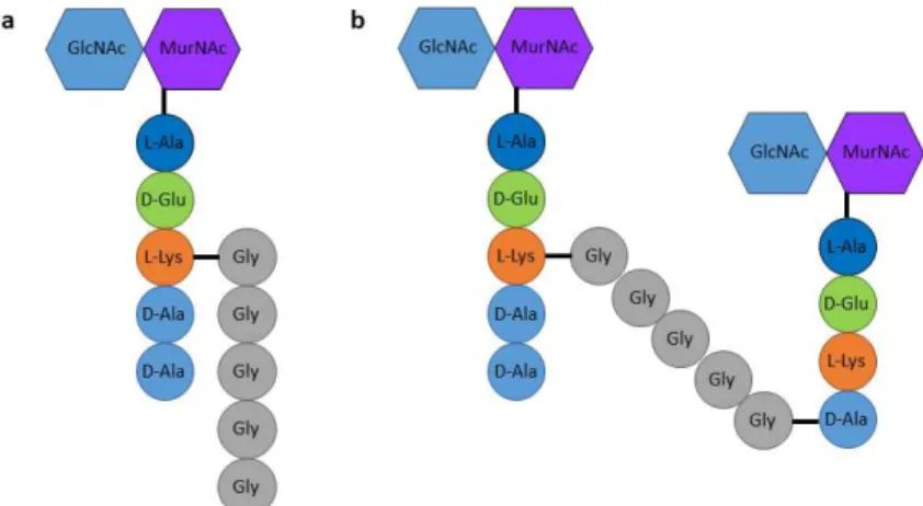
aureus is L-Ala(1)–D-Glu(2)–L-Lys(3)–D-Ala(4)–D-Ala(5) and its
cross-linking occurs via an amino acid bridge composed of five glycines (Fig. 2)10. The disaccharides in S. aureus PGN adopt a 4-fold screw helical
symmetry, resulting in stem peptides with 90o angles to one another,
allowing cross-linking in all directions19. Moreover, long bridge
26 which justifies the high percentage of cross-linking observed in S.
aureus strains in comparison with other gram-positive bacteria such as Bacillus subtilis or Streptococcus pneumoniae19, and can probably
compensate for S. aureus shorter glycan strains.
Figure 2. Building block and structure of S. aureus peptidoglycan. a, S.
aureus PGN basic unit consists on N-acetylglucosamine(GlcNAc)-N-acetylmuramic acid(MurNAc)-pentapeptide with L-Lys on the third position, connected to a pentaglycine cross bridge. b, Cross-linking between PGN glycan strands occurs through a transpeptidation reaction in which the D-Lys(3)
of one stem peptide is connected to the D-Ala(4) of a second stem peptide
through a pentaglycine bridge.
PGN synthesis occurs in three stages that take place in three different cellular locations: the cytoplasm, the cytoplasmic membrane and the extracellular space (Fig. 3). This process culminates with the polymerization of the newly synthesized disaccharide-peptide units into growing PGN chains.
27
Figure 3. Synthesis of PGN in S. aureus. PGN is synthesized in three stages
that occur in three different locations. The first stage is the synthesis of UDP-N-acetylglucosamine (UDP-GlcNAc) and UDP-N-acetylmuramic acid (UDP-MurNAc)-pentapeptide precursors, in the cytoplasm. The second stage comprises the synthesis of lipid I and lipid II precursors at the inner leaflet of the cytoplasmic membrane and the flipping of lipid II across the membrane to the outer leaflet. The final stage is the incorporation of the disaccharide-peptide moiety of lipid II into nascent PGN chains through reactions of transglycosylation and transpeptidation, leading to polymerization and cross-linking of the glycan strains, respectively. Reproduced from 20.
Cytoplasmic steps of peptidoglycan synthesis
In S. aureus, the first stage of PGN synthesis is the synthesis of the uridine diphosphate-GlcNAc (UDP-GlcNAc) and uridine diphosphate-MurNAc (UDP-MurNAc)-pentapeptide precursors in the cytoplasm. Conversion of fructose-6-phosphate into UDP-GlnNAc occurs in four successive steps, catalyzed by the GlmSMU enzymes21–
23. Following these reactions, a two-step process catalyzed by MurA and
MurB leads to UDP-GlnNAc conversion into UDP-MurNAc24. Five amino
28 in UDP-MurNAc-pentapeptide. Amino acids L-Ala, D-Glu, L-Lys and the dipeptide D-Ala-D-Ala are added in a sequential manner by MurC, MurD, MurE and MurF, respectively, elements of the Mur-ligase family which are believed to have a common mechanism of catalysis25–27. For these reactions to occur it is necessary to maintain appropriate levels of D-amino acids in the cell, controlled by the specific racemases MurI and Alr, which convert L-Glu to D-Glu and L-Ala to D-Ala, respectively28,29.
The D-Ala-D-Ala dipeptide is then formed by the Ddl ligase, prior to incorporation by MurF30.
Lipid II synthesis and flipping across the membrane
In the second stage of S. aureus PGN synthesis, UDP-MurNAc-pentapeptide is connected to lipid-carrier undecaprenyl phosphate, or bactoprenol. Bactoprenol is a 55-carbon long-chain isoprene lipid that, because of its lipophilic nature, allows the transport of hydrophilic precursors like sugar intermediates from the cytoplasm to the extracellular space, across the plasma membrane31. The transfer
reaction is catalyzed by MraY in the inner leaflet of the membrane, with the formation of lipid I28,32. The coupling of UDP-GlnNAc to lipid I by
MurG with elimination of UDP and formation of a -1,4 glycosidic bond originates lipid II [GlcNAc- -(1,4)-MurNAc-(pentapeptide)-pyrophosphoryl-undecaprenol]24,33. In S. aureus, lipid II is further altered
by the peptidyltransferases FemX, FemA and FemB, which sequentially add five glycines to the L-Lys amino acid in the third position of the stem peptide34. During this membrane associated stage, D-Glu on the second
position of the stem peptide is amidated by the GatD/MurT enzyme-complex35,36. In S. aureus, this amidation reaction is necessary to
maintain a high degree of PGN cross-linking, and occurs in vitro with all bactoprenol-linked PGN precursors, including lipid I and lipid II with or
29 without the five glycines bridge, suggesting that GatD/MurT complex might have different substrates in vivo35.
The percentage of lipid II in gram-positive bacteria is predicted to be less than 1% of the total amount of phospholipids in the cytoplasmic membrane37. At the end of the second stage, lipid II has to
be flipped from the inner to the outer side of the membrane, where polymerization of new PGN strands occurs. In protein-free artificial membranes, no spontaneous translocation of fluorescently 7-nitro-2,1,3-benzoxadiazol-4-yl (NBD)-labelled lipid II is observed, but it does occur in E. coli membrane vesicles38, indicating that this flipping process
is performed by an enzyme. In the last decade there has been a debate regarding which enzyme was responsible for this step, with the two strongest candidates being FtsW and MurJ. FtsW is a membrane protein with 10 transmembrane domains39 belonging to the SEDS (shape,
elongation, division and sporulation) family40. The activity of E. coli FtsW
as the lipid II flippase was shown using in vitro biochemical studies41,
although it was later observed that this activity was not specific for lipid II42. Contrary to the biochemical evidence, in vivo studies were never
able to show FtsW flipping activity43. Moreover, recent studies in the
gram-positive bacteria B. subtilis, S. aureus and Streptococcus
thermophilus, and in the gram-negative bacteria Pseudomonas aeruginosa, have identified SEDS proteins as a new class of
transglycosylases able to work in complexes with other peptidoglycan synthases, as will be discussed in the next section44–46.
MurJ is a protein mostly embedded in the membrane, with 14 transmembrane domains, and belongs to the MOP (multidrug/oligosaccharidyl-lipid/polysaccharide) exporter superfamily47.
This family of transporters include other putative flippases, homologous to MurJ, capable of translocating undecaprenyl diphosphate-linked oligosaccharides across the cytoplasmic membranes of bacteria48–50. A
30 reductionist bioinformatics study has identified MurJ as the best candidate for lipid II flipping in E. coli51. This prediction has been
corroborated by several in vivo studies in E. coli, B. subtilis and S.
aureus, showing that MurJ is essential for cell viability, for PGN
biogenesis and for the maintenance of cellular shape and integrity51–54. It was also shown that in the absence of MurJ, synthesis of PGN was reduced and lipid-linked PGN precursors were accumulated in the cytoplasmic side of the membrane43,51–53. These results strongly support the idea that MurJ is the flippase of lipid II, although in vitro data in E.
coli failed to support the in vivo observations41. Last year however,
biochemical assays showed that E. coli MurJ has a much higher affinity for lipid II binding than FtsW, forming a complex that is relatively stable55.
A recent study by Liu and collegues showed that in E. coli, FtsW presence and activity was necessary for MurJ to be correctly localized at midcell, suggesting an interdependence between these two proteins and the possibility that they may work together during cell division56.
Because of these conflicting data, and despite the fact that MurJ is currently mostly accepted by the scientific community as the lipid II flippase, some doubts remain and further work is necessary.
Peptidoglycan polymerization and cross-linking steps
Once lipid II is translocated to the outer leaflet of the cytoplasmic membrane, the third and final stage of PGN synthesis starts. Here, the newly synthesized disaccharide-peptide hydrophilic precursors are polymerized and cross-linked into new PGN by transglycosylation (TG) and transpeptidation (TP) reactions, respectively. In the TG reaction, the reducing end of the MurNAc present in the nascent PGN strain is linked to the C-4 carbon of the GlnNAc in the lipid-linked precursor57. This
31 dephosphorylation restores the bactoprenol molecule that can be re-used in another round of lipid II transport. In S. aureus, the TG reaction is performed by the transglycosylase (TGase) domain of PBP258,59, by
the monofunctional transglycosylases MGT and SgtA58,60–62 and by the SEDS protein FtsW, whose TGase activity was recently identified46. In
recent studies in B. subtilis, RodA, another member of the SEDS family, has also been shown to have TG activity44,45.
The TP reaction first cleaves the bond between the two last amino acids of one stem peptide (D-Ala-D-Ala). In S. aureus, the released energy is then used to link the D-Ala on the fourth position to the last glycine of the pentaglycine bridge on a second stem peptide. In bacteria that do not possess a pentaglycine bridge, like E. coli or B.
subtilis, a direct cross-link is performed between the D-Ala on the fourth
position of the first stem peptide and the dibasic amino acid m‐A2pm on the third position of the second stem peptide24. This reaction is
performed by the transpeptidase (TPase) domain of penicillin-binding proteins (PBPs)58.
PBPs are the most well studied family of proteins involved in last stages of PGN synthesis, as they are the target of -lactam antibiotics, such as penicillin. These proteins can be divided into high-molecular-weight (HMW) PBPs and low-molecular-high-molecular-weight (LMW) PBPs. HMW PBPs are attached to the cytoplasmic membrane by an N-terminal transmembrane anchor and have two domains on the outer surface of the cytoplasmic membrane10. The C-terminal domain possesses TPase
activity and is inhibited in the presence of penicillin, due to structural similarities between this -lactam antibiotic and the D-Ala-D-Ala end of the stem pentapeptide precursor, the natural substrate of this domain63.
HMW PBPs can be divided into class A or class B depending on the structure and catalytic activity of their N-terminal domain. In class A HMW PBPs (aPBPs), the N-terminal domain has TGase activity, making
32 these PBPs capable of performing both TG and TP reactions10.
PBP-like TG active sites are inhibited by the antibiotic moenomycin64. Class
B HMW PBPs (bPBPs) only possess TPase activity and their N-terminal domain is believed to establish protein-protein interactions with cell division proteins, contributing for cell morphogenesis58,65. The LMW
PBPs are monofunctional enzymes that can have different activities including DD-carboxypeptidase or transpeptidase, leading to a reduction or increase in the degree of PGN cross-linking, respectively58,66.
S. aureus has only 4 native PBPs, of which PBP2 is the only
aPBP. PBP1 and PBP3 are bPBPs and PBP4 is a LMW PBP with TPase activity. In methicillin-resistant S. aureus (MRSA) strains, a fifth bPBP, PBP2a, from an extra‐species source, is present67,68. PBP1 primary
amino acid structure is highly similar to PBP2b from B. subtilis, PBP2x from S. pneumoniae, and PBP3 from E. coli69, enzymes shown to be
involved in cell division70–72. PBP1 localizes to the division site and is an
essential enzyme for the viability of S. aureus cells. In its absence the population of bacteria becomes morphologically heterogeneous, with larger and more elongated cells showing difficulties in completing septum synthesis, and eventually dying69,73. Inactivation of PBP1 TPase
domain results in problems in cell separation and decreased PGN cross-linking, but does not impair bacterial growth or septal localization, suggesting a second essential function for this enzyme, independent of its TPase activity74. PBP2 is a bifunctional PBP responsible for both TG
and TP reactions. Its TPase domain is essential in methicillin-sensitive
S. aureus (MSSA) strains but not in MRSA strains, due to the presence
of PBP2a75,76. It has been suggested that PBP2 TPase domain is
responsible for the production of the initial cross-linked muropeptides, up to pentamers, with progressively decreasing efficiency77. PBP2
33 substrates in MSSA strains or on a possible interaction with PBP2a in MRSA strains78,79. PBP3 primary amino acid structure is similar to
PBP2a from B. subtilis, PBP2b from S. pneumoniae and PBP2 from E.
coli, proteins involved in cell elongation80–82. Little is known about the role of this protein in S. aureus since it is not essential for cell viability or growth and its absence does not affect PGN muropeptides composition or resistance to -lactam antibiotics83. The only minor alterations
observed in the absence of PBP3 were a small decrease in the autolysis rate and the appearance of cells with abnormal size and shape and disoriented septa in the presence of sublethal concentrations of methicillin83. PBP4 is the only LMW PBP in S. aureus and although it
localizes mainly to the septum it can also be found in the cell periphery84,85. PBP4 is not essential for cells survival but its TPase
activity is responsible for most of the peripheral incorporation of PGN precursors and for the high degree of PGN cross-linking observed in S.
aureus77,85. PBP2a has a TPase domain with very low affinity to -lactam
antibiotics, which allows its function when the TPase domains of the remaining PBPs are inactive86,87. In the presence of -lactams, the
TPase domain of PBP2a and the TGase domain of PBP2 can work together in a possible complex for the synthesis of PGN76.
Besides aPBPs and monofunctional transglycosylases, a new class of TGases has been suggested based on the fact that bacteria lacking these enzymes were able to survive. B. subtilis encodes four aPBPs and does not encode other enzymes with similar TGase domains. However, cells lacking all four aPBPs are viable and are able to synthesize PGN and to maintain a rod morphology88. These cells
have a reduced growth rate when compared to cells lacking only 3 aPBPs and display some morphological defects, like an increased cell length, and division problems, with mislocalization of the division septa88. In the presence of moenomycin, cells from the parental strain
34 show similar alterations but the quadruple mutant cells are not affected88. Analogous observations were performed in Enterococcus
faecalis, in which deletion of all 3 aPBPs resulted in viable cells with
reduced growth rate and PGN cross-linking level, and resistant to moenomycin89. These studies, performed over a decade ago,
suggested the existence of a different class of TGase proteins, not sensitive to moenomycin and able to synthesize PGN in the absence or upon inactivation of all aPBPs.
As referred above, the SEDS family has been recently identified as a new class of TGases, remotely similar to the O-antigen ligase from gram-negative bacteria, which is a membrane TGase that also uses lipid-linked precursors as substrates44. RodA is a member of the SEDS
family involved in cell elongation. In rod-shaped bacteria like E. coli or
B. subtilis, RodA is essential for viability and its depletion results in
defects in the elongation process with the formation of round cells that eventually lyse40,90. Overexpression of RodA can lessen the growth
defect of the B. subtilis strain lacking all four aPBPs, and almost suppress its morphological defects and high levels of lysis44.
Additionally, RodA shows TGase activity in vitro and is responsible for moenomycin resistance in the four aPBPs mutant, confirming that its TGase domain is different from the one of aPBPs which is inhibited by this antibiotic44,45. A similar role was proposed for the SEDS protein
FtsW in the divisome44 and its TGase activity has been recently
identified in vitro for the B. subtilis, S. aureus, S. thermophilus and P.
aeruginosa proteins and shown to be dependent on the interaction with
a cognate bPBP46. SEDS proteins and bPBPs have been shown to
interact and to form complexes involved in cell growth and division91–93. In many bacteria, members of these two families are genetically linked and are often found in the context of the mreBCD operon (cell elongation) or in the cluster of cell wall synthesis and cell division44,94.
35 With the discovery of SEDS TGase activity, the hypothesis that these proteins formed functional TGase-TPase pairs with bPBPs was postulated (Fig. 4)44,45,95.
Figure 4. SEDS proteins and class B PBPs (bPBPs) can work together in a TGase-TPase functional complex. Lipid II precursor is flipped across the
cytoplasmic membrane by a lipid II flippase, probably MurJ, polymerized into nascent PGN strains by a newly identified transglycosylase SEDS protein and cross-linked by its bPBP cognate pair. Adapted from 96.
FtsW was shown to be part of the division machinery together with PBP3 in E. coli, PBP2b in B. subtilis or PBP2x in S. pneumoniae
71,97–99. In E. coli, localization of FtsW is necessary for the recruitment of
PBP3100, while in B. subtilis, FtsW and PBP2b require the presence of
each other for proper localization at the division site, evidencing the interdependency between them101. These localization dependencies
suggested the existence of a pre-formed complex between FtsW and bPBPs involved in cell division. The existence of this complex was later
36 proved by the identification of direct interactions between FtsW and PBP3 in E. coli, both in vivo and in vitro by Förster resonance energy transfer (FRET) and co-immunoprecipitation experiments91. Similarly,
co-immunoprecipitation assays were used to show interactions between FtsW and PBP2x in S. pneumoniae, PBP1 in S. aureus and PBP3 in P.
aeruginosa46,99.
The second SEDS protein, RodA, is part of the elongation machinery in rod-shaped and ovococcoid bacteria102–104. In B. subtilis, proteins from the elongation complex organize in patches that move processively along the lateral cell wall, perpendicularly to the longer cell axis104,105. RodA and PBP2a/PbpH in B. subtilis or PBP2 in E. coli show
a similar movement pattern, moving slowly and directionally around the cell circumference102,104 and contributing to the idea of a functional
connection between SEDS and bPBPs. Additionally, in E. coli cells lacking PBP1b, the main PGN synthesizing enzyme in normal E. coli membranes, high levels of both RodA and PBP2 are necessary to perform TGase and TPase reactions essential to PGN synthesis, with RodA being required for the proper function of PBP2106. Besides the
functional association, a direct physical interaction between RodA and PBP2b was identified in vitro in S. pneumoniae, by two-step pull-down and bacterial two-hybrid assays, confirming the formation of RodA-PBP2b complex103,107.
Although only one PGN synthetic machinery has been identified in S. aureus, this species possesses two SEDS proteins, FtsW and RodA and two bPBPs, PBP1 and PBP3. Previous work by Monteiro and Fernandes et al. has shown that S. aureus cells slightly elongate during specific phases of the cell cycle85. This raised the hypothesis of the
existence of two functional TGase-TPase pairs, involved in PGN incorporation, similar to what was observed in other species, a question addressed in chapter 3 of this thesis.
37
Bacterial Divisome
Most bacteria divide by binary fission, a process in which the mother cell divides in two equal and genetically identical halves. In this process, DNA is replicated and segregated into opposing cell poles followed by synthesis of a division septum at the equatorial plane, that upon constriction and splitting originates two daughter cells. The formation of the division septum involves the cooperation of over 30 proteins, collectively known as the divisome, which are responsible for coordinating chromosome segregation, membrane invagination and PGN synthesis and hydrolysis108. In most bacteria, divisome assembly
depends on FtsZ, a cytoskeletal protein homologue to tubulin109. In
terms of structure the two cytoskeletal proteins are very similar, containing two domains connected by a central helix110,111. FtsZ also
contains a C-terminal tail responsible for its interaction with other divisome proteins112–115. FtsZ polymerizes into protofilaments in a GTP-dependent reaction and assembles into a ring-shaped structure (Z-ring) next to the inner side of the cytoplasmic membrane at the future division site116. This structure functions as a scaffold for the other members of
the divisome that are subsequently recruited. The assembly of Z-ring is very accurate, both in time and space, guaranteeing a precise division of the mother cell and avoiding bisection of bacterial genetic material117,118.
Positioning of FtsZ ring
The location of the new division site is determined by the positioning of FtsZ ring, which can be regulated by both negative and positive mechanisms. The negative mechanisms prevent FtsZ polymerization away from midcell and include the Min system and nucleoid occlusion proteins. The Min system was initially identified in E.
38
coli and B. subtilis and is based on the inhibition of FtsZ polymerization
by the MinCD protein complex. MinC binds and disrupts FtsZ polymers and is recruited and activated by MinD, which binds to the membrane in an ATP dependent manner119. In E. coli, MinCD oscillates between cell
poles resulting in a protein gradient where higher concentrations are present at the poles and lower concentrations at midcell120. On the other
hand, no oscillatory movement is observed in B. subtilis, and MinCD complex is recruited to both cell poles by DivIVA119. DivIVA senses
regions of high membrane curvature, localizing to the cell poles and to the nascent division septum (future cell pole) during membrane invagination119,121. This localization at midcell prevents the formation of
further Z-rings on both sides of the division septum. After cell division, the division septum becomes the new cell pole and DivIVA localization changes from midcell rings into polar patches121, recruiting Min
proteins119. In both Min systems, MinC and MinD are concentrated on
cell poles and the midcell becomes the preferential location for Z-ring assembly.
Nucleoid occlusion proteins bind specific DNA sequences scattered throughout the chromosome, but essentially absent from the terminus region, and inhibit the formation of the Z-ring in the proximity of the nucleoid122–125. This effect is mediated by different proteins like SlmA in E. coli123 or Noc in B. subtilis122,125 and S. aureus124. After
binding to the nucleoid, SlmA affinity for FtsZ filaments increases, leading to filament disruption near the nucleoid126. On the contrary, no
interaction has been identified between Noc and FtsZ in B. subtilis and Noc is believed to physically inhibit the assembly of the division machinery by binding directly to the cytoplasmic membrane and recruiting the DNA sequences to the periphery125. In both E. coli and B.
subtilis, the combination of nucleoid occlusion and the Min system leads
39 only mechanism identified for determining Z-ring positioning124 and is
also involved in the control of DNA replication initiation127. In the
absence of Noc, a small percentage of S. aureus cells form multiple FtsZ rings that are placed over the nucleoid124. The fact that Noc depleted S.
aureus cells are viable, and that E. coli and B. subtilis cells without both
nucleoid occlusion and Min system, despite having several septation problems and aberrant FtsZ structures, can still correctly position FtsZ, suggests that additional regulators are likely to exist128,129.
Positive mechanisms controlling Z-ring positioning were identified more recently and include proteins that localize at midcell and recruit FtsZ to that localization. One example is MapZ in S. pneumoniae, which can interact with newly synthesized PGN on the extracellular space and with FtsZ on the cytoplasm, functioning as a physical anchor and stabilizer of the Z-ring at midcell130. Other examples of positive
regulators are PomZ in Myxococcus xanthus131 or SsgAB complex in
Streptomyces132.
Assembly of FtsZ ring
In E. coli and B. subtilis, divisome assembly can be temporally divided in two stages and was proposed to follow a two-step assembly model. The first step consists on the formation of the Z-ring at midcell, and the second on the recruitment of downstream division proteins98,133.
In E. coli, formation of the Z-ring occurs without visible cell constriction and coincides with preseptal elongation134,135, a phase when cells
elongate at midcell in an FtsZ-dependent manner136. Although FtsZ
does not possess any membrane-binding domain, FtsZ filaments attach to the inner side of the cellular membrane via FtsA and ZipA137,138. These
are two essential proteins that localize to midcell through interaction with FtsZ138–140 and anchor it to the membrane. Although each of these
40 proteins alone is able to support the formation of Z-rings, both of them are necessary for the recruitment of later division proteins137. Moreover,
the equilibrium between FtsZ, FtsA and ZipA is of extreme importance for proper cell division, since alteration of the proportions of these proteins, mainly FtsZ and FtsA, can lead to problems in Z-ring formation and septation141. In S. aureus, FtsA can also associate with FtsZ and
this interaction enhances FtsZ GTPase activity113,142. While FtsA is
widely conserved among bacteria, ZipA can only be found in -proteobacteria143. An alternative membrane anchor for FtsZ, found in
gram-positive bacteria is SepF, originally named YlmF. In B. subtilis cells, FtsA depleted cells are viable but display severe cell division problems. Overexpression of SepF can compensate for the lack of FtsA in ftsA-null mutants and the two genes are synthetic lethal144.
An important regulator of FtsZ polymerization is EzrA, an FtsZ inhibitor which controls the frequency and location of Z-rings. Depletion of EzrA leads to abnormal FtsZ polymerization away from midcell in rod-shaped cells like B. subtilis110 or its mislocalization in S. aureus with
occasional formation of multiple complete FtsZ rings145,146. EzrA
localizes to the division site in an FtsZ-dependent manner110,146,
interacts with several division proteins and its absence leads to a mislocalization of PBPs in S. aureus145,146. EzrA structure is similar to
that of proteins of the eukaryotic spectrin family147. Spectrins act as a
scaffold connecting actin filaments to the membrane and coordinate membrane proteins interactions148. However, if EzrA can perform a
similar function in bacteria is still an open question.
Recruitment of late division proteins
The second step of divisome assembly occurs after FtsZ ring assembly, with a time delay corresponding to 20% of the cell cycle in B.
41
subtilis98 or 15% to 34% in E. coli133. This step is characterized by the
recruitment of a variety of proteins, called “late” division proteins, including SEDS protein FtsW, PGN synthases like E. coli PBP3 (FtsI) or
B. subtilis PBP2b, DNA-binding proteins FtsK and SpoIIIE, and the
structural and regulatory proteins FtsN and complexes FtsB/FtsL/FtsQ (FtsBLQ) in E. coli or equivalent DivIC/FtsL/DivIB in B. subtilis143,149.
DivIC, FtsL and DivIB are bitopic membrane proteins with one transmembrane segment and a C-terminal extracellular region. The three proteins interact to form a ternary complex, stabilizing each other against proteolysis, and localize to the division septum in an interdependent manner150–152. In E. coli, the equivalent FtsBLQ complex has been identified by immunoprecipitation experiments and was shown to preassemble prior to septal localization153. Localization of
DivIC/FtsL/DivIB and PBP2b are interdependent in B. subtilis72 and a
strong interaction between DivIB and PBP2b has been identified154.
More recently, FtsW localization was also shown to be necessary for the localization of FtsL and PBP2b, and to require these two proteins for its own localization, evidencing an interdependent interaction101. These
observations evidence a cooperative assembly of late division proteins in B. subtilis, since DivIB, FtsL, DivIC, PBP2b and FtsW assemble interdependently and the absence of any of these proteins prevents the localization of all the others101,150,155.
Contrary to what is observed in B. subtilis, the localization of the different division proteins in E. coli seems to occur in a linear sequential manner, as the presence of each protein is necessary for the recruitment of the following one156. However, the fact that FtsQ, FtsL and FtsB form
a complex before they localize to the Z-ring153, that late proteins can
back-recruit upstream proteins93,157 and that some essential division
proteins can be bypassed by overexpression or mutations of other divisome members108 point to a non-sequential assembly of the
42 divisome in E. coli and demand a careful interpretation of the hierarchical assembly pathway model. Also, both early and late division proteins contribute to the regulation of divisome assembly, since the cooperative assembly of divisome proteins was suggested to depend on interactions between the early protein FtsA and the late protein FtsN158,159. In these studies, a model was proposed in which at low FtsN
concentrations both FtsA and FtsBLQ subcomplex are in an “off” conformation, suppressing PGN synthesis. Once FtsN concentration increases, it changes FtsA into an “on” conformation, which will in turn activate FtsBLQ subcomplex that may stimulate PGN synthesis158,159.
Despite the important role of FtsN in E. coli, this protein is poorly conserved in bacteria160 and no homologues have been found in
gram-positive bacteria, implying a different regulatory mechanism to initiate PGN synthesis. One possible regulatory mechanism, proposed for B.
subtilis, is through FtsL, due to its high instability and rapid protein
degradation upon repression of gene transcription. FtsL depletion prevents DivIB and DivIC septal localization, leading to the degradation of DivIC protein and to a rapid arrest of cell division150,161.
In S. aureus, it was recently shown that divisome assembly occurs in a progressive way, with FtsZ being recruited earlier, followed by DivIB, FtsW and PBP1, approximately at the same time, and later by MurJ, the putative lipid II flippase20. MurJ localization to midcell, but not
FtsW or PBP1 localization, is dependent on the presence of the complex DivIB-DivIC-FtsL20. S. aureus DivIB is capable of binding PGN through
its extracellular domain and is essential for cell growth, since its depletion inhibits the completion, but not initiation, of septum synthesis162.
FtsW has been suggested to play a role in Z-ring stabilization in
E. coli, since its absence leads to a reduction in septation frequency163,
43 formation of long filamentous cells. In B. subtilis, the assembly of Z-ring is dependent on the presence of FtsW101,164 and in Streptomyces
coelicolor, disruption of ftsW or ftsI (bPBP) genes inhibits Z-ring
formation in aerial hyphae and prevents reorganization of spiral polymers of FtsZ into rings165. This indicates an early role for SEDS
proteins and their cognate bPBPs in cell division. Also, a triple interaction between FtsW, PBP3 and FtsZ was identified in
Mycobacterium tuberculosis, where FtsW silencing prevents PBP3
localization and cell septation, contributing to the idea that these proteins are involved in the regulation of septal PGN biogenesis166,167.
Coordination of different peptidoglycan synthesis
machineries
A wide number of proteins involved in the processes of PGN synthesis and cell division organize in functional machineries that allow cells to grow and divide, while maintaining their characteristic morphology. The number, identity and function of these machineries varies according to the shape of the bacterial cells and the mechanisms that control these multi-protein complexes are not fully understood.
The Rod shape
The majority of rod-shaped bacteria combine PGN synthesis at the septum with elongation of the lateral sidewall, along the longitudinal cell axis (Fig. 5, top panel). Septal PGN synthesis is responsible for generating the new cell poles of the daughter cells during cytokinesis, whereas lateral PGN insertion allows the establishment and propagation of the rod shape. While septal growth is dependent on the activity of the
44 divisome machinery directed by FtsZ, lateral wall elongation is mainly oriented by MreB.
Figure 5. PGN incorporation during growth and cell division in different bacteria. Rod-shaped bacteria (top panel) divide by septal PGN synthesis
performed by the divisome complex at midcell. Elongation results from insertion of new PGN along the lateral wall by the MreB-dependent elongasome and from FtsZ-dependent pre-septal PGN incorporation at midcell. Ovococcoid cells (middle panel) combine septal and peripheral PGN incorporation, performed by the divisome and elongasome, respectively. Both machineries are probably dependent on and coordinated by FtsZ. Coccoid cells (bottom panel) divide by PGN incorporation at midcell by the divisome complex. Slight cell growth and elongation occurs before and after septum synthesis.